Lichen (mycosis) is an infectious disease of the human skin. Appears as a result of contact of microspores of various types of fungi on the skin. It is a mistake to think that lichen planus is an exclusively childhood disease. Adults are no less susceptible to it than children.
The main factors that influence the appearance of lichen are visiting crowded places with high humidity, for example, swimming pools, saunas, baths, gyms. Domestic and street animals are also often carriers of infections. Often, pathologies of the thyroid gland, diabetes mellitus, and hormonal imbalances in the body can contribute to infection of a person’s skin with lichen.
Symptoms of lichen
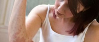
What symptoms indicate that you have contracted lichen planus? It should be noted that lichen affects not only the skin of the body, but also mucous membranes, nails and even human hair. As a rule, any type of this disease begins with the appearance of the following symptoms:
- minimal increase in body temperature;
- the appearance of chills, mild headache, and sometimes nausea;
- later the temperature rises, and itching appears in certain places (face and body, less often - limbs);
- after a few hours, bubbles will appear at the site of redness, which soon burst and only a crust remains.
Pityriasis versicolor
Pityriasis versicolor (syn.: versicolor, pityriasis versicolor, pityriasis furfuracea) is a fungal skin disease characterized by damage to the stratum corneum of the epidermis. The causative agent of this disease is the fungus Pityrosporum orbiculare or P. ovale. However, the question of whether both of these forms represent one organism at different stages of its development or are separate species has not been fully resolved. It is currently accepted that both micromorphological varieties P. orbiculare and P. ovale represent different stages in the life cycle of the fungus. Moreover, its oval shape - P. ovale - is more often found on the skin of the scalp, and its round shape - P. orbiculare - on the skin of the body [25]. The correct taxonomic identification of the lipophilic yeasts causing this disease is still a matter of debate. Some scientists prefer the name Pityrosporum orbiculare, while others prefer Malassezia furfur. Thanks to molecular technologies, 10 species of the genus Malassezia have now been identified. Ogunbiyi AO and George AO (2005) identified the most common Malassezia species: M. furfur, M. symboidalis, M. obtusa, M. globosa, M. restricta, M. slooffie and M. pachydermatis [12, 15, 17].
As a result of their analysis, it was established that the cause of pityriasis versicolor in humans is most often M. globosa. Hort W. et al. (2006), having examined 112 patients diagnosed with seborrheic dermatitis, atopic dermatitis, lichen versicolor and AIDS, found that these patients had various types of Malassezia fungi. According to the authors, M. globosa was the most pathogenic species and was more often recorded in HIV-positive patients and patients with lichen versicolor. In the group of patients with seborrheic dermatitis, along with M. globosa, M. sympodialis was isolated.
The issue of the contagiousness of the fungus has been discussed in the scientific literature for quite a long time. Previously, it was believed that transmission of infection occurs either through direct contact with a sick person or through contaminated underwear. This observation was based on the high prevalence of this disease among individuals in the same family. However, it has now been proven that lichen versicolor is not contagious, and a genetic predisposition to the development of keratomycosis has been established: known cases of familial disease are explained by similar skin type in family members who are consanguineous [6, 25, 28].
The causative agent of lichen versicolor is isolated from 10–15% of the population, and 2 times more often in men. The disease mainly develops between the ages of 15 and 40 years. This mycosis is characterized by deterioration in the summer, cases of spontaneous recovery are possible. In some patients, the disease becomes chronic and prone to relapse. However, in most cases, P. orbiculare (ovale) exhibits its pathogenic properties only in adolescence [1, 2, 6, 9]. P. orbiculare (ovale) is a lipophilic fungus, so the intensity of skin colonization is related to the function of the sebaceous glands. In particular, in children under 5 years of age the fungus is not detected at all, while in 15-year-olds it is detected in 93% of cases. Further, with age, the percentage of detection of P. orbiculare decreases, which once again confirms the assumption of a relationship between the presence of the fungus and the functional activity of the sebaceous glands [6, 26]. The primary location of keratomycosis is the mouth of the pilosebaceous follicles; here the fungus multiplies, forming colonies in the form of yellowish-brown dots. Fungi concentrate around the sebaceous glands, using their secretions as a source of fatty acids necessary for their growth and development. Increased air humidity also contributes to the pathogenicity of the fungus, as evidenced by the high prevalence of lichen versicolor among the population of tropical and subtropical countries. Thus, the incidence rate in temperate climates is 2%, in tropical and subtropical climates - up to 40% of cases [6, 16, 19, 24, 26].
Being a yeast-like fungus, P. orbiculare has many of the qualities inherent in this group of fungi. In particular, the disease develops when a saprophytic form is transformed into a pathogenic one under special, favorable circumstances. The development of the disease is promoted by: increased sweating, seborrhea, decreased physiological peeling of the skin, decreased nutrition, and pathology of internal organs. Lichen versicolor is a unique marker of diabetes mellitus, tuberculosis, rheumatism, and AIDS. In these diseases it is found in 52–63% of patients. If pityriasis versicolor develops against the background of tuberculosis, lymphogranulomatosis and other diseases accompanied by sweating, there are no age restrictions and clinical signs of this dermatosis can be observed at any age [6]. The literature describes a case of lichen versicolor diagnosed in a three-month-old child with leukemia [23].
In recent decades, hematogenous infections caused by M. furfur have been observed; they have been described in weakened and immunosuppressed patients, especially after organ transplantation, as well as in neonatal children receiving lipids through a central venous catheter [1, 11, 13, 24].
The presence of cross-reacting antigens in P. orbiculare with fungi of the genus Candida provokes the development of allergic reactions of immediate, immunocomplex and delayed types. Studies conducted by various authors have proven that one of the main risk factors for the formation of complicated forms of atopic dermatitis at an early age in children is the predominance of fungi of the genus Malassezia. The addition of an associated fungal infection changes the clinical picture of atopic dermatitis, which is characterized by a more severe course, widespread process and resistance to traditional therapy [10, 16, 27]. According to the observations of Mayser P. et al. (2000) in patients with P. orbiculare colonization on the scalp and neck, specific IgE antibodies were more often recorded than in patients with localization on the skin of the trunk. In addition, the author found that patients with Malassezia more often complained of diffuse hair thinning.
There is evidence to support the role of P. orbiculare in the development of seborrheic dermatitis. As a result of their vital activity, these fungi break down sebum triglycerides into free fatty acids, and these, in turn, are the direct cause of dermatitis on the surface of the skin, but this assumption still requires proof [8]. In particular, an experimental model of seborrheic dermatitis was obtained by rubbing a P. orbiculare culture into the skin of subjects. Subsequently, regression of experimental dermatitis was noted under the influence of various antimycotic drugs, which confirmed the etiological role of P. orbiculare in seborrheic dermatitis. Moreover, if normally the microflora of the scalp contains 46% of this fungus, then with dandruff it consists of 74% of them, and with seborrheic dermatitis the number of fungi reaches 83% [6, 28].
It must be remembered that seborrheic dermatitis associated with P. orbiculare (ovale) may be the single earliest manifestation of AIDS. According to foreign authors, from 30% to 80% of patients with HIV infection have seborrheic dermatitis, compared to healthy young people in whom this dermatosis was diagnosed only in 3–5% of cases. AIDS-associated seborrheic dermatitis is characterized by resistance to therapy and papular rashes resembling psoriasis [1, 8, 9, 13].
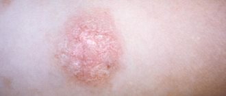
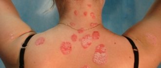
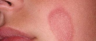
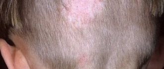
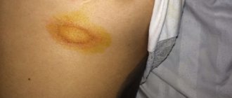
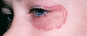
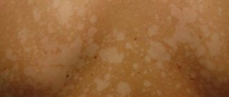
Clinical picture of the disease. The onset of the disease is characterized by the appearance of yellowish dots confined to the mouth of the hair follicles. The primary morphological element is a pink-yellow spot, gradually changing to brown-yellow, on the surface of which there are pityriasis scales. The elements are characterized by peripheral growth and, subsequently, fusion into larger lesions with scalloped edges. With a long course of mycosis, the lesions can occupy large areas of the skin. Over time, the color of the lesions can vary from white to dark brown, this served as the basis for the second name for lichen - multi-colored. The surface of the rash is covered with pityriasis-like scales, which are hardly noticeable upon superficial examination, but when scratched, peeling easily occurs (Beignet's symptom). Since the favorite localization is confined to the “seborrheic zones,” the upper half of the body and the scalp are affected. Additional diagnostic criteria, well known to doctors, are the Balzer iodine test and a golden-yellow or brownish glow under a Wood’s lamp, as well as detection of the pathogen during microscopic and cultural studies [1, 2, 5, 6, 9].
A feature of the clinical picture of pityriasis versicolor is the presence of pseudoleukoderma. Scientists have divided opinions about the causes of uneven skin coloring in this dermatosis. Some researchers believe that P. orbiculare (ovale) inhibits tyrosinase activity during the oxidation process, which leads to a decrease in melanin synthesis and is clinically manifested by the occurrence of true leukoderma [6]. According to other authors, the loosened stratum corneum on the lesions prevents the penetration of ultraviolet rays into the depths of the epidermis. Therefore, after removal of scales during water procedures, the affected lesions become lighter than the surrounding healthy skin [2, 9].
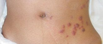
As a rule, diagnosis is not difficult, but there are several points that the practitioner needs to pay attention to. Firstly, in individuals who received treatment and were irradiated with ultraviolet rays, the Balzer test will be negative. Secondly, identifying affected areas on the scalp is of practical importance: if the doctor does not pay attention to this localization of keratomycosis, then there is a high probability of relapse. To diagnose lesions on the scalp, a Wood's lamp is used (the lesions have a greenish-yellow, yellowish-brown or brownish glow). Thirdly, pinkish-brown spots are barely noticeable on white skin, so they are often ignored when examined, but in the summer, if you have a tan, they become more noticeable. And the last nuance: in people without immune deficiency, isolated spots do not exceed, as is known, 1–1.5 cm in diameter, but with severe immunodeficiency they can reach 5 cm in diameter. In this case, not only a disseminated character is noted, but also rich pigmentation and infiltration of elements of multi-colored lichen. In patients with AIDS, lichen versicolor more often manifests itself as seborrheic dermatitis, less often as atopic dermatitis and multiple folliculitis in areas of the skin with symptoms of vasculitis and a necrotic component.
The atypical course of lichen versicolor was described by various authors [11, 14]. The rarest manifestations of lichen versicolor include lesions on the skin of the soles. In the domestic literature, such localization is reported by V. M. Rukovishnikova (1999), who in her monograph refers to the observations of V. P. Zhirkova (1977) of a 16-year-old boy with hyperhidrosis, who, along with rashes of multi-colored lichen in typical places (chest , back, neck, face) there were foci of mycosis on the soles. A pronounced torpidity of the lesions of this unusual localization was noted. Even after four months of treatment, non-inflammatory brownish spots of irregular shape and outline remained on the heels and in the transitional fold from the toes to the sole.
Differential diagnosis of pityriasis versicolor is carried out with syphilis (with syphilitic roseola and leucoderma). The diagnosis of syphilis is confirmed by positive results of classical serological tests (CSR), Treponema pallidum immobilization test (TPI), and immunofluorescence test (RIF). In addition, roseola in secondary syphilis has a pinkish-livid tint, disappears with diascopy, does not peel off, does not fluoresce in the light of a fluorescent lamp, and the Balzer test is negative.
In syphilitic leukoderma there are no confluent hyperpigmented spots and micropolycyclic edges. Syphilitic roseola is characterized by a predominant localization on the trunk and upper extremities, which determines some similarity in the clinical picture, however, with syphilitic lesions there is no tendency to growth and fusion of elements. The roseola spot is of vascular origin, there is no peeling, it is not accompanied by subjective sensations, the Balzer test is negative.
Lichen versicolor should be distinguished from pink lichen of Zhiber, in which erythematous spots are acutely inflammatory, round or oval in shape, with a peculiar peeling in, there is a “maternal” plaque. The rashes are located symmetrically along Langer's lines. The edges of the central, flaky part of the plaque are surrounded by a collar of scales. Numerous secondary rashes are visible around the maternal plaque.
Pityrosporum folliculitis (Malassezia folliculitis) is an infection of the hair follicle caused by yeasts, the same ones that cause pityriasis versicolor. This disease is a separately located, sometimes itchy papulosquamous rash, localized mainly on the upper half of the body and shoulders. Pityrosporum folliculitis most often affects young and middle-aged people and women. Follicular occlusion appears initially, which is secondarily accompanied by increased growth of the fungus. Predisposing factors are diabetes mellitus, as well as taking broad-spectrum antibiotics or corticosteroids. The condition can appear on the forehead and mimic persistent acne. The clinical picture consists of asymptomatic or slightly pruritic dome-shaped follicular papules and pustules with a diameter of 2–4 mm. This dermatosis is more common in the tropics, where it manifests itself as follicular papules, pustules, nodules and cysts. A distinctive feature of Pityrosporum folliculitis is the absence of comedones, torpidity to therapy and localization in the forehead. According to Thomas P. Habiff (2006), very often patients with Pityrosporum folliculitis are mistaken for acne patients. According to the author, this disease should be suspected in young and middle-aged patients with follicular lesions located on the trunk and complaints of itching.
Treatment. Since this disease develops when the saprophytic form of the fungus is transformed into a pathogenic one under special, favorable circumstances, it is necessary, first of all, to identify the provoking factors. Lichen versicolor is a marker of diabetes mellitus, tuberculosis, rheumatism, and AIDS. Therefore, when examining a patient, it is necessary to conduct appropriate studies. Particular attention should be paid to persons who do not fall into the age category from 15 to 45 years. Often the development of a persistent clinical picture of pityriasis versicolor is caused by chemotherapy in cancer patients. As a rule, after completing the course of intensive treatment, such patients undergo spontaneous self-healing.
As mentioned above, pityriasis versicolor is characterized by damage to the surface layer of the epidermis - the stratum corneum. Therefore, treatment of keratomycosis should begin with external means. Medicines for the treatment of pityriasis versicolor can be divided into several groups:
- keratolytic agents;
- fungicidal preparations;
- products containing zinc pyrithioneate;
- combined means.
Therapy for lichen versicolor depends on the prevalence and location of the lesions. Previously, keratolytic agents were used in the treatment of this mycosis: 2–5% salicylic alcohol or an alcohol solution of resorcinol 2 times a day. Modern methods of therapy include fungicidal drugs from the azole group. It has been established that under the influence of antimycotic drugs, after 24 hours, dehydration and vacuolization of the cytoplasm in the fungal cell occurs, the cell wall clears, from which after 48 hours only a shadow remains. Therefore, specific antifungal treatment is often preferred over keratolytic drugs.
Considering the superficiality of skin lesions with keratomycosis, it is preferable to use fungicidal agents in the form of solutions (clotrimazole, bifonazole, ciclopirox, naftifine (Exoderil), terbinafine (Lamisil)) or econazole in powder form, sold under the trade name "Ifenek", which is applied to the affected areas of the skin and rub lightly. A more convenient form of using the drug is a spray (Lamisil, Thermikon). All antimycotic solutions for the treatment of pityriasis versicolor are prescribed 2 times a day for 1 week. Ketoconazole has higher activity against P. ovale, inhibiting its growth in concentrations 25–30 times lower than other antifungal drugs and several times lower than any systemic antimycotics. When topical forms of ketoconazole are applied to the skin, effective concentrations remain inside and on the surface of the epidermis for 72 hours after discontinuation of the drug, which is explained by the affinity of the drug for keratinized tissues.
Zinc pyrithione also has a direct antifungal effect. Today, the mechanism of therapeutic effects of zinc pyrithione is associated not only with cytostatic, but also with antifungal and antimicrobial effects. The effectiveness of drugs containing zinc pyrithione against yeast-like fungi has been studied by many authors [1, 3, 4]. These drugs include Psorilom and Skin-cap, produced in two forms: spray and cream. In the future, these drugs can be used by patients as prophylactic agents at least once every 2 weeks. While inferior to ketoconazole, zinc pyrithione has superior antifungal activity to other drugs, including selenium sulfide and some imidazoles. In the last decade, zinc-based shampoos (Head and shoulders, Friederm zinc) have been widely used in the treatment of dandruff. Thus, the most effective drugs in the treatment of pityriasis versicolor are antifungal agents and zinc pyrithione.
When the scalp is affected by fungus, medicated shampoos containing fungicidal agents (Nizoral, Sebazol, Ducre Quelual DS, Ketoconazole) are used, which are prescribed daily with an exposure of 2–5 minutes for 7–10 days. Treatment with shampoos containing tar (Psoril) is effective in the presence of seborrheic dermatitis to eliminate such manifestations as infiltration, swelling, peeling, and erythema.
Combined preparations include shampoos: Node DS plus, which contains salicylic acid, climbazole, zinc pyrithione, and Keto plus based on ketoconazole and zinc pyrithione.
Systemic treatment is indicated for patients with advanced disease who do not respond to topical therapy or who experience frequent relapses. Intraconazole is prescribed at a dose of 200 mg 2 times a day for one day or 200 mg every day for 5 days. The drug is taken with food to improve absorption. Ketoconazole is taken in a dose of 400 mg once or 200 mg daily for 5 days at breakfast with fruit juice. Fluconazole is prescribed at a dose of 150 mg (2 capsules per week for 4 weeks or 2 capsules as an initial dose, repeated after 2 weeks). If the process resolves slowly, the course of systemic antimycotics can be repeated after 2 weeks. The patient is not recommended to take a bath for 12 hours after treatment, since abstaining from water procedures allows the medicine to accumulate in the skin. Some authors recommend changing clothes daily for one month to prevent relapses. Patients must accept that residual hypopigmentation, as a consequence of pseudoleukoderma, lasts for quite a long time.
In the treatment of Pityrosporum folliculitis, one should adhere to the same principles as in the treatment of pityriasis versicolor, but it is preferable to combine systemic ketoconazole (200 mg daily for 4 weeks) with external antifungal agents.
Literature
- Skin Diseases: Diagnosis and Treatment / Thomas P. Habiff; lane from English; Under general ed. acad. RAMS, Prof. A. A. Kubanova. M.: MEDpress-inform, 2006. 672 p.
- Skin and sexually transmitted diseases: Handbook / Ed. O. L. Ivanova. M.: Medicine, 1997. 352 p.
- Mokronosova M.A., Pyzh V.V., Kashaeva O.V., Reznikov O.V. Therapeutic effect of activated zinc pyrithione in patients with atopic dermatitis / eczema syndrome with sensitization to yeast-like fungi // Russian Journal of Allergology. 2004, no. 3. pp. 83–87.
- Moshkalova I. A., Mikheev G. N., Sokolovsky E. V. et al. Blistering dermatoses. Psoriasis. Modern methods of treatment. St. Petersburg: Sotis, 1999. 133 p.
- Novikov A.I., Loginova E.A. Skin diseases of infectious and parasitic origin. M.: Medical book, N. Novgorod: Publishing house of the NGMA, 2001. 283 p.
- Potekaev N. N., Novikov A. G. Multi-colored lichen. A modern look at an old problem // Russian Journal of Skin and Venereal Diseases. 2004, no. 2. pp. 42–45.
- Rukovishnikova V. M. Mycoses of the feet. M.: MSD, 1999. 317 p.
- Sukolin G.I. Seborrheic dermatitis: new in etiology and treatment // Russian Medical Journal. 1998; 6:382–384.
- Khabib O. N. Mycoses of smooth skin // Consilium Medicum, 2002, volume 2, no. 4.
- Khaertdinova L. A. Medical and social aspects of atopic dermatitis in children complicated by secondary infection. Author's abstract. dis. ...cand. honey. Sci. 2006. 22 p.
- Aljabre SH Intertriginous lesions in pityriasis versicolor // J Eur Acad Dermatol Venereol 2003? 17(b): 659–662.
- Crespo-Erchiga V., Florencio VD Malassezia yeasts and pityriasis versicolor // Curr Opion Infect Dis. 2006; 19 (2): 139–147.
- Christian Schnake S., Hector Gutierrez B., Marcos Saez G., Mario Becker C. Tinea versicolor. Pitiriasis versicolor en lactantes menores Rev // Chil. Pediatr. 1988, 59 (1); 50–52.
- Darling M. J, Lambiase M. C, Young RJ Tinea versicolor mimicking pityriasis rubra pilaris // Gutis. 2005; 75(5):265–267.
- Gemmer CM, DeAngelis YM, Theelen B., Boekhout T., Dawson Jr. TL Differentiation of three biotypes of Malassezia species on human normal skin. correspondence with M. globosa, M. sympodialis and M. restricta // Mycopathologia. 1999; 145(2):69–74.
- Hort W., Nilles M., Mayser P., Edward M., DeSimone R. Ph. Common Superficial Fungal Infections // US Pharmacist. 1999, 24 (4).
- Mayser P., Gross A. IgE antibodies to Malassezia furfur, M. sympodialis and Pityrosporum orbiculare in patients with atopic dermatitis, seborrheic eczema or pityriasis versicolor, and identification of respective allergens // Acta Derm Venereol. 2000; 80(5):357–361.
- Naseri M., Namazi MR Fast, noninvasive method for molecular detection and differentiation of Malassezia yeast species on human skin and application of the method to dandruff microbiology // J Clin Microbiol. 2002; 40(9):3350–3357.
- Nematian J., Ravaghi M., Gholamrezanezhad A., Nematian E. Isolated scalp involvement with pityriasis versicolor alba (pityrias versicolor albus capitis) in a patient from a dry, temperate region // Dermatol Online J. 2003; 9 (3): 17.
- Nenoff P., Haustein UF Effect of anti-seborrhea substances against Pityrosporum ovale in vitro // Hautarzt, 1994, v. 45 (7), p. 464–467.
- Ogunbiyi AO, George AO Pityriasis versicolor: Current concepts in Aetiology and Management // Niger Postgrad Med J. 2005; 12 (3): 183–188.
- Parry ME, Sharpe GR Seborrhoeic dermatitis is not caused by an altered immune response to Malassezia yeast // Br. J. Dermatol. 1998; 139:254–263.
- Schoepfer C., Carla H., Bezou MJ // Arch. Pediatr. 1995. Vol. 2, No. 3. P. 245–248.
- Silverberg NB, Sidbury R., Mancini AJ Childhood molluscum contagiosum: experience with cantharidin therapy in 300 patients // J Am Acad Dermatol. 2000; 43:503–507.
- Shuster S. The aetology of dandruff and the mode of action of therapeutic agents // Br. J. Dermatol. 1984; 111:235–242.
- Tarazooie B., Kordbacheh P., Zaini F., Zomorodian K., Saadat F., Zeraati H., Hallaji Z., Rezaie S. Study of the distribution of Malassezia species in patients with pityriasis versicolor and healthy individuals in Tehran, Iran // BMC Dermatol. 2004; 4:5.
- Thoma W., Kramer HJ, Mayser P. Pityriasis versicolor alba // J Eur Acad Dermatol Venereol. 2005; 19 (2): 147–152.
- Wikler JR, Nieboer C., Willemze R. Quantitative skin cultures of Pityrosporum yeasts in patients seropositive for the human immunodeficiency virus with and without seborrhoeic dermatitis // J. Am. Acad. Dermatol. 1992; 27:37–39.
Yu. A. Gallyamova, Doctor of Medical Sciences, Associate Professor
GOU DPO RMAPO, Moscow
Contact information for authors for correspondence
Treatment of lichen in adults
The diagnosis of lichen planus is made by a dermatologist; only in some cases additional tests are necessary. If the diagnosis is made incorrectly, then treatment can bring a negative result, which can lead to the transition of the disease to the chronic stage.
Treatment of lichen planus should be comprehensive. Only a doctor can select a remedy that will help to cope with the disease as much as possible, taking into account the stage of infection and its extent within certain areas of the body.