What is ringworm?
Ringworm is an infectious skin disease caused by the parasitic fungi Microsporum canis and Trichophyton tonsurans, it is part of the group of ringworms that primarily affect the skin, hair and nails. This is an infectious dermatological pathology caused by dermatophyte fungi. It is also called dermatomycosis, scab, micro- and dermatophytosis.
Doctors distinguish several types of ringworm, depending on the location, nature of the course and the causative agent.
The main types of the disease are:
- chronic or ringworm of smooth skin;
- surface;
- ringworm of the nail plate;
- infiltrative-suppurative.
Since ringworm is caused by two types of fungi, in medicine it is diagnosed as microsporia and trichophytosis. These fungi have persistent pathogenicity, pronounced virulence and often cause acute diseases with varied clinical manifestations.
A disease caused by fungi of the genus Microsporum canis is microsporia. Depending on the location, there is microsporia of smooth skin and microsporia of the scalp.
Trichophytosis is caused by the activity of the dermatophyte Trichophyton tonsurans, which, through the anthroponotic route of infection, causes inflammation of the scalp and smooth skin, and through the zootroponotic route, inflammation in the deep layers of the dermis, accompanied by suppuration.
The presence of fungal spores does not always provoke damage to the skin and its appendages. The transition of the fungus from an inactive state to a pathogenic state is facilitated by metabolic and endocrine disorders, immune diseases, hypovitaminosis or vegetative-vascular dystonia. This increase in the pathogenicity of dermatophytes provokes an exacerbation of inflammatory processes, the spread of the fungus through the blood and lymph flow.
The disease is weakly expressed during the development of a latent infection and when localized in previously affected areas, and the high virulence of the fungus is formed during a relapse, gradually decreasing during the course of the infection.
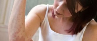
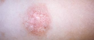
Symptoms of ringworm: At the end of the incubation period, itchy ring-shaped spots of red-pink color appear on the skin, on the surface of which bubbles appear, after which a crust and peeling remain. The hair at the site of the lesion breaks and falls out.
Normally, the immune system is able to cope with the pathogen and prevent its activity, but there are a number of factors that increase the risk of ringworm, namely:
- skin microtraumas;
- lack of vitamins and microelements;
- prolonged contact with the source of infection;
- high humidity and air temperature;
- exacerbation of chronic diseases, etc.
In a situation where there is an adult or child with lichen at home, it is necessary to take preventive measures to reduce the likelihood of its spread, namely:
- use separate towels and bed linen;
- wash your hands thoroughly with soap, especially if you touched the patient’s things;
- use gloves when touching areas of disease;
- undergo examination and take tests to identify pathogens, etc.
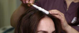
The main method for diagnosing ringworm is the use of a Wood's lamp, under the light of which the affected areas acquire a characteristic greenish glow. Microscopic examination and culture of skin and hair scales are effective to confirm the diagnosis.
Timely diagnosis and a properly selected treatment regimen can effectively eliminate ringworm in children and adults, preventing its spread. The pathology is prone to relapse, since the presence of spores of viable fungi can cause repeated damage. It is extremely important to complete the course of treatment and follow all doctor’s recommendations.
Microsporia
Potekaev N.N.
Central Research Institute of Dermatovenerology, Ministry of Health of the Russian Federation, Moscow
Microsporia is a fungal disease from the group of dermatophytosis, which affects the skin and hair, and in extremely rare cases, the nail plates . The name of this mycosis comes from the name of its causative agent - a fungus of the genus Microsporum, which belongs to the dermatophytes. The disease is also known as “ringworm” (the term combines microsporia and trichophytosis), which is due to the peculiarities of its clinical picture.
Etiology
The causative agent of microsporia was first described by Gruby in 1843. The scientist discovered a sheath of small spores on the surface of the affected hair and gave the fungus the name Microsporum audouinii in honor of the late Dr. Audouin. However, the author’s discovery was not appreciated, and highly respected dermatologists (in particular, Bazin) identified microsporia with trichophytosis. Sabouraud managed to restore the truth in 1893, who, having carefully studied the biology of the causative agent microsporia , indicated the signs that distinguish this mycosis from trichophytosis. In Russia, microsporia was first described by S.L. Bogrov in 1912.
Currently, more than twenty species of the Microsporum fungus are known. Of these, the following are identified as pathogens:
- M. distorum, M. rivalieri, M. langeronii.
- Zoophilic group —
- M. canis, M. nanum, M. persicolor.
- Geophilic group - M. gypseum, M. cookeii, Keratynomyces ajelloii.
Of the listed species, only M.canis (seu lanosum) has become practically the only pathogen of microsporia in recent years. It is no coincidence that it is called a cosmopolitan mushroom.
Once on the skin, the pathogen penetrates it and begins to multiply. When located near the mouths of hair follicles, fungal spores germinate, leading to hair damage. Quite quickly spreading over the surface of the latter, the mycelial hyphae destroy the cuticle, between the scales of which spores accumulate. Thus, the fungus surrounds the hair, forming a sheath, and tightly fills the follicular apparatus.
Epidemiology
Microsporia is the most common mycotic infection among dermatophytoses, not counting mycoses of the feet. The disease occurs everywhere. In Russia, up to 100 thousand patients with microsporia are registered annually. Mycosis is highly contagious, children are more often affected. In the last two decades, there has been an increase in the incidence of microsporia in newborns [1]. Adults rarely get sick—mostly young women. The rarity of the disease with microsporia in adults, especially with damage to the scalp, and the usually occurring spontaneous recovery at the beginning of puberty is explained by the presence of fungistatic organic acids (in particular, undicylenic acid) in the hair of adults. Patients with lesions of the scalp pose a particular danger in epidemiological terms. This is due to the fact that this form of mycosis, firstly, is most often diagnosed untimely, and, secondly, its therapy is associated with certain difficulties. Unfortunately, data from recent epidemiological studies conducted in Russia indicate an increase in the number of patients with hair damage [2].
As already indicated, the most common pathogen of microsporia is Microsporum canis , a zoophilic fungus that is found in 90-97% of patients. The main source of the disease is cats (usually kittens), less often dogs. Infection occurs through direct contact with a sick animal or objects contaminated with fur or scales. Once in the soil with an affected hair or scale, M.canis remains viable only for 1-3 months. Thus, soil is only a factor in the transmission of infection and does not serve as its natural reservoir [3]. Intrafamily spread of infection is common. In this case, infection usually occurs from one animal. Transmission of zoonotic microsporia from sick family members is possible, but this is quite rare. There are isolated observations of families in which three generations were sick with this mycosis. It should be emphasized that in such situations, women and children of younger age groups, including newborns, are at greatest risk of infection.
Clinical manifestations in animals are characterized by areas of baldness on the face, the outer surfaces of the ears, as well as on the front, less often the hind, legs. Under Wood's lamp a green glow is detected. Often, clinically healthy cats can be mycocarriers, and then only a luminescent study helps to identify the fungus. However, situations are possible when the fact of carriage cannot be confirmed either by clinical or luminescent examination. In such cases, and they are observed in 2-3% of carriers, wool is sown from various areas [4].
The incidence of zoonotic microsporia varies throughout the year. Seasonal fluctuations are associated with litters in cats, as well as more frequent contact of children with animals in the summer. The rise in incidence begins at the end of summer, the peak occurs in October-November, and the decrease to a minimum occurs in March-April. The emergence of epizootics of microsporia in cats and kittens in a number of regions and cities leads to the formation of epidemic foci among children.
Clinic
Since the main causative agent of microsporia in our time is Microsporum canis, when describing the clinical picture of the disease, more attention will be paid to the zoonotic form rather than the anthroponotic one.
The incubation period for zoonotic microsporia is 5-7 days . The nature of the clinical picture of the disease is determined by the localization of the lesions and the depth of penetration of the pathogen. There are microsporia of smooth skin and microsporia of the scalp.
Microsporia of smooth skin
, a swollen, raised erythematous spot with clear boundaries appears . Gradually the spot increases in diameter and infiltrates. A continuous raised ridge is formed along the periphery, represented by small nodules, bubbles and crusts. In the central part, the inflammatory phenomena resolve, as a result of which it acquires a pale pink color, with pityriasis-like peeling on the surface. Thus, the focus has the appearance of a ring. As a result of autoinoculation of the fungus in the central part and repeated development of the inflammatory process, bizarre foci of the “ring within a ring” type are formed. Such iris-like figures are more common with anthroponotic microsporia. Vellus hair is often involved in the process, which complicates the treatment of the disease. The number of foci with microsporia of smooth skin is usually small (1-3). Their diameter ranges from 0.5 to 3 cm. The rash can be located on both open and closed areas of the skin, since a sick animal is often warmed under clothes and taken to bed. However, the most common lesions are located on the skin of the face, neck, forearms and shoulders. There are no subjective sensations or moderate itching.
In persons with a reduced delayed-type hypersensitivity reaction, an abortive course of microsporia is possible. In this case, the lesion has the appearance of a pale pink scaly spot without clear boundaries, along the periphery of which there are no nodular and vesicular elements. In 1957, J. Esteves first described parasitic achromia , a rare variant of microsporia characterized by minimal symptoms [5]. With parasitic achromia, along with damage to the scalp, there are numerous depigmented spots of round shape, with slight peeling on the surface.
In newborns and young children, as well as in young women, due to a hyperergic reaction, an erythematous-edematous form of microsporia is often observed, in which pronounced inflammatory phenomena and minimal peeling are noted.
The papular-squamous form occurs when microsporia is localized in seborrheic areas of the skin - on the face, chest and back. The lesions are characterized by infiltration and lichenification, accompanied by significant peeling and itching. Since this form of microsporia is usually observed in individuals with signs of atopy (in particular, in patients with atopic dermatitis), mycosis is often masked by manifestations of the underlying process and is not always diagnosed in a timely manner. The use of local corticosteroid drugs only increases the spread of mycotic infection.
In young women with hypertrichosis, follicular-nodular elements with a diameter of 2-3 cm may appear in the lower leg area - the so-called deep form of microsporia of smooth skin.
Localization of single foci of microsporia in places atypical for it can sometimes lead to difficulties in diagnosing the disease. T.I. Meerzon, in particular, described an isolated focus of zoonotic microsporia on the skin of the shaft of the penis in an 18-year-old patient [6].
A rare type of microsporia includes damage to the skin of the palms, soles and nail plates . On the palms, and less often on the soles, dyshidrotic and/or squamous-keratotic rashes are observed. Microsporic onychomycosis is characterized by isolated lesions of the nail, usually its proximal part [7]. Initially, a dull spot is formed, which becomes white over time. The nail in the area of leukonychia becomes softer and more fragile, and subsequently can collapse as onycholysis. When examining the affected nail under a Wood's lamp, a bright green glow is detected. Microsporic onychomycosis not diagnosed in time can cause reinfection and further spread of the disease among others.
Microsporia of the scalp
Damage to the scalp occurs mainly in children 5-12 years old . Over the past 20 years, there has been a 20-fold increase in the incidence of this form of microsporia in newborns [1]. It is generally accepted that the rarity of this form in adults is explained by the presence of fungistatic organic acids in their hair and water-lipid mantle of the skin. This fact indirectly confirms the spontaneous recovery of children during puberty, when the composition of sebum changes. Perhaps the difference in hair thickness between children and adults matters. It is noteworthy that microsporia of the scalp practically does not occur in children with red hair [8].
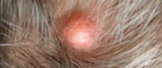
Foci of microsporia of the scalp are located mainly on the crown, in the parietal and temporal regions (Fig. 2). Usually there are 1-2 large lesions ranging from 2 to 5 cm in size, with round or oval outlines and clear boundaries. Along the periphery of large lesions there may be screenings - small lesions with a diameter of 0.5-1.5 cm. At the beginning of the disease, a peeling area forms at the site of infection. In the first days, the fungus is localized only at the mouth of the hair follicle. Upon closer inspection, you will notice a whitish ring-shaped scale surrounding the hair like a cuff. On the 6-7th day, the process spreads to the hair itself, which becomes fragile, breaks off above the level of the surrounding skin by 4-6 mm and looks as if it has been trimmed (hence the name “ringworm”). The remaining stumps look dull and are covered with a grayish-white sheath, which is the spores of a fungus. If you “stroke” the stumps, they deviate in one direction and, unlike intact hair, do not restore their original position. The skin in the lesion, as a rule, is slightly hyperemic, edematous and moderately infiltrated, its surface is covered with grayish-white small scales.
With microsporia of the scalp caused by anthropophilic fungi , numerous small foci with minimal inflammation and unclear boundaries are observed. A characteristic feature of anthropophilic microsporia is its localization in the marginal zone of hair growth, when one part of the lesion is located on the scalp, and the other on smooth skin.
Atypical, rare variants of microsporia of the scalp include infiltrative, suppurative (deep), exudative, trichophytoid and seborrheic forms.
In the infiltrative form of microsporia, the lesion on the scalp rises somewhat above the surrounding skin, is hyperemic, and the hair is often broken off at a level of 3-4 mm. It should be especially emphasized that with this type of microsporia, the sheath of fungal spores at the root of broken hair is weakly expressed.
In the suppurative form, against the background of significant inflammation and infiltration, soft bluish-red nodes form, the surface of which is covered with pustules. When pressed, pus is released through the follicular openings. Such clinical manifestations correspond to the picture of kerion Celsi (Celsius honeycomb) - infiltrative-suppurative trichophytosis. The formation of infiltrative and suppurative forms of microsporia is facilitated by irrational (usually local) therapy, the presence of serious concomitant diseases, and late consultation with a doctor.
Exudative microsporia of the scalp is characterized by severe hyperemia and swelling, with small bubbles located against this background. Due to the constant impregnation of the scales with serous exudate and gluing them together, dense crusts are formed, which, when removed, exposes the moist, eroded surface of the lesion.
The listed three forms of microsporia of the scalp are often complicated by regional lymphadenitis, and patients with suppurative microsporia may also experience symptoms of intoxication.
With the trichophytoid form of microsporia, numerous small foci with weak pityriasis-like peeling are scattered on the scalp. The boundaries of the lesions are unclear, there are no acute inflammatory phenomena, the hair is broken off at a level of 1-2 mm above the surrounding skin. Along with broken hair, there are healthy hairs. Trichophytoid microsporia is more common in older age groups with serious concomitant diseases.
With seborrheic microsporia of the scalp, thinning of the hair is mainly observed. The areas of rarefaction are abundantly covered with yellowish scales, upon removal of which a small amount of broken hair can be found.
Late diagnosis and inadequate treatment of atypical forms of microsporia lead to further changes in clinical symptoms, dissemination of rashes and chronicity of the process, irreversible alopecia in the patient and dissemination of infection in the environment.
Diagnostics
To confirm the clinical diagnosis of microsporia, fluorescent, microscopic and cultural studies are used.
Luminescent study
The method is based on identifying a bright green glow in hair affected by fungi of the genus Microsporum when examined under a Wood's lamp. At the same time, both long and vellus hair glows. The reason for this phenomenon has not yet been established. Luminescence testing must be carried out in a darkened room. The lesions are first cleaned of crusts, ointments, etc. When examining fresh lesions, there may be no glow, which is due to insufficient hair damage. In such situations, the hair should be epilated from the suspected site of fungal penetration, and the glow can be detected in its root part. When the fungus dies, the glow in the hair remains.
The luminescent method is used for:
- pathogen identification;
- identifying affected hair;
- evaluation of therapy results;
- control over persons in contact with the patient;
- determination of infection or mycocarriage in animals.
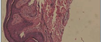
Microscopic examination
To confirm the fungal etiology of the disease, scales from lesions of smooth skin lesions are subjected to microscopic examination, and if the scalp is involved in the process, hair fragments are subjected to microscopic examination. Immediately before collecting pathological material, the lesion on smooth skin must be treated with 960 alcohol. Then, using a blunt scalpel, carefully scrape off the scales from the periphery of the lesion. On the scalp, using tweezers, hair fragments are also removed from the marginal zones of the lesion. Then the pathological material is placed on a glass slide in a drop of 20% potassium hydroxide solution. Microscopic examination is carried out after 30-40 minutes.
In scales from lesions on smooth skin, twisted threads of septate mycelium are found. Microscopic examination of the affected hair reveals many small spores (2-3 microns) on its surface (ectothrix-type lesion). In this regard, the boundaries of the hair appear blurred. The spores surrounding the hair are arranged chaotically, like a mosaic.
Cultural examination
Carrying out cultural diagnostics with positive results of luminescent and microscopic studies is required to identify the causative fungus. The method allows you to determine the genus and type of pathogen and, therefore, carry out adequate therapy and prevention of the disease. Pathological material (scales, hair) is placed on Sabouraud's medium. The growth of Microsporum canis colonies (the main pathogen of microsporia) is observed on the 3rd day after sowing. By the 10th day, the colony reaches a diameter of 4-5 cm and is represented by a flat disk covered with a whitish, delicate fluff, which spreads like rays along the walls of the test tube. The reverse side of the colony is yellow in color.
Treatment
When treating microsporia of smooth skin without affecting vellus hair, external antimycotic drugs are used. Apply 2-5% iodine tincture to the foci of mycosis in the morning, and apply antifungal ointment in the evening. Use traditional 10-20% sulfur, 10% sulfur-3% salicylic or 10% sulfur-tar ointments. Modern ointments are used twice a day: clotrimazole, ciclopirox, isoconazole, bifonazole, etc. The allylamine drug terbinafine (Lamisil) , produced in the form of 1% cream and spray, has proven itself well.
Terbinafine has a fungicidal effect (i.e., it leads to the death of the fungus) and is the most active antimycotic agent against dermatophyte fungi. The drug inhibits the functions of svalene epoxidase, resulting in disruption of the synthesis of ergosterol, the main component of the fungal cell membrane. At the same time, the amount of squalene, a high molecular weight hydrocarbon, increases inside the cell. These disturbances lead to the death of the fungal cell. The sensitivity of svalene epoxidase in fungi is 10,000 times higher than in humans, which explains the selectivity and specificity of the action of terbinafine in relation to the fungal cell. The drug can be used once a day. It should be emphasized that, having a keratophilic ability, Lamisil accumulates in the stratum corneum of the epidermis and is present here for a long time in fungicidal concentrations. This circumstance explains the persistence of a pronounced antifungal effect even after discontinuation of the drug. The convenient dosage form of terbinafine spray ensures contactless application of the drug to large areas of affected skin. Terbinafine cream and spray are quickly absorbed and do not leave marks on clothing.
In case of a pronounced inflammatory reaction, it is advisable to prescribe combination drugs containing additional corticosteroid hormones. Similar products include mycozolon and travocort .
When a secondary bacterial infection occurs, Triderm cream is useful . With severe infiltration of the lesion, as well as with deep forms of microsporia, preparations containing dimexide , which is known to have conductive properties, are indicated. In particular, in such situations, a 10% solution of quinosol is widely used (quinosole and salicylic acid 10.0 each, dimexide 72.0, distilled water 8.0). The solution should be applied 2 times a day until clinical manifestations resolve and the fungi disappear.
When vellus hair, and especially long hair, is affected, systemic antimycotic therapy for microsporia is necessary.
In the treatment of microsporia of the scalp, griseofulvin , a chlorine-containing antibiotic produced by the mold Penicillium nigricans, remains the drug of choice. Griseofulvin, available in the form of 125 mg tablets, is prescribed at the rate of 22 mg per 1 kg of patient body weight. The drug is taken daily in 3-4 doses during meals with a teaspoon of vegetable oil, which is necessary to increase the solubility of griseofulvin and increase the duration of its action (a-tocopherol contained in oils delays the metabolism of griseofulvin in the liver). For children under 3 years of age, it is preferable to prescribe griseofulvin in the form of a suspension, 8.3 ml of which corresponds to 1 tablet (125 mg) of the drug. Continuous therapy is carried out until the first negative test result for fungi, after which griseofulvin is taken at the same dose every other day for 2 weeks, and then for another 2 weeks, 2 times a week. The general course of treatment is 1.5-2 months. During therapy, it is necessary to shave your hair weekly and wash your hair 2 times a week . It is recommended to simultaneously rub any antifungal ointment into the affected area. In parallel with oral administration of the antimycotic, manual hair removal can be performed with preliminary application of a 5% griseofulvin patch to the lesion.
Side effects of griseofulvin include headache, allergic rashes, and discomfort in the epigastrium; Granulocytopenia and leukopenia are less common. Unfortunately, due to hepatotoxicity, griseofulvin is contraindicated in children who have had hepatitis or suffer from liver disease. The drug is also not prescribed for kidney diseases, gastric and duodenal ulcers, neuritis, blood diseases, and photodermatoses.
In recent years, terbinafine (Lamisil) . Local forms of the drug have already been mentioned earlier. In the treatment of microsporia of the scalp, terbinafine is used in the form of tablets, available in doses of 125 and 250 mg. The drug has a high safety profile, which is largely due to the peculiarities of its mechanism of action. Squalene epoxidase, which is inhibited by terbinafine, is not associated with the cytochrome P-450 system, so the drug does not affect the metabolism of hormones and other drugs. Since terbinofine is lipophilic, after oral administration it quickly reaches the dermal layer of the skin, overcomes it and accumulates in the lipids of the stratum corneum of the epidermis, hair follicles and hair.
When treating microsporia of the scalp in children, the dose of terbinafine is determined depending on body weight. The manufacturer recommends prescribing the drug for a child weighing less than 20 kg at a dose of 62.5 mg per day; children weighing from 20 to 40 kg - 125 mg; more than 40 kg - 250 mg. However, our experience shows that these doses are often insufficient, since we obtained the maximum therapeutic effect by changing the officially recommended treatment regimens [9]. In this regard, the doses of terbinafine we offer are 50% higher than those recommended by the manufacturer: 94 mg/day (3/4 tablets of 125 mg) for children weighing 10-20 kg and 187 mg/day (1.5 tablets in 125 mg) - 20-40 kg. For body weight over 40 kg, terbinafine is prescribed at a dose of 250 mg/day. For adults, terbinafine is prescribed at a dose of 7 mg per 1 kg, but not more than 500 mg per day.
Terbinafine is taken once a day. The drug is well tolerated. Patients may be bothered by a feeling of fullness in the stomach, minor abdominal pain. Following a diet aimed at relieving flatulence relieves patients from unpleasant sensations.
Prevention
Prevention of microsporia consists of timely identification, isolation and treatment of patients with microsporia. In children's institutions, periodic medical examinations should be carried out. A child diagnosed with microsporia must be isolated from other children and sent for treatment to a specialized mycological hospital. For each sick person, a notification is filled out according to registration form 281. Things belonging to a patient with microsporia are subject to disinfection. Relatives and people in contact with the patient must be examined. Particular attention should be paid to pets, since they are often the source of infection. Animals with microsporia are either destroyed or given full antifungal treatment. An important role in the fight against microsporia is assigned to health education authorities, as well as veterinary supervision of stray animals.
Literature
1. Mohammad Yusuf. Clinical and epidemiological features of microsporia in modern conditions and the development of treatment with new medications. Author's abstract. diss...candidate of sciences. M., 1996
2. Fakhretdinova Kh.S. Clinical and epidemiological features of modern microsporia. Author's abstract. diss... doc. med. sc. M., 1999.
3. Sheklakov N.D., Andriasyan S.G. Some ecological features of Microsporum canis and the incidence of zooanthroponotic microsporia. Vestn dermatol. 1979; 2: 18-23.
4. Stepanova Zh.V., Davydov V.I. On the carriage of fluffy microsporum by clinically healthy animals. Vestn dermatol. 1970; 3:42-6.
5. Esteves J. Acromia parasitaria devida ao M. Felineum. Trab. Soc. Derm. Vener. 1957; 15:43.
6. Meerson T.I. Atypical localization of smooth skin microsporia caused by Microsporum canis. Vestn dermatol. 1985; 5:70.
7. Stepanova Zh.V., Klimova I.Ya., Shapovalova F.S. Onychomycosis caused by fluffy microsporum. Vestn dermatol. 1997; 4:37-9.
8. Feyer E., Olah D., Szatmari S. et al. Medical mycology and fungal diseases. Budapest. 1966.
9. Potekaev N.S., Kurdina M.I., Potekaev N.N. Lamisil for microsporia. Vestn. Dermatol. 1997; 5: 69-71.
How is ringworm transmitted?
Ringworm is transmitted in two ways:
- from a sick animal – zoonotic with an incubation period of one week;
- from an infected person – anthroponotic with an incubation period of up to 6 weeks.
Children are at risk because they most often come into contact with the carriers of the pathogens of this disease - cats and dogs. Children's skin is characterized by low density of the stratum corneum and weak protective properties of the water-lipid mantle. The stratum corneum prevents the penetration of the fungus into the layers of the skin, since its spores are located between the horny scales, and the water-lipid mantle forms a barrier to the penetration of pathogens.
The infection is also transmitted by non-compliance with personal hygiene standards and the use of other people's personal belongings containing fungal spores (hats, combs, bed linen, etc.).
Routes of infection
Infection occurs through contact and household contact from contaminated objects or living beings:
- tactilely from a sick child or adult;
- contact from communication with domestic animals (dogs, cats, rodents, cattle);
- in contact with affected grass, hay, soil;
- if hygiene rules are not followed, using the same items: towels, combs, scissors, bed linen.
The incubation period lasts about a week. This is the time from infection by the spores to the onset of symptoms. In some cases, this requires from 2 to 7 weeks.
Based on the routes of infection by the fungus, ringworm is divided into 3 types:
- anthropophilic – occurs after contact with a sick person or household objects;
- zoophilic – develops against the background of infection from animals;
- Haemophilus influenzae - the cause is contaminated soil.
How does ringworm begin?
Favorable conditions contribute to the introduction and reproduction of a pathogenic fungus in the skin, triggering the infectious process. In the initial stages, this period is asymptomatic. After which the development of the pathology acquires clinical manifestations, the signs of which depend on the stage of the pathology.
The stages of the infectious process during the development of ringworm are:
- incubation period;
- period of mushroom growth;
- refractory stage (period of insensitivity, rest);
- regression.
In the skin, the fungus forms branched mycelium, which gradually invades new areas of the skin, while old lesions become the location of its spores. This period of fungal growth is determined by the rapid increase in the colony of the pathogen, active division of skin cells and the high rate of exfoliation of the affected epidermis.
Since most often the growth of the colony outstrips the rate of change of the stratum corneum, the infectious process spreads. An intense immune reaction (inflammation) in the lesion is displayed in the form of a red ring, which, in the chronic course of mycosis, becomes the permanent residence of the pathogen.
What does ringworm look like?
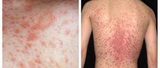
Clinical manifestations of ringworm depend on the type of pathogen, course and stage of the disease. Dermatophytes secrete enzymes and toxins that destroy keratin protein (they feed on it), so the severity of symptoms directly depends on the ability of fungi to produce these substances.
Common symptoms of ringworm on the scalp are:
- hair thinning;
- the appearance of localized peeling of the skin;
- redness of the epidermis;
- hair breaking off at the root;
- the appearance of small bubbles with cloudy contents along the edges of bald patches, after opening which yellow crusts form.
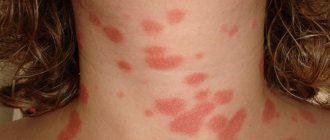
Ringworm of smooth skin includes the following symptoms:
- pronounced itching;
- the appearance of round red spots;
- light epidermis at the site of damage, surrounded by gray scales;
- the edges of the damaged area consist of pink-red bubbles;
- growth of the lesion in diameter.
The chronic form of the disease is accompanied by the presence of microscopic blisters on the affected areas, the formation of scars after their opening and severe itching. In a situation where the site of the lesion is the nail plate, its separation, discoloration and thickening are observed.
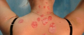
Ringworm of the infiltrative-suppurative form is characterized by the appearance of large bright red spots up to 10 cm in diameter, the outer surface of which is lumpy and uneven, and purulent follicles form along the edges. Pain, swelling and hyperemia of the affected areas are also observed.
The appearance of the first signs of the disease requires a mandatory visit to a dermatologist. The lack of qualified treatment can lead to the rapid spread of infection throughout the body and cause a number of complications: tissue scarring, death of hair follicles, allergic reactions and the addition of a secondary infection.
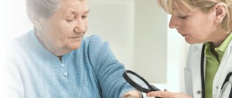
How to recognize lichen in a child?
Parents are advised to pay attention to:
- any rash and peeling;
- redness of the skin that does not go away for a long time;
- complaints of itching;
- pain in the area of the rash;
- pain when eating hot food (if the rash is in the mouth);
- swollen lymph nodes in areas near the rash;
- temperature increase;
- uneven tan: affected areas remain whitish;
- change in the nature of hair (appearance of scales, fragility, loss, etc.).
It is difficult to make a diagnosis on your own. If you suspect lichen, it is better to contact a pediatric dermatologist [1].
How is ringworm treated?
In each case, treatment for ringworm should be selected individually, since its composition and duration have their own specifics and are determined by the type, location and stage of the pathology.
Only prescriptions from a dermatologist are effective; they take into account the current clinical picture, the characteristics of the patient’s physical health and his age.
Therapy is aimed not only at relieving the symptoms of the pathology, but also at inactivating the pathogen. The chronic form of the disease also provides support for the immune system, normalization of metabolism and hormonal levels.
The following groups of drugs are used to treat ringworm:
- systemic and local antimycotics;
- antiseptic solutions;
- multivitamin complexes.
How to cure lichen in a child?
Since the word “lichen” refers to a fairly large group of skin diseases, there is no clear answer to this question.
In any case, treatment should be recommended by a specialist after examination and diagnosis.
- If the cause of the appearance of lichen is known, then the treatment is etiotropic - that is, it affects the pathogen. Thus, for fungal and viral lichens, antifungal and antiviral agents are recommended.
- In cases where the mechanism of formation of a pathological reaction is known, pathogenetic treatment is prescribed: anti-inflammatory drugs, hormonal drugs, antiallergic drugs, etc.
- To alleviate the condition and reduce symptoms, symptomatic therapy is prescribed: painkillers, itching agents, moisturizing ointments, etc. [1,2]
How long does it take to treat ringworm?
The treatment process for ringworm is lengthy, usually taking at least six weeks, with systemic therapy lasting mainly 15-25 days.
The effectiveness of therapy is assessed using laboratory tests. In a situation where tests indicate a significant decrease in the number of fungal spores after 2 weeks of a medication course, it is necessary to continue the chosen treatment. In some cases, fungal shedding can last for several months.
After the signs of pathology disappear, you need to scrape the epidermis three times in previously damaged areas:
- immediately after completion of therapy;
- 7 days after therapy;
- after 2-3 months.
The result of therapy is considered successful if all three tests show negative results.
Prevention measures
To prevent your child from contracting ringworm, follow these guidelines:
- do not allow contact with strangers and street animals;
- Have your pets vaccinated against infections from a veterinarian;
- avoid sunburn and injury;
- ask your child to wash their hands more often;
- moisten the floor in the apartment using antiseptic disinfectants;
- Provide your child with a well-balanced diet, which will strengthen the immune system.
If the child has already suffered an infection, you will also have to adhere to certain rules, since the spores remain viable in the environment for 24 months. To prevent re-infection you should:
- Disinfect furniture, carpets, mattresses with antiseptics. Use steam generators. Treat bed linen and clothes (boil, iron with a hot iron on both sides).
- For 2-3 weeks after recovery, wash your hair and body with antifungal shampoos.
- Ventilate rooms more often.
- Use separate comb, towel and other household items.
- Postpone visiting the pool and open water bodies for a while.
Ringworm in children spreads quickly, so it is important to consult a doctor in a timely manner, and after diagnosis, adhere to the prescribed treatment regimen and follow all instructions.
How to treat ringworm in children?
Treatment of ringworm in a child is the responsibility of a pediatric dermatologist, since the characteristics of the child’s body require special doses and composition of medications.
Hygiene and child care are also important elements of pathology therapy:
- boiling and ironing clothes in contact with affected areas of the skin;
- treating household surfaces with disinfectants to prevent the spread of spores;
- frequent change of bed linen;
- washing in the shower rather than the bath, etc.
Diagnostics
Ringworm can masquerade as other skin pathologies: dermatoses, other types of lichen, allergies. Therefore, if even one sign of infection is detected, you need to go to the hospital for examination. After visiting the pediatrician, the child is referred to a dermatologist or infectious disease specialist.
Initially, the doctor examines the patient using a black lamp. Features of this diagnostic procedure:
- The lights in the office turn off.
- The affected area is illuminated with a device (Wood's lamp) that emits long-range ultraviolet waves.
- If a green tint appears, it means trichophytes are present. If the color is blue, we are talking about microsporums. A white glow indicates lichen planus.
There is a risk of obtaining a false positive result. This is influenced by the presence of tetracycline and other similar substances on the skin, if the skin was lubricated with ointments before visiting the doctor.
Laboratory research must be carried out, including the following activities:
- Microscopy of biomaterial . The specialist takes a scraping of scales from the affected area of skin and/or hair and sends the samples to the laboratory. The degree of spore activity is studied using a microscope.
- Bacteriological or cultural culture . After collecting the biological material, the spores are placed in a nutrient medium for 10 days for cultivation. The purpose of the examination is to determine the type of pathogen.
If a child is infected with ringworm, the rest of the family members and everyone who came into contact with him are examined. Examinations are carried out in the clinic every day for 3 days, after which you need to visit the doctor another 3-5 times within 3 weeks.
Can I go to work or school with ringworm?
The presence of this diagnosis requires isolation of the patient from the team for at least two weeks. In schools and other children's institutions, it is mandatory to notify parents for timely detection of the disease in other children.
The disappearance of ringworm symptoms and 3 negative tests for the presence of fungus are a reason to return to school or work.
If a dermatomycosis infection has been detected in a team, then to prevent pathology, you can use antifungal shampoos for some time, carry out wet cleaning with the addition of antiseptic solutions, and strictly observe personal hygiene.
- Treatment of lichen in Kharkov;
- treatment of lichen in Uzhgorod;
- treatment of deprivation in Sumy;
- treatment of lichen in Poltava;
- treatment of lichen in Odessa;
- treatment of deprivation in Nikolaev;
- treatment of lichen in Mariupol;
- treatment of deprivation in the Dnieper.
3
1
1
Article rating:
3.8 out of 5 based on 5 ratings
Author: Mangusheva Victoria Yurievna
Dermatovenerologist, trichologist. Candidate of Medical Sciences, doctor of the highest category. Work experience more than 10 years.
Hygiene measures when in contact with a sick person
If you or your child have touched a sick animal or interacted with a sick person, then you need to take immediate action.
- The sooner you wash away particles containing fungus from your skin, the less likely you are to become infected.
- Wash your hands several times with antifungal soap. The simplest remedy, which is available in almost every store, is cinnamon laundry soap, or better yet, soap with birch tar.
- Wash your entire body with this soap. Suddenly, particles of the patient’s skin got under his clothes. Do not use a hard washcloth. It leaves micro-scratches on the skin, into which fungus easily penetrates.
- To wash your hair, you must use an antifungal shampoo. For example, Nizoral. You can also use it as a shower gel.
- A modern remedy with a powerful antifungal effect is Citeal. Dilute it in a small container five times. You will end up with a foaming liquid that can be used to wash your hands and entire body.
- Lavender oil, tea tree oil and turpentine have an antifungal effect. They can be used to treat small areas of skin.
Also, five days after contact, it is advisable to consult a dermatologist. He examines the body with a Wood's lamp. If you do become infected, the disease can be detected in the early stages. This will help you quickly treat her at home and avoid going to the hospital.