Red spots and small rashes on the head of the penis can be the main symptoms of many diseases. Often such rashes are the only thing that bothers a man.
The most common cause is balanitis (inflammation of the head of the penis). Less commonly - dermatological diseases, allergic reactions, oncology.
Table 1 - Main causes of symptoms
| Infectious | |
| Bacterial infection (nonspecific balanoposthitis) | Streptococci, staphylococci, Gardnerella vaginalis |
| Protozoal infection | Trichomoniasis |
| Viruses | Papillomaviruses, genital herpes |
| Fungi | Candidiasis |
| Non-infectious | |
| Benign dermatoses | Plasmacytic balanitis Circinar balanitis Lichen planus Psoriasis |
| Precancerous conditions | Bowenoid papulosis Erythroplasia Queyrat |
| Penis cancer | |
| Taking a number of medications (salicylates, warfarin, etc.) | |
| Mechanical injury | |
| Allergic balanitis | |
Candidiasis
Infection of a man with Candida fungi usually occurs during unprotected sexual intercourse with a woman suffering from thrush. The disease develops over several days and is accompanied by redness of the head of the penis, the appearance of small red dots or, conversely, red spots with a whitish coating, itching (optional), dryness and flaking.
A man may be bothered by swelling of the head, difficulty and pain when retracting the foreskin, discomfort during sex and pain when urinating. Candidiasis balanoposthitis is well treated with local antifungal drugs (ointments and creams based on clotrimazole, miconazole).
In addition, daily genital hygiene, changing underwear, and abstaining from sex during the treatment period are recommended. If the infection persists and relapses frequently, it is necessary to exclude diabetes mellitus (glucose profile, glucose tolerance test, determination of glycated hemoglobin level), immunodeficiencies and STIs.
When to go to the doctor
By taking a shower or changing synthetic underwear to clothes made from natural fabrics, you can get rid of non-infectious stains in a few days. If the measures taken do not help, the spots do not disappear, and additional symptoms appear, then contacting a doctor becomes the only right decision. Remember that infections can spread to internal organs, so do not hesitate to visit the clinic. If you feel slightly unwell, you can go to a urologist. The specialist will diagnose and prescribe competent treatment, and if necessary, refer you to doctors of another profile - a venereologist, allergist or dermatologist.
Genital herpes
This is a viral infection transmitted from an infected person during unprotected sex and is most common among young men 14-49 years old. Infection often occurs from a virus carrier who has no symptoms. The first signs may appear several weeks after infection and include fever, general weakness, and decreased vitality.
First, red spots appear on the head, on which, after a few hours, vesicular rashes filled with clear liquid appear. At the moment of their appearance, a man may feel pain and burning, they often itch.
The blisters burst and turn into superficial erosions (similar to herpes on the lips). They become crusty and gradually heal. Therapy includes the prescription of antiviral drugs: acyclovir, valacyclovir or famciclovir.
Papillomas and condylomas
Papillomas and genital warts are benign neoplasms that occur when infected with papillomavirus. It is transmitted during sex (oral, vaginal, anal). The maximum incidence is recorded at the age of 15-25 years among sexually active persons.
The first signs of the disease appear several weeks or months after infection. Papillomas are pink, dome-shaped protrusions of skin. The growths may be flesh-colored, red, or brown. Genital warts with their growths resemble cauliflower.
Formations can occur on the shaft of the penis, foreskin, scrotum, perineal skin and around the anus. In most cases, their appearance is not accompanied by any symptoms; less often, a man may be bothered by itching and discomfort.
Treatment is complex and includes removal of tumors using local drugs or surgical microsurgery and systemic immunotherapy (Panavir, Interferon-alpha).
Rice. 1 – Multiple genital warts of the glans penis
results
Detailed clinical examination results were available for 38 of the 41 study participants. In 17 cases, the genital lesions were irregular in shape and color. In 22 cases, the lesions had at least two colors, including several shades of brown and black, zones of white or gray regression, and mixed zones of hyper- and hypopigmentation.
The size of 12 lesions exceeded 1.5 cm. 22 patients had multiple lesions, and 19 patients had only one.
The researchers found no significant correlations between clinical manifestations and histological results.
The researchers also checked for a history of human papillomavirus (HPV) infections and inflammatory dermatoses. HPV and inflammatory dermatoses are considered risk factors for the development of genital melanosis. During the analysis, no connection was found between HPV infection and inflammatory dermatoses with clinical or histological manifestations of genital melanosis.
The history of melanoma in close relatives and the person himself was analyzed in 32 and 34 patients, respectively. 7 out of 32 had melanoma in relatives, and 5 out of 34 had melanoma in their personal medical history. Only one patient was diagnosed with genital melanosis and genital melanoma simultaneously. Those. on the genitals of one person in different places (!) both genital melanosis and melanoma were present (approx. D.S.)
No correlation was found between family history of melanoma and clinical manifestations of genital melanosis.
In patients with melanoma, a tendency towards an increase in the number of suprabasally located melanocytes was revealed. The average number of melanocytes per square millimeter was 53.6. In patients without melanoma, this figure was 35.5.
Of the three patients with nuclear atypia in melanocytes, two had a history of melanoma.
Follow-up history is known for 30 of the 41 patients. None of the 30 participants developed genital melanoma. In one patient, genital melanosis developed 10 years after identification and successful removal of genital melanoma.
Detailed descriptions of clinical examination findings are available for 23 patients. Nine of the 23 study participants had unchanged genital lesions over 33 months. Ten of the 23 patients had no evidence of residual lesions 56 months after diagnosis of genital melanosis. Only 2 out of 23 people had relapses after excision of the formations. No correlation was found between the recurrence rate and any histological or clinical features.
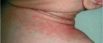
Contact dermatitis
The appearance of red dots on the head may be a sign of contact dermatitis (simple or allergic). Allergies can develop to almost any known chemical substance.
Most often, the reaction occurs to spermicides (components in contraceptives), lubricants, washing powders, men's shower gels, and soap. Contact dermatitis can also develop when wearing tight, synthetic underwear, after violent or prolonged sex (especially if the vulva and vagina of the partner are dry).
In addition to redness and a small rash, a man is bothered by moderate pain, dry skin and swelling. The described symptoms disappear by limiting contact with the allergen, prescribing sexual rest and prescribing reparative ointments (Dexpanthenol, Bepanten). Allergic reactions are quickly relieved by ointments with glucocorticoids (Advantan, Elokom, etc.)
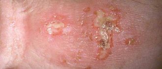
Syphilis
The most dangerous disease that causes red dots on the head is syphilis. Its treatment is the longest and most difficult. If you do not contact a doctor in a timely manner, cardiovascular pathologies, brain damage, blindness, mental disorders, and even death may occur. To avoid these consequences, you should immediately consult a doctor if characteristic symptoms occur. The disease has an incubation period of 3–7 weeks. Its first manifestation is the appearance of a red dot, similar to a pimple, which gradually turns into a hard chancre. The ulcer does not itch or cause pain, but it leads to enlargement of the lymph nodes in the groin area. After about 2 months, the pathology enters the second stage with the following symptoms:
- spots and sores all over the body
- temperature increase
- painful urination
- headache
- hair loss
- weakness
- cough, runny nose
Gradually these symptoms disappear, and the disease enters a latent phase. If syphilis was not treated at the second stage, after some time the third phase of its course begins. It is characterized by pathological changes in all body systems with damage to bones, kidneys and other organs. Treatment of pathology is carried out using penicillin derivatives.
Plasmacytic (Zoon) balanitis
Rice. 2 – Zoon-balanitis
A benign non-venereal disease of the penis, which is characterized by the appearance of a single red-orange, well-circumscribed plaque on the head. It most often develops in middle-aged and elderly men who have not undergone circumcision.
The exact cause of the pathology has not been established, but the main predisposing factors are phimosis, constant irritation and mechanical damage to the skin.
Apart from the plaque, a man may not be bothered by anything; pain and burning during urination are less likely to occur. Circumcision eliminates the manifestations; local ointments based on glucocorticosteroids and 5% imiquimod are also used in treatment.
Circinar balanitis
It occurs in 12-70% of men with Reiter's syndrome (inflammation of the urethra, joints, conjunctiva of the eye due to chlamydia) and is characterized by the appearance of well-demarcated, moist, red spots with an uneven contour on the head of the penis.
In circumcision patients, the lesions have a dry, scaly surface and can mimic psoriasis lesions. Most often, the skin around the external opening of the urethra is involved in the pathological process.
Rice. 3 – Circinar balanitis
Prevention
As part of preventive procedures, doctors recommend:
- Take vitamins 2 times a year to strengthen your immune system.
- Use barrier contraception.
- Wear underwear and clothes made from natural fabrics (cotton).
- Follow the rules of hygiene, take a shower and change your underwear once a day.
- Visit a doctor once every 6 months for preventive examinations.
- Take tests and undergo scheduled diagnostic procedures once a year (including donating blood for ELISA, RIT and RIF, and for the Wasserman test).
Notifying partners about the presence of certain diseases of the intimate area can be regarded as prevention or treatment. Since therapy for some diseases is carried out in patients simultaneously.
Psoriasis
Psoriasis is a chronic disease. The lesions appear as well-demarcated areas of bright red color with flakes of exfoliated skin.
The process can involve both individual areas of the penis and the entire skin of the perineum. The diagnosis is usually established after examination by a dermatologist (typical signs are the psoriatic triad, localization of plaques, progressive nature of the disease).
The treatment is combined, long-term, and includes mandatory correction of lifestyle and nutrition, local use of glucocorticoids, salicylates, ultraviolet radiation and other techniques.
Rice. 4 - Psoriatic rash on the penis
SUMMARY FROM D.S.:
This study shows that age spots on the genitals are, in the vast majority of cases, NOT DANGEROUS. However, researchers note a possible increased risk of developing skin melanoma (not just the genitals, but also other areas) in people with genital melanosis. This means that if you have similar pigment spots, they must be examined annually with a dermatoscope by an oncologist or dermatologist, just like ALL other pigmented skin formations.
Lichen planus
In approximately 25% of patients, the skin of the penis may also be involved in the process. The head is most often affected: pale pink or violet-pink papules with a flat surface and shine are identified on it.
They tend to merge and form round or ring-shaped plaques. Scales are visible on their surface, and when treated with vegetable oil, dots and stripes appear (Wickham's symptom).
Usually, rashes are found on other parts of the body: torso, arms, forearms, legs, and in the mouth. In rare cases, ulceration occurs.
The diagnosis is based on examination data; if there is doubt about the diagnosis, a biopsy of the papules is performed.
Rice. 5 – Lichen planus
Methods
The study involved 41 patients with genital melanosis. The criterion for participation was the presence of a mass larger than 1 cm or multiple associated lesions whose total size exceeded 1 cm.
The patients were divided into two groups according to the size of the formation. The first included participants with formations larger than 1.5 cm, the second included participants with formations less than 1.5 cm.
Biopsies were performed in 35 of the 41 patients. During the histological examination, pathologists assessed, among other things, the following data:
- The number of melanocytes per square millimeter of the epidermis.
- Presence of nuclear atypia.
- Location of melanocytes in the epidermis.
Five patients were biopsied multiple times. Samples with the highest number of melanocytes per square millimeter of epidermis were used for overall statistical analysis.
Taking medications
The appearance of red dots and spots on the head may be associated with constant use of antibiotics (especially tetracycline, sulfonamides), salicylates, Phenacetin, Co-trimoxazole and some other drugs.
The rashes have a clearly defined round or oval shape and a bright red color; a bubble of liquid may form in the center of the spot.
In most cases, discontinuation of the drug leads to gradual resolution of the rash. Sometimes the prescription of glucocorticoid ointments is required. Healing is accompanied by the formation of pigment spots.
Precancerous conditions
10.1. Bowenoid papulosis
The disease develops against the background of infection with papillomavirus types 16, 18, 34, 39, 40, 42, 45. Rashes in the form of papules, multiple, small, appear on the head and foreskin and gradually merge into one large, flat plaque, their color is red-brown or gray-brown. Bowenoid papulosis can transform into cancer.
The main method of treatment is surgical excision/cauterization of the affected areas (CO2 laser, cryodestruction).
10.2. Erythroplasia Keira
Malignant tumor of the head of the penis in situ (non-invasive tumor). The foreskin may also be involved in the pathological process (single or multiple red neoplasms with clear edges are detected on the skin).
The disease affects men who have not undergone circumcision. Over time, the tumor may grow deeper into the tissue. The main treatment is surgical excision. The patient is also recommended to undergo circumcision.
Squamous cell skin cancer of the penis
This form accounts for 95% of all malignant tumors of the penis. Peak incidence is 40-50 years. Risk factors are phimosis, balanitis obliterans and other precancerous conditions, frequent change of sexual partners, infection with HPV types 16 and 18.
Squamous cell carcinoma practically does not occur in men after circumcision. Red, compacted areas with unclear contours may appear on the skin of the head; in some cases, the tumor protrudes significantly above the skin.
Tumor germination is accompanied by superficial ulceration and bleeding. Examination and treatment are prescribed by an oncologist. The treatment regimen depends on the stage of the tumor and may include surgical excision, radiation, chemotherapy, or laser removal of the tumor.
Rice. 6 – Squamous cell carcinoma of the head of the penis
The discussion of the results
During the study, doctors studied data from clinical observation and histological analysis of 41 patients with genital melanosis. In 22 patients, the formations had several colors; in 11 patients they were more than 1.5 cm.
Researchers considered the average age of onset of this condition to be one of the clinical signs of benign genital melanosis. Among the study participants, it was 41 years. Genital melanoma develops on average in the sixth decade of life.
The researchers concluded that it is necessary to avoid unnecessary surgical intervention for genital melanosis. Clinicians consider it necessary to excise and send for histological examination all formations that seem suspicious to them. In the case of genital melanosis, this approach is not necessary. Most of the lesions examined during the study with clinical atypia, such as multiple colors and larger than 1.5 cm in size, were found to be benign.
Excisional biopsy in the genital area not only causes discomfort for the patient. The operation can harm sexual health and disrupt urinary function. The cosmetic result of the operation may be unfavorable.
Researchers have found that in most cases of genital melanosis, it is sufficient to actively monitor patients. Only one of the 41 study participants had both genital melanosis and genital melanoma. In this patient, genital melanosis was discovered almost 11 years after the diagnosis of vulvar melanoma. The formation was excised, and according to the results of histological examination it was recognized as benign.
During 30.5 months of follow-up, none of the study participants developed genital melanoma due to genital melanosis. In 19 patients who came for examination, the formations stabilized, decreased or disappeared. One patient was diagnosed with recurrence of genital melanosis after excision of the lesion.
Based on these data, the researchers concluded that the risk of genital melanosis transforming into melanoma is so small that it is difficult to measure.
The incidence of genital melanosis in the population is unknown. According to studies [1, 2], this condition occurs in 0.011% of patients who come to see a dermatologist. The incidence of genital melanoma is 0.19 cases per 1 million men and 1.8 cases per 1 million women. [4]
The study found that 15% of the experiment participants had a history of melanoma. In only one out of five cases the tumor was found in the genital area. The average age of patients with melanoma was 39 years.
Scientists have confirmed an increased incidence of genital melanosis in patients with a history of melanoma. Such patients have a suprabasal distribution of melanocytes, as well as an increased number of melanocytes in the epidermis.
Genital melanosis with clinical signs of atypia is recognized as an indication for active monitoring of patients, including examination of the whole body with a dermatoscope. However, according to the study, genital melanosis cannot be considered a precursor to genital melanoma. Therefore, in most cases, in patients with genital melanosis, radical surgery with excision of formations is not indicated.