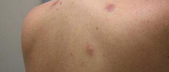
For the first time, it is impossible to take your eyes off your newborn baby. After counting all the fingers and toes several times, the mother notices that the baby has spots on his face, head and body. Immediately after birth, babies often have many spots on their skin. They pass quickly, and those that remain are called moles, or birthmarks.
Not all moles last a lifetime. Some fade over time, but there are also some that remain forever, and sometimes even require medical intervention. After reading this article, you will learn about which birthmarks appear on a child’s body from birth, and which ones appear in later life.
Why does a child develop congenital moles?
Congenital melanocytic nevi (CMN) are benign pigmented tumors consisting of nevus cells that arise as a result of impaired melanoblast differentiation between the second and sixth months of intrauterine life.
UMN occurs in 1% of Caucasian newborns of both sexes.
Visually, such formations have clear boundaries, round or oval outlines, and the surface can be lumpy, wrinkled or folded. Color varies from brown to blue or black. The consistency is usually like that of healthy skin, and hair may grow on the surface.
The reason for their development is always genetic. They can be transmitted from parents or appear on their own, completely by accident.
The time of appearance of such nevi is nonspecific. They are formed during fetal development, but are not always noticeable at birth. In the literature you can find the terms “late UMN” or “with congenital features” - such formations are no different from congenital melanocytic ones, they just appear later.
Typically, UMN are those elements that develop in the first 2 years of life. An important distinguishing feature is that a child’s moles grow with him, in proportion to the baby’s growth.
LNs are often classified as melanoma-dangerous formations, but according to the latest data, such moles are potential precursors of melanoma only in 0.7% of cases.
Pigment formations
All pigmented formations on the skin can be divided into two groups: non-dangerous (melanoma-non-dangerous) and dangerous (melanoma-dangerous) pigmented nevi (Table).
Table 1. Classification of skin tumors of melanocytic origin
| Skin tumors of melanocytic origin | |
Melanomic nevi
| Melanoma-dangerous nevi
|
What are the types of congenital moles in a child?
VMN come in various sizes, and depending on the diameter they are divided into:
- small - up to 1.5 cm;
- medium - from 1.5 to 10 cm;
- large - from 10 to 20 cm;
- giant - more than 20 cm.
One of the most typical symptoms of VN is hypertrichosis, that is, hair is present on the surface of the formation, most often dense and black. It is worth noting that the appearance of some VN is formed gradually, the nature of the coloring may become uneven, tubercles may appear on the surface, and over time, hair begins to grow.
For VN, such changes are a variant of the norm, but they require monitoring by a doctor.
The structure of the skin
The skin is the largest integral multifunctional organ, interconnected with all other organs and systems of the body. In direct contact with the external environment, it performs a barrier-protective function. On the surface of the skin there is a complex pattern in the form of triangular and rhombic fields, formed by numerous grooves. Coarser grooves form folds in the palms, soles, scrotum, and facial wrinkles.
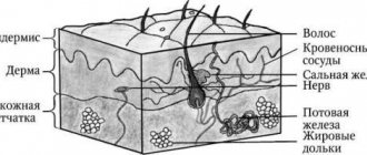
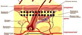
Histologically, three layers of skin are distinguished (Fig. 1):
- epidermis;
- derm (dermis);
- subcutaneous fatty tissue (subcutis), or hypodermis (hypodermis).
Rice. 1. Skin structure
The epidermis is the epithelial part of the skin, and the dermis and hypodermis are connective tissue. The border zone between the epidermis and dermis has the appearance of a wavy line due to the presence of outgrowths in the dermis - papillae, which cause the formation of ridges and furrows on the surface of the skin, forming a skin pattern. The connective tissue part of the skin (dermis and hypodermis) contains nerves, blood and lymphatic vessels, and muscles. In addition, the skin has its own appendage structures, which include hair, sebaceous and sweat glands, as well as nails.
Important features of congenital melanocytic nevi
- Small-sized UMNs are more common and may not appear until age two.
- Small and medium-sized nevi grow more slowly than the child himself and tend to darken and become hairy.
- Large and giant nevi usually occupy part of an anatomical area or all of it, for example an entire limb, neck, part of the back.
- Congenital giant melanocytic nevi transform into melanoma in 6–10% of cases.
Giant VN exceeds 20 cm in size, and such formations are sometimes compared to clothing, called “shirt” or “swimsuit” type. According to statistics, they are rare, affecting 1 in 500,000 newborns.
Giant VNs are only diagnosed at birth. Unfortunately, there is no prenatal screening to detect them, and they are not visualized on ultrasound.
The main medical problem with congenital giant melanocytic nevus is the high risk of developing melanoma, and the tumor can appear anywhere and anytime. What to do in this case? At the moment, the main method is staged excision.
The child's mole is growing. What to do?
Acquired melanocytic nevi (AMN) are benign tumors that arise from melanocytes that have migrated into the skin. They usually appear after six months of life, and reach their maximum size and number at a young age. Subsequently, they may regress or disappear altogether.
The localization of acquired formations is varied. They can be on the skin of the scalp, palms, feet, and also come from the nail matrix, creating difficulties for diagnosis and observation.
Factors influencing the appearance of nevi:
- genetic predisposition;
- level of ultraviolet radiation in childhood;
- phenotypic features of the child’s skin (fair skin, eyes, blond or red hair).
The classification of acquired nevi is varied and includes typical and atypical forms. They are also classified based on the location of the melanocytes.
PMNs are characterized by a round or oval shape and have clear boundaries. Normally they are symmetrical in color, structure and shape.
Most PN are benign and do not require any intervention, but only require lifelong monitoring.
The frequency of malignant transformations in PMN is low, since melanomas most often develop on clean skin, i.e. beyond previous melanocytic nevi. Therefore, their removal for preventive purposes is impractical.
It is worth noting that the tactics for managing patients with melanocytic formations in childhood can be implemented in three ways: surgical excision of the element, dynamic observation of the formation, and zero-intervention tactics, when further observation or surgical intervention is not required. The doctor makes a decision based on an analysis of all factors characterizing the formation: the child’s age, morphology, location, size and melanoma danger of the element.
An important diagnostic point when examining skin formations is to perform dermatoscopy.
Modern technologies have allowed doctors and patients to “monitor” the formation using its mapping. This is a procedure for fixing moles, which makes it possible to evaluate the dynamics of changes in structures, sizes and the appearance of new formations.
Melanoma-dangerous nevi
Phenotypically, these nevi do not reveal clinical signs of malignancy, but are distinguished by melanocyte dysplasia and a tendency to malignancy, thus giving reason to consider them as premalignant or borderline formations. These are the only tumors that require mandatory prophylactic removal with morphological verification of the pathological process. If it is multiple in nature, it is advisable to classify this group of patients as a risk group and subject them to mandatory clinical examination with dynamic monitoring by an oncologist.
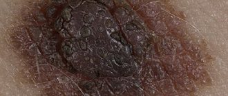
Dysplastic nevus
Elements of the rash. A spot with a separate slightly raised area, usually in the center, above the skin level. The shape is round, oval or irregular with “ragged” edges. The boundaries are irregular and blurred.
Color. Various shades of black, brown, reddish, light red.
Localization. The most common are the torso, arms, buttocks, dorsum of the feet, and less commonly, the face.
Course and prognosis. A dysplastic nevus may not undergo any transformations throughout a person’s life, but may transform into a superficial melanoma; Complete regression of education is also possible. Against the background of a dysplastic nevus, melanoma develops in 9% of cases. It differs in clinical, histological, and biological manifestations from the known forms of melanoma, and is considered by some authors as “minimal melanoma.” The appearance of these nevi in young patients is not an alarming syndrome; they, as a rule, do not transform into melanoma.
With dysplastic nevus syndrome, the prognosis is unfavorable, and the risk of developing malignant melanoma increases significantly.
Treatment. Not all dysplastic nevi require immediate removal. Dynamic observation with photography and measurement of the size of the formation is necessary. Changing, suspicious, and traumatizing nevi are subject to excision.
Rice. 11. Dysplastic nevus
Red, hanging moles - which nevi can scare parents?
Nevus Spitz
Histological examination of UMN shows that this nevus accounts for 1–2% of all UMN. This pink-red or deep black mole has a flat, hemispherical shape. The boundaries are clear, even, and the outlines are regular.
The mole grows, changes quickly, initially appears as a small speck, and mothers often confuse it with dirt on the child’s skin.
Spitz nevus is a benign structure, but clinically and histologically it is the main simulant of melanoma. That is why doctors recommend removing it immediately, or taking it under total control and observation.
Red flags for Spitz nevus:
- size greater than 8 mm;
- rises strongly above the skin level, resembles a knot, a hanging mole;
- independent ulceration, i.e. the appearance of a crust on the surface for no reason;
- pronounced clinical or dermoscopic asymmetry.
Galonevus
This is an acquired melanocytic nevus surrounded by a depigmented rim. It is represented by a red-brown nodule that rises slightly above the skin level. The diameter of the nevus itself is small and is approximately 0.2–1.2 cm, but the whitish rim around it exceeds the size of the nodule itself.
This formation is usually not dangerous and does not require treatment, but dynamic observation and mapping of the element is recommended.
Nevus Reed
A black mole with clear, smooth edges. It is small in size, no more than one centimeter, and is a benign formation.
Nevi of Oto and Ito
They are bluish-gray formations with disturbed pigmentation, located in certain areas. The localization of Oto's nevus is characterized by the periorbital region (in other words, these moles appear on the face, around the eyes), and for Ita - the skin of the neck and shoulders.
Most often, these formations require constant monitoring; laser lightening may be used due to a pronounced cosmetic defect.
Types of moles
The following types of birthmarks are found in newborns:
Pigmented birthmarks. Vascular birthmarks.
Pigmented birthmarks
Pigmented moles are usually brown, yellowish, or gray in color. They form in areas where there is a higher concentration of cells responsible for skin coloring.
Pigmented birthmarks include:
"Coffee" stains. Cutaneous melanocytosis. Nevi (warts).
"Coffee" stains
These are oval, yellowish or light brown (cafe-au-lait) spots that may be present on a child's skin from birth or appear during the first few years of life.
If there are only a few coffee stains, there is no reason to worry. However, if your baby has several spots larger than a 25-cent coin (about 25 millimeters) on his body, it could be a sign of neurofibromatosis, a genetic disorder that causes abnormal growth of nerve cells.
A large “coffee” spot with jagged edges sometimes indicates the presence of a rare hereditary disease called McCune-Albright-Braitsev syndrome.
At the slightest suspicion, we advise you to show the baby’s moles to a pediatrician and, possibly, a dermatologist and geneticist.
Cutaneous melanocytosis (“Mongolian” spots)
The so-called “Mongoloid” spots are flat moles of various sizes, brown, gray and even bluish in color.
Areas of cutaneous melanocytosis are usually located in the lower back or on the baby’s buttocks. Such birthmarks are very common in dark-skinned, Mongoloid babies. Usually these spots fade before school age.
Nevi
Nevi can be either congenital or acquired. Nevi can grow with the child.
The color of nevi varies from yellowish to black. This is a relatively common occurrence - about 1 percent of all newborns are born with nevi.
As a rule, nevi are not dangerous and are benign skin formations, but sometimes after a long enough time they can turn into a malignant skin tumor (melanoma). It is recommended to periodically show the baby's pigment elements to the pediatrician. If the size of the wart has changed, be sure to tell your doctor about it.
In extremely rare cases (1 in 20,000 children), a nevus can grow to a size of over 20 centimeters in diameter. Such nevi can be either flat or convex, and hair can grow on them. Consult a doctor - he will conduct an examination and advise what to do in this case.
Vascular birthmarks
Vascular birthmarks are collections of blood vessels. For this reason, such spots may be red or even purple.
These spots can be either flat or convex in shape. The following types of vascular birthmarks are distinguished:
Infantile hemangioma. Simple nevus. "Wine" stains.
Infantile hemangioma (“Strawberry” spots)
So-called “strawberry” spots, or hemangiomas, are collections of small blood vessels under the skin.
Usually these spots are not visible immediately after birth. In the first weeks they may be flesh-colored or even white, and only turn red later. "Strawberry" spots occur in 10 percent of babies aged 2 months, most often appearing by 2-3 weeks.
Such spots can appear on any part of the skin, but, as a rule, they are concentrated on the head or neck. They are more common in Caucasians, female babies, twins, premature babies and low birth weight babies.
Hemangiomas often grow with the baby and reach their largest size by three or four months of age. These spots gradually disappear by school age.
If your baby’s hemangioma worries you, grows too quickly, begins to bleed, or bothers your child, then let a doctor examine it. Typically, hemangiomas do not require treatment and are self-necrotic, but they are sometimes removed if they are causing serious problems, such as being located near a child's eye, in the mouth or throat. They are also removed if there is bleeding or infection.
Most often, hemangioma is single. In rare cases, a child may have several hundred small hemangiomas throughout the body. Sometimes hemangioma lies in the deep layers of the skin.
In the latter case, local treatment, oral medications, injections, laser treatment or surgery are prescribed. Discuss all your options with your doctor.
Simple nevus
Popularly, these pink spots on a child's neck, forehead, upper eyelids, nose or upper lip are called "salmon" spots, "stork bites" and "angel kisses".
Such spots are very common - in more than 80 percent of babies, most often in fair-skinned children. As a rule, they merge with the skin after a few years.
Wine stains
These are large, flat, uneven patches of dark red or purple color on a child's face or neck. They appear due to disturbances in the development of blood vessels under a specific area of the skin. Such spots do not disappear, but, on the contrary, darken over time. Port-wine stains do not go away on their own and require treatment.
To reduce the likelihood of darkening of port-wine stains during adolescence and adulthood, pulsed dye laser treatment is sometimes prescribed to young children. We recommend discussing all possible treatment options with your doctor.
If a port-wine stain is located near the eye, forehead, or scalp, it may indicate an eye and brain disorder called Sturge-Weber disease. This very rare neurological disease causes glaucoma, seizures, developmental delays and learning delays.
The pediatrician will be able to make the correct diagnosis and prescribe the appropriate treatment, including laser lightening of the spot.
What to do if a child scratches or tears off a mole?
In most cases, nevi are not a cause for concern. The appearance of moles in a child of any age is absolutely normal. They will gradually increase along with the growth of the child himself, and the location of the moles can be anywhere, including on the head, soles and even the genitals. Moles in children can easily be irregular in shape, have uneven borders, varied color patterns and large size, especially for congenital nevi.
It is also normal that moles will arouse your child's curiosity. If a child scratches or even picks off a mole, do not panic. This is only a reason to contact a dermatologist for dermatoscopy and further observation. So the answer to the question of what to do if your baby picks a mole is very simple: don’t worry and make an appointment with a specialist. ⠀
Parents need to be wary only in the following cases:
- rapid, sudden growth of formation in volume or diameter;
- the appearance of bleeding or a crust on the surface of the mole without previous injury;
- a rare type of mole;
- a large number of moles (>50) or cases of melanoma in the family;
- positive “ugly duckling” symptom (one formation is very different from other moles).
If you or your child just touched a mole, there is no need to worry.
How to accurately determine whether a mole is growing or not?
Sometimes it may seem that the mole that we have been carrying all our lives has grown in size. Sometimes it may seem that it has increased greatly. Unfortunately, the words “seems to have increased” will not help the doctor make a diagnosis. Accurate data required. There is an easy way to get them:
- Take a good photo of the mole against the background of a ruler with millimeter divisions. It's better to use a camera with a flash in macro mode. The photo must be taken by another person.
- If you have the slightest doubt about the size of the mole, you need to attach a ruler to it and compare the size with that in the original photo.