Types of skin cancer
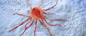
There are 3 types of common malignant skin tumors. They differ both in incidence (i.e., the chance of getting sick) and in the degree of danger to life - basal cell carcinoma, squamous cell carcinoma and melanoma.
Melanoma is one of the rare and dangerous skin tumors. It accounts for only 4% of the total number of malignant skin tumors, but is the cause of almost 80% of deaths in this localization. You can read more about melanoma here.
Sign up for the webinar “Carcinogens in cosmetics: truth, lies and... marketing”
Diagnostics
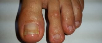
To establish an accurate diagnosis of cylindroma, a tumor biopsy is necessary, since clinically (visually) the tumor is very easily confused with other types of pathology. Moreover, a biopsy with excision of a tumor site is the diagnostic method of choice, since puncture of a tumor with a needle can give false results.
On biopsy, cylindroma is described as a benign tumor with eccrine and apocrine differentiation. Usually making a diagnosis is not difficult.
In case of ulceration of the neoplasm or the presence of an inflammatory focus directly on the cylindroma or on adjacent areas of the skin, it is necessary to conduct a bacteriological examination. Such diagnostics allows you to identify the infectious pathogen and select the necessary antibacterial treatment if necessary.
Other diagnostic methods, such as ultrasound or dermatoscopy, may be prescribed by a doctor to determine the extent of tumor growth, but they are optional. The results obtained are of an auxiliary nature and are not used to confirm the diagnosis of cylindroma.
Basal cell skin cancer
Basalioma is the most common, but at the same time the safest type of skin cancer. Death from basal cell carcinoma is possible only in very advanced cases or with aggressive forms (basosquamous) tumor. The favorable course of basal cell carcinoma is due to the fact that it almost never metastasizes (only 0.5% of cases).
Symptoms and signs
Most often, basal cell carcinoma occurs on the skin of the nose, a little less often on the face and much less often on other parts of the body.
The peak incidence occurs over the age of 40 years. The youngest patient diagnosed with basal cell carcinoma by histology was 39 years old.
What basal cell skin cancer looks like depends on the form:
- Nodular form (synonymous with nodular). The tumor is presented in the form of a nodule. It can be distinguished from other skin formations by an increased number of vessels on the surface, a waxy sheen and small gray-blue inclusions. All these signs are visible in the photo.
Nodular form of basalioma
In addition, on the surface of nodular basalioma there may be another characteristic sign - ulceration.
Nodular basal cell carcinoma with ulceration
- The superficial form of basal cell carcinoma in most cases is presented as an area of redness on the skin. Elements of peeling and the waxy sheen already mentioned above are also possible.
Superficial form of basalioma
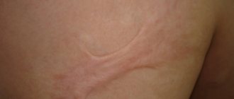
- Scleroderma-like form of basalioma is very rare and often presents difficulties in diagnosis. It is characterized by a lighter and harder seal compared to the surrounding skin.
Scleroderma-like form of basalioma
- The pigmented form of basal cell carcinoma makes up a very small part of the total number of these tumors. It is distinguished by a large amount of pigment. In this regard, basal cell carcinoma is often mistaken for melanoma when examined without a dermatoscope.
Pigmented form of basalioma
- The ulcerative form of basalioma can reach very large sizes and in advanced cases is practically untreatable.
Ulcerative form of basalioma
Photos in the initial stage
Unfortunately, basal cell skin cancer is extremely difficult to diagnose in the early stages, i.e. when it is minimal in size. Here are some photos:
Basalioma of the skin of the nose, nodular form, size 5 mm
Basalioma, nodular form, 3 mm in diameter
Nodular basalioma of the temporal region, diameter 2 mm
Diagnosing basal cell carcinoma in the early stages, when the tumor is small, can present significant difficulties. Only a combination of a comprehensive examination of the entire skin, a thorough determination of the history of the existence of the formation and dermatoscopy will help in establishing the diagnosis of basal cell carcinoma at an early stage.
Basaliomas with high and low risk of recurrence (NCCN, 2018)
Area H: Facial mask (including eyelids, eyebrows, skin around eyes, nose, lips [skin and red border of lips], chin, lower jaw, skin/grooves in front and behind the auricle, temples, ears), genitals, palms and feet .
Area M: cheeks, forehead, scalp, neck and legs
Region L: trunk and limbs (excluding shins, palms, feet, nails and ankles)
Notes
- Localization, regardless of size, may be a sign of high risk
- Histological forms of low risk: nodular (nodular), superficial, keratotic, piloid, with differentiation towards skin appendages, Pincus fibroepithelioma
- Area H means high risk regardless of size
- Morphea-like, basosquamous (metatypical), sclerosing, mixed infiltrative, micronodular in any part of the tumor
To assign a tumor the status of “high risk of recurrence”, only one of the factors from the right or left column is sufficient.
Treatment of basal cell carcinoma
The main goal of treatment for basal cell carcinoma is complete removal of the tumor while maximally preserving the cosmetic properties and functions of those parts of the body where this tumor has developed.
As a rule, the best results are achieved by surgical methods. However, the desire to maintain functionality and cosmetic properties may lead to the choice of radiation therapy as the primary treatment modality.
Depending on the degree of risk of relapse (see above), the approach to treating basal cell carcinoma may vary.
In patients with superficial basal cell carcinoma and a low risk of recurrence, when surgery or radiation therapy is contraindicated or inappropriate, the following treatments may be used:
- 5-fluorouracil ointment;
- Imiquimod ointment (Aldara, Keravort);
- photodynamic therapy;
- cryodestruction.
Mohs micrographic surgery may be recommended for patients at high risk of recurrence.
Chemotherapy for basal cell carcinoma includes drugs that are inhibitors of the hedgehog signaling pathway – vismodegib (Erivedge) and sonidegib (Odomzo). These drugs can help in cases where surgical methods, like radiation therapy, are not applicable or contraindicated.
What you need to know about basal cell carcinoma?
- In the vast majority of cases, basal cell carcinoma does not pose a threat to life.
- If a histological examination of a distant formation results in basal cell carcinoma, there is nothing to worry about. It is important to make sure that the formation is completely - be sure to consult with an oncologist.
- If, after removal of a basal cell carcinoma, the histological examination contains the phrase “tumor cells in the resection margin” or something similar, further treatment in order to completely remove the tumor.
- strongly do not recommend removing basal cell carcinoma without histological examination, since even a very typical-looking formation may not be what it seems at first glance.
- Basalioma needs to be treated . Observation is a bad option for a diagnosis like this. Treatment of advanced forms (see photo of ulcerative form) is extremely difficult and expensive.
- If you have already had a basal cell carcinoma removed, you should regularly have your entire skin examined by an oncologist in order to possibly identify another such tumor.
- The likelihood of metastasis in the metatypical (basosquamous) histological type is higher than in other types.
Rehabilitation period after removal
The duration of the rehabilitation period after laser removal depends on the area of intervention and averages 7 days. After the procedure, a scab forms at the site of laser treatment, which cannot be removed prematurely. Therefore, the treated area of skin should not be scratched/damaged in any other mechanical way. During the rehabilitation period, sun exposure to the laser treatment area should be avoided and visits to the solarium, swimming pool and sauna should be avoided.
As a rule, the rehabilitation period after surgery to remove moles on the head is quite short, since it is a low-traumatic intervention. The duration of complete recovery depends on the size of the removed lesion and on average takes from 7 to 14 days. During this period, it is necessary to regularly treat the suture in accordance with the doctor’s recommendations, eliminate any mechanical impact on the operated area, sleep in a position in which the pillow does not injure the suture, and avoid visiting the sauna, bathhouse, solarium and swimming pool.
Squamous cell carcinoma
It is less common than basal cell carcinoma, the second most common type of skin cancer, and has a slightly less favorable prognosis. However, it should be noted that the course of the disease much less malignant than that of melanoma.
Metastases occur relatively rarely - on average in 16% of cases [1]. In patients with squamous cell skin cancer less than 2 cm in size, the 5-year survival rate is about 90%; for larger sizes and tumor invasion into the underlying tissue, it is less than 50% [1].
It can occur on any part of the body, including the genitals and mucous membranes, but most often in places exposed to sunlight.
Symptoms and signs
What squamous cell skin cancer looks like depends largely on the clinical form of the disease.
The keratinizing form is a raised or flat surface covered with horny scales that can grow and fall off. If damaged, it may bleed.
Keratinizing form of squamous cell skin cancer
It must be remembered that it is the keratinizing form of squamous cell carcinoma that may be hiding under the mask of the cutaneous horn . In this regard, such formations should always be removed only with histological examination:
The cutaneous horn should be removed with histology - a keratinizing form of squamous cell carcinoma may be hidden under its mask
Non-keratinizing endophytic form (growing towards surrounding tissues). Most often it looks like a long-term non-healing wound or ulcer, which can deepen and expand over time.
Non-keratinizing endophytic form of squamous cell skin cancer
The exophytic non-keratinizing form of squamous cell skin cancer appears as a nodule that rises above the level of the skin. The surface of the node may be eroded or wet.
Exophytic nonkeratinizing form of squamous cell skin cancer
Photos in the initial stage
The initial stage of squamous cell carcinoma refers to a condition when the malignant process is limited to the epidermis - the outermost layer of the skin. It is referred to in the diagnosis as in situ or intraepidermal squamous cell carcinoma. This disease is not life-threatening if completely removed.
There are 2 forms of this phase of the disease:
Bowen's disease
Most often it is represented by single flat plaques, with clear boundaries, an asymmetrical shape, and uneven edges. The size reaches 7–8 mm. The formation may gradually increase, and peeling or crusting is often observed on the surface.
The color is red or brown, located on any part of the body. [3]
On my own behalf, I will add that in my practice, histologically confirmed Bowen’s disease occurred only once. It looked like a small (3 x 4 x 3 mm) flesh-colored lump with a smooth surface on the skin of the shaft of the penis in a 43-year-old man.
Bowen's disease
Erythroplasia Keira
The second form of early-stage skin cancer, which develops most often on the skin of the foreskin of the penis or the glans. Much less commonly, the disease affects the female external genitalia.
The most common appearance of Queyre's erythroplasia is a bright red spot with clear boundaries and a moist, shiny surface [3].
Erythroplasia Keira
Treatment of squamous cell skin cancer (NCCN, 2018)
As in the case of basal cell carcinoma, squamous cell carcinoma is divided into groups of high and low risks of recurrence and metastasis.
Area H: Facial mask (including eyelids, eyebrows, skin around eyes, nose, lips [skin and red border of lips], chin, lower jaw, skin/grooves in front and behind the auricle, temples, ears), genitals, palms and feet .
Area M: cheeks, forehead, scalp, neck and legs
Region L: trunk and limbs (excluding shins, palms, feet, nails and ankles)
Notes
- The rim of hyperemia should be taken into account when measuring size.
- Excisional biopsy is preferred over incisional biopsy.
- The modified Breslow thickness measurement should exclude parakeratosis and crusting and should be taken from the base of the ulcer, if present.
- Localization, regardless of size, may be a sign of high risk.
- Area H implies high risk regardless of size.
The basic principles and methods of treatment for squamous cell carcinoma are the same as for basal cell carcinoma.
The main goal is to maintain functionality and cosmetic qualities. method is considered to be removal of the tumor, including 4–6 mm of healthy tissue with a low risk of recurrence and metastasis. For high-risk tumors, Mohs micrographic surgery or wider excision is recommended than for low-risk tumors.
Radiation therapy is useful in cases where other methods cannot be used. Platinum drugs (cisplatin, carboplatin) as well as EGFR inhibitors (cetuximab) can be used in chemotherapy for squamous cell carcinoma.
Stages
Staging is a determination of where the cancer is located, whether and where it has spread, and its effect on other parts of the body.
Doctors use diagnostic tests to determine the stage of cancer; they may need information based on tissue samples obtained at surgery, so staging may continue until all tests are completed. Information about the stage helps the doctor determine which treatment is optimal and predict the possibility of recovery.
TNM tumor classification system
One way to determine the stage of cancer is the TNM system. Doctors use the results of diagnostic tests and scans to answer questions such as:
- Tumor (T): its size and location?
- Lymph nodes (N): extent of spread of the process to the lymph nodes?
- Metastasis (M): Has the cancer spread to other parts of the body?
The results are combined to determine each patient's cancer stage. Stage provides a common way to describe cancer so doctors can work together to plan the best treatment.
How to avoid getting skin cancer? What to avoid?
Sunlight. The most proven cause of both types of skin cancer, as well as melanoma, is exposure to sunlight. If you like to travel to hot countries, have fair hair and skin, or your work involves prolonged exposure to the sun, you should seriously consider UV protection.
Precancerous skin diseases are the next factor that may precede the development of the squamous cell form: actinic (solar) keratoses and cheilitis, leukoplakia, human papillomavirus infection of the mucous membranes and genitals. This type of tumor can also develop against the background of scar changes after burns or radiation therapy.
Contact with carcinogens
Various chemicals can lead to the development of skin cancer: arsenic and petroleum products.
Weakened immune system. People taking immunosuppressive drugs after an organ transplant or people living with HIV have an increased risk of developing squamous cell skin cancer.
Treatment of brain tumors at the Yusupov Hospital
Treatment of brain tumors at the Yusupov Hospital Oncology Center is carried out using the most advanced non-surgical techniques:
- stereotactic radiosurgery – which allows you to remove small tumors in one session;
- radiation therapy using the latest generation installations, the latest linear accelerators and systems that ensure maximum safety and effectiveness of therapy, etc.
If surgical treatment is unavoidable, it is performed by the best specialists who masterfully master all the technologies and methods of interventional neuroradiology and modern neurosurgery. Doctors at the Yusupov Hospital Oncology Center make every effort to use gentle, minimally invasive methods and operations with transnasal access, which do not require an incision in the skull.
Thanks to the innovative equipment of the oncology center, doctors have the opportunity to perform high-quality diagnostics and carry out effective treatment of brain tumors using non-surgical or minimally invasive methods.
In parallel with neurosurgeons, neurologists, oncologists, and radiologists take part in the treatment of patients with brain tumors. The patient is provided with supportive therapy and qualified psychological support.
You can make an appointment with an oncologist at the oncology clinic and find out the conditions of hospitalization by calling the Yusupov Hospital or online on the website from the coordinating doctor.