Hallus valgus is a condition in which damage occurs to the joint of the big toe. This condition is commonly called bunion. Bunions are caused by the formation of a bone spur in response to injury. In reality, Hallus valgus is not just a reaction to injury. Interestingly, this disease is almost never found in cultures that do not wear shoes. Tight shoes, such as high heels and cowboy boots, can contribute to the development of hallux valgus. Wide shoes with room for the toes reduce the chances of developing deformities and help reduce irritation to the area of the bunion, if present.
The term hallus valgus actually describes what happens to the big toe. Hallus is the medical term for the big toe, and valgus is an anatomical term that means that the deformity occurs away from the midline of the body. So valgus deformation of the big toe beyond the foot. As this process progresses, other changes appear. One of those changes is that the bone that sits above the big toe, the first metatarsal bone, begins to deviate more in the other direction. This bone is called primus varus. Primus means the first metatarsal bone, and varus is a medical term that means that the deformity is a deviation from the midline of the body. This creates a situation where the first metatarsal bone and big toe now form an angle with respect to a line running along the inner edge of the foot. Bunion, which develops first, is actually a reaction to pressure from shoes. Initially, the reaction to trauma is an area of irritated, swollen tissue that constantly rubs between the shoe and the bone located under the skin. Over time, constant pressure can cause the bone tissue to thicken, which increases swelling and further friction from the shoe.
Causes
Many problems that arise in the legs are the result of abnormal pressure or friction. The easiest way to determine the presence of the consequences of pathological pressure is to examine the leg. The leg is a hard bone covered with skin. In most cases, symptoms develop gradually as the skin and soft tissues absorb the excess impact on the leg. Any protruding bone or injury aggravates the already existing consequences of the injury. The skin reacts to friction and pressure by forming a callus. The soft tissues located under the skin react to excess stress. Both the callus and the thickened soft tissue underneath the callus become painful and inflamed. Reducing pain helps to reduce pressure. Pressure can be reduced externally through looser shoes or internally through surgery and removal of excess tissue.
Risk factors
- Shoes influence the incidence of hallux valgus (it is lower in adults who do not wear shoes). However, this does not mean that only shoes cause this disease. Tightening shoes can cause pain and pinching of the foot nerves along with the formation of hallux valgus. Fashionable shoes may be too tight and too narrow for the 'foot to look aesthetically pleasing.' High heels increase stress on the foot, further exacerbating the problem. Moreover, fashion trends are followed not only by young people, but also by people in the older age group. Risk factors can be divided into the main ones:
- There is a high probability of hallux valgus deformity in females. The cause may be shoes.
- Ballerinas who spend a lot of time on blocks dancing on their toes and thus may be expected to have a higher likelihood of developing hallux valgus
- Age. The incidence of deformity increases with age, with rates of 3% in people aged 15–30 years, 9% in people aged 31–60 years, and 16% in those over 60 years of age 1
- Genetic factors play a role
- Associated diseases
There are specific causes of biomechanical instability, including neuromuscular disorders. This may be associated with various types of arthritis. These associated diseases include:
- Gout.
- Rheumatoid arthritis.
- Psoriatic arthropathy.
- Joint hypermobility associated with diseases such as Marfan syndrome, Down syndrome.
- Multiple sclerosis.
- Sharot's disease
- Cerebral paralysis.
Reasons for appearance
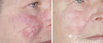
The exact causes of the disease have not yet been established. The main one is regular exposure to ultraviolet radiation. It affects the dermis, epidermal layers, blood vessels, sebaceous glands, and melonocytes.
Gradually, under the influence of sunlight, the disturbances increase, reaching the peak of the disease.
The following factors contribute to the development of pathology:
- genetic predisposition;
- weakened immune system;
- influence on the skin of chemicals (resinous substances, oil, sand, etc.);
- past infections;
- age-related changes (the disease most often affects people over 50 years of age).
Due to weak immunity, AIDS carriers, people with problems with the nervous and endocrine systems, as well as patients who have undergone chemotherapy or complex operations are more prone to the appearance of keratosis.
Some types of keratoses often affect young people. This usually applies to red-haired or fair-haired people with gray, blue or green eyes. Research shows that by the age of 40, 60% of the population has at least one element of keratosis.
Over the age of 80, everyone has some type of this pathology.
Symptoms
Symptoms of hallux valgus are mainly caused by bunions. Bunion is quite painful. With severe hallux valgus, a cosmetic problem also appears. Moreover, choosing shoes becomes difficult, especially for women who want to be fashionable and for them, wearing fashionable shoes becomes a real challenge. Finally, increasing deformity begins to move the second toe and may create conditions for the second toe to rub against the shoe.
Forms
In normal condition, the metatarsal bones are parallel to each other. Under the influence of certain reasons, the first metatarsal bone deviates outward and because of this, a protruding small bump appears on the foot, ligaments and tendons lose their elasticity, and their dysfunction develops.
The disease goes through several stages:
- Early.
At this stage, the deviation of the big toe is less than 15 degrees.
- Average.
The deviation of the first finger to the side is from 15 to 20 degrees. At the same time, deformation of the second finger is observed. It rises above the thumb and becomes shaped like a hammer.
- Heavy.
The thumb deflection is 30 degrees. All the toes are already deformed, and a large bone growth is observed at the base of the first phalanx. In places where there is a lot of stress on the foot, rough calluses appear.
Diagnostics
Disease history
- The patient may experience pain in the big toe when walking or making any movements. This may indicate degeneration of intra-articular cartilage.
- The pain may be aching in the metatarsal area due to wearing shoes. Possible increase in deformation. .
- It is necessary to find out what physical activities increase the pain and what relieves the pain (maybe just taking off your shoes).
- History of injury or arthritis.
- It is quite rare to experience sharp pain or tingling in the dorsal region of the bursa of the thumb, which may indicate traumatic neuritis of the middle dorsal cutaneous nerve.
- The patient may also describe symptoms caused by the deformity, such as a painful second toe, interdigital keratosis, or ulcer formation.
Visual inspection
- It is necessary to observe the patient's gait. This will help determine the degree of pain and possible gait disturbances associated with problems in the legs.
- The position of the big toe in relation to the other toes. Distortion of the joint can be in different projections.
- Prominent joint position. Erythema or swelling indicates shoe pressure and irritation.
- Range of motion of the big toe at the metatarsal joint. Normal posterior flexion is 65-75° with plantar flexion less than 15°. Moreover, it is necessary to pay attention to whether pain and crepitus are present. Pain without crepitus suggests the presence of synovitis.
- The presence of any keratosis, which suggests pathological chafing from abnormal gait..
- Associated deformities may include hammertoes and flexible or rigid flat feet. These deformities can cause hallux valgus to progress more rapidly as the lateral support of the foot is reduced.
Changes in movement of the thumb joint:
- Increased abduction of the big toe in the transverse and frontal planes.
- Increased average toe prominence.
- Change in backward flexion of the joint.
In addition, it is necessary to pay attention to the condition of the skin and peripheral pulse. Good blood circulation is especially important if surgical treatment is planned and normal healing of the postoperative wound is necessary.
Treatment
Conservative treatment
Treatment of hallux valgus almost always begins with the selection of comfortable shoes that do not cause friction or stress. In the early stages of Hallus valgus, wearing shoes with a wide front can stop the progression of the deformity. Since the pain that results from bunions occurs due to pressure from shoes, treatment focuses on relieving the pressure that shoes place on the deformity. Wider shoes reduce pressure on bunions. Bunion pads can reduce pressure and friction from shoes. There are also numerous devices, such as spacer orthotics, that can splint the toe and change the load distribution on the foot.
Drug treatment and physiotherapy
Nonsteroidal anti-inflammatory drugs and physical therapy may be prescribed to reduce inflammation and relieve pain. In addition, corticosteroid injections are possible. Long-term physical therapy has not proven to be therapeutically effective.
Orthopedic products
It is possible to use various orthopedic products (instep supports, toe correctors, interdigital rollers). The use of orthopedic devices helps to stop further deformation in the early stages. With severe deformation, the use of orthopedic products can only slightly reduce pain. Custom insoles help correct damaged arches.
If the deformity is caused by a metabolic disorder or a systemic disease, then it is necessary to carry out treatment aimed at correcting the underlying disease with the involvement of a rheumatologist or endocinologist.
Prevention
After treatment of corns, a relapse can easily occur, because it is not enough to relieve inflammation and accelerate tissue regeneration - you need to 100% eliminate the factors that provoke the development of the disease. To avoid relapse, follow 7 rules:
- Wear only comfortable shoes that do not cause pain or discomfort. Avoid high heels and very hard soles.
- Never skimp on shoes and prefer models made from natural materials,
- Use soft orthotics.
- Alternate heavy physical work and sports with periods of rest. Be sure to undergo a professional pedicure and foot massage, and take medicinal baths.
- Get regular medical examinations to identify problems with the endocrine and cardiovascular systems at an early stage.
- Avoid sudden weight gain (adjust your diet and exercise).
- If you experience itching, redness or severe pain, immediately make an appointment with a dermatologist. This will eliminate the disease at an early stage and avoid complications.
The multidisciplinary CELT clinic employs specialists with extensive experience. Here they will be able to determine the cause of the appearance of pathological formations on the soles and prescribe effective treatment. If the problem has already arisen, then you need to contact a specialist as soon as possible.
Make an appointment through the application or by calling +7 +7 We work every day:
- Monday—Friday: 8.00—20.00
- Saturday: 8.00–18.00
- Sunday is a day off
The nearest metro and MCC stations to the clinic:
- Highway of Enthusiasts or Perovo
- Partisan
- Enthusiast Highway
Driving directions
Surgery
If all conservative measures are not effective, then a decision is made on surgical treatment. Currently, there are more than 100 surgical techniques for the treatment of Hallus valgus. The main tasks in surgical treatment are as follows:
- remove bunion
- reconstruct the bones that make up the big toe
- balance the muscles around the joint so that there is no recurrence of the deformity
Removing the “growth”
In some mild cases of bunion formation, only the growth on the joint capsule may be removed during surgery. This operation is performed through a small incision on the side of the foot in the area of the bunion. Once the skin is cut, the growth is removed using a special surgical chisel. The bone is aligned and the skin incision is closed with small sutures.
It is more likely that reconstruction of the big toe will also be necessary. The main decision that must be made is whether the metatarsal bone needs to be cut and also reconstructed. To resolve this issue, the angle between the first metatarsal and the second bone is important. The normal angle is approximately nine or ten degrees. If the angle is 13 degrees or greater, the metatarsal bone will most likely need to be cut and reconstructed. When the surgeon cuts and repositions the bone, it is called an osteotomy. There are two main techniques used to perform osteotomy and reconstruction of the first metatarsal bone.
Distal Osteotomy
In some cases, the distal end of the bone is cut and moved laterally (this is called a distal osteotomy). This effectively reduces the angle between the first and second metatarsals. This type of surgery usually requires one or two small incisions in the leg. Once the surgeon has achieved satisfactory bone alignment, the osteotomy is followed by fixation of the bones using metal pins. After surgery and healing, the pins are removed (usually they are removed 3-6 weeks after surgery).
Procymal osteotomy
In other situations, the first metatarsal bone is cut at the proximal end of the bone. This type of surgery usually requires two or three small incisions in the leg. Once the skin is cut, the surgeon performs an osteotomy. The bone undergoes reconstruction and is temporarily fixed with metal pins. This operation also reduces the angle between the metatarsal bones. In addition, the tendon of the adductor big toe muscle is released. Therefore, after the operation, a special bandage is put on.
Rehabilitation after surgery
It takes an average of 8 weeks for the soft tissue and bones to heal. During this period, it is better to place the foot in shoes with a wooden sole or a special bandage in order to prevent trauma to the operated tissues and allow normal regeneration. Immediately after surgery you may need crutches.
In patients with severe bursitis, physiotherapy (up to 6-7 procedures) may be prescribed a certain time after surgery. In addition, you must wear shoes with wide fronts. It is also possible to use correctors. All this can allow you to quickly return to normal walking.
Hyperkeratosis. Horny skin and calluses (corns)
21.07.2018
| no comments
The skin change that most often occurs on the feet is hyperkeratosis, or calositas: excessive formation of keratinized skin. It comes in two forms, namely callus and callus.
Unlike callus, which is only superficial keratinization, in the case of callus (Latin: clavus; Greek: heloma), the deeper layers of the skin also change. A callus usually has a clearly defined, more or less hard “core”, which is located in the depths and clearly appears on the surface of the skin (if it is not covered by a callus). In addition, a callus is recognized by its soreness, caused by the pressure of the “core” on the lower layers of the skin through which the nerves pass, or by pressure on the periosteum (periosteum).
Experience shows that excessively thickened keratinized skin and calluses primarily appear in those places where the skin is subject to increased stress and a particular irritant. This irritant is not always pressing. We can talk about chafing (a double irritant due to the heat generated by friction), damage, as well as the influence of heat or cold, acids or alkalis.
There are:
- mechanical irritant;
- thermal stimulus;
- chemical irritant.
We must assume that the increased formation of keratinized skin is a measure of the body's defense. The irritant first causes a strong blood supply to the dermis and inner layer of skin to release excessive heat, as the destroyed parts of the tissue must be expelled. Finally, the germinal layer, due to this blood supply to the lower layers of the skin, is stimulated to accelerate cell division, the thickened stratum corneum protects the affected area. This is how callus appears.
The patient needs to realize that he must adapt not the foot to the shoe, but the shoe to the foot. Give your clients the following advice:
- Wear shoes with good support, insoles and the right size (optimal height, width and length, without pressure on the seams or in the toe area). You should see for yourself what the quality of the shoes is, since it is unlikely that the patient considers his shoes to be too narrow or small.
- Change your shoes daily to avoid constant pressure on the same place.
- Show toes or foot deformities that are causing pressure to a specialist
- ·If shoes put pressure on bone or joint protrusions, the patient needs special shoe inserts.
Unfortunately, there are cases when hyperkeratoses constantly form in the same place, although the cause has been eliminated. Here, obviously, the skin’s “memory” of the original place of compression has been preserved, or the reason is a violation of the reflex zone. This should not be an excuse to consider the specialist's work superficial or poorly executed. You need to understand that areas of compression or callus will return in any case until the cause is eliminated.
Forms of hyperkeratosis
Cutaneous horn (Cornu cutaneum)
The skin grows in a certain place (arm, face, foot), like the horn of an animal. Sometimes it is combined with a common wart (Verrucae vulgaris) or squamous cell carcinoma. The cutaneous horn should be removed due to possible degeneration into a malignant neoplasm.
Keratinized skin infected with a fungus (keratomycosis)
In this case, the keratinized skin is affected by a fungus. Possibly due to foot fungus (Tinea pedis).
Palmoplantar keratosis (Keratosis palmo plantaris)
Dyskeratosis, or the tendency for keratinization of the palms and soles. Sometimes it is caused by a fungal infection or psoriasis.
Keratoma
Limited, tumor-like dyskeratosis of the skin, the occurrence of which is associated with a combination of many factors.
Keratoderma blenorrhagicum
Increased formation of keratinized skin, mainly on the edge of the forefoot, on the toes. Cause: Reiter's disease (appears under the influence of gram-negative bacteria or chlamydia). We are talking about seronegative joint disease (without positive rheumatic factor). It can be combined with conjunctivitis, urinary tract infection and inflammation of the knee joint (gonitis) or another joint - Reiter's triad.
Pressure callus (calositas)
This is the body's defense mechanism. We are talking about a limited compaction of keratinized skin on an area of the skin that is particularly stressed or exposed to specific irritants. On the foot, it appears primarily in the area of the forefoot, on the balls of the feet, heels, with too narrow shoes or foot deformities: anywhere where the skin is subjected to strong pressure or irritation. Calluses and keratinized skin are not limited to the feet; they occur wherever the skin experiences increased stress (palms, workers, etc.).
Callus in diabetic neuropathic injury
This callus is characterized by an ulcer crater with a necrotic (dead), infected bottom, which is surrounded by severe callus. Such callus is usually macerated (softened). It occurs primarily on the toes, heels and under the transverse arch.
Keratoma climacterium
Increased keratinization on the palms and soles shortly before or after menopause, often in combination with obesity
Most patients complain that calluses constantly appear in the same place. They indirectly blame the pedicurist for this, citing his allegedly unprofessional actions. However, most often the root of the problem lies in themselves, since the cause of calluses and calluses is too narrow shoes.
In young people they are caused by a variety of fashion trends, in older people they are often caused by painful swelling of the feet. Both lead to the same result: increased pressure and rubbing of certain unprotected areas of the foot. Pressure relief patches only help for a short time, in most cases they increase the pressure even more as they increase the size of the foot. In this case, callus around the callus may increase.
Latest Posts

