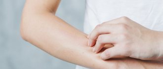
Any surgical intervention is a big test for the patient’s body. This is due to the fact that all his organs and systems experience increased stress, no matter whether the operation is small or large. It especially affects the skin, blood and lymphatic vessels, and if the operation is performed under anesthesia, then the heart. Sometimes, after everything seems to be over, a person is diagnosed with “seroma of the postoperative suture.” Most patients do not know what it is, so many are frightened by unfamiliar terms. In fact, seroma is not as dangerous as, for example, sepsis, although it also does not bring anything good with it. Let's look at how it happens, why it is dangerous and how it should be treated.
What is it - postoperative suture seroma?
We all know that many surgeons perform “miracles” in the operating room, literally bringing a person back from the other world. But, unfortunately, not all doctors conscientiously perform their actions during the operation. There are cases when they forget cotton swabs in the patient’s body and do not fully ensure sterility. As a result, in the operated person, the suture becomes inflamed, begins to fester or separate.
However, there are situations where problems with a stitch have nothing to do with medical negligence. That is, even if 100% sterility is observed during the operation, the patient suddenly accumulates liquid in the incision area that looks like ichor, or pus of a not very thick consistency. In such cases, they speak of seroma of the postoperative suture. What it is, in a nutshell, can be said this way: it is the formation of a cavity in the subcutaneous tissue in which serous effusion accumulates. Its consistency can vary from liquid to viscous, the color is usually straw-yellow, sometimes supplemented with blood streaks.
Seroma formation
Seroma is not related to malignant neoplasms, but requires removal. It increases the risk of infection entering the operated area, so it requires timely treatment.
The formation of seroma is possible from a hematoma. Sometimes it is formed from lymphatic fluid due to capillary leakage. “Leakage” from blood vessels is also possible if there are breaks in them. Another risk factor is dying or damaged cells, which can cause inflammation.
At-risk groups
Theoretically, seroma can occur after any violation of the integrity of lymph vessels, which do not “know how” to thrombose quickly, as blood vessels do. While they are healing, lymph continues to move through them for some time, flowing from the rupture sites into the resulting cavity. According to the ICD 10 classification system, seroma of the postoperative suture does not have a separate code. It is assigned depending on the type of operation performed and the reason that influenced the development of this complication. In practice, it most often occurs after such cardinal surgical interventions:
- abdominal plastic surgery;
- cesarean section (this postoperative suture seroma has ICD 10 code “O 86.0”, which means suppuration of the postoperative wound and/or infiltration in its area);
- mastectomy.
As you can see, it is mainly women who are at risk, and those who have solid subcutaneous fat deposits. Why is that? Because these deposits, when their integral structure is damaged, tend to peel off from the muscle layer. As a result, subcutaneous cavities are formed, in which fluid begins to collect from the lymph vessels torn during the operation.
The following patients are also at risk:
- those suffering from diabetes;
- elderly people (especially overweight);
- hypertensive patients.
Seroma after abdominoplasty
Seroma is an accumulation of serous fluid in the area of a surgical wound.
Serous fluid is a straw-yellow liquid of varying degrees of viscosity, which consists of two main parts: the liquid fraction and formed elements.
Formed elements include leukocytes, mast cells, macrophages. And the liquid fraction is represented by albumins, globulins, i.e. protein fractions contained in the blood.
Causes of seroma formation:
The main reason for the formation of seroma is the detachment of large surfaces of the subcutaneous tissue, a large wound surface.
A large wound surface is associated with damage to a large number of lymphatic vessels. Lymphatic vessels cannot thrombose as quickly as blood vessels, causing a buildup of serous fluid, which is mostly lymph. The presence of blood gives the seroma a reddish color.
Other causes of seroma formation may be:
- Traumatic work with tissues.
The surgeon must work with soft tissues as delicately as possible. Do not roughly grab fabrics or use tools with a crushing effect. The cuts must be made carefully and in one motion.
Numerous incisions create a “vinaigrette” effect, significantly increasing the area of damaged tissue, which leads to an increased risk of seroma formation.
- Excessive use of coagulation.
Coagulation is a tissue burn. Any burn is accompanied by necrosis with the formation of inflammatory fluid (exudate). Coagulation should be used in isolation only to cauterize a bleeding vessel.
- Large thickness of subcutaneous fat.
The thickness of the subcutaneous fat is more than 5 cm, which is almost always guaranteed to lead to the formation of a seroma. Therefore, if the thickness of the subcutaneous fat is more than 5 cm, it is recommended to perform liposuction first. Then after three months you can return to the issue of abdominoplasty.
This solution is always more effective both from the point of view of health and from the point of view of the aesthetic result of the operation.
What does seroma look like?
As a rule, seroma does not hurt. Only in rare cases, when the volume of serrous fluid is large, pain may occur.
Quite often, because of this, seroma remains unrecognized for a long time.
If the seroma is small, there is no significant pain.
The main manifestations of seroma are as follows:
- The patient has a sensation of fluid transfusion in the lower abdomen.
- Swelling and bulging may appear in the lower abdomen. Often patients say that their stomach has suddenly become enlarged, although a couple of days ago everything was normal.
With large volumes of seroma, the following symptoms may occur:
- Pain or tension in the area of seroma accumulation, usually the lower abdomen;
- Unpleasant pulling sensations that intensify when standing;
- Redness of the skin in the area of greatest accumulation of seroma;
- Increase in body temperature to 37-37.5, general weakness, fatigue.
Diagnosis of seroma
Diagnosis of seroma is based on examination and instrumental research methods.
- Inspection.
During the examination, the surgeon will notice the presence of swelling in the lower abdomen. On palpation, fluid flows from one side to the other, a fluctuation indicating that there is an accumulation of fluid.
In addition, the presence of seroma symptoms will leave no doubt for making the correct diagnosis.
- Instrumental research methods - ultrasound of the soft tissues of the abdomen.
With ultrasound, the accumulation of fluid between the muscles of the anterior abdominal wall and subcutaneous fat is very clearly visible.
Taking into account all the symptoms and ultrasound scan results, it does not seem difficult to diagnose seroma.
Treatment of seroma
Seroma treatment includes two types of treatment:
- surgery
- conservative, drug treatment
Surgical treatment includes:
Removal of seroma using punctures. This is the easiest way to remove serous fluid. In 90% of cases this is enough.
The surgeon uses a syringe to remove liquid, the volume of which can be from 25-30 ml to 500-600 ml.
Gray needs to be pumped out regularly, every 2-3 days. As a rule, 3 to 7 punctures are sufficient until the seroma is completely eliminated. In some particularly stubborn cases, 10, 15, and sometimes more punctures may be required.
After each puncture, a decrease in the amount of serous fluid is observed, i.e. each time it becomes less and less.
In patients with a large thickness of subcutaneous fat or after surgery combined with liposuction with a large amount of soft tissue trauma, the seroma reaches large sizes, and puncture is not enough.
In this case, the surgeon installs a drainage with active aspiration. The fluid constantly drains away through the drainage, it becomes less and less and gradually the cavity closes, grows together and the seroma disappears. At the same time, it is necessary to carry out drug treatment.
Drug treatment is based on the use of:
- broad-spectrum antibiotics to prevent suppuration.
- non-steroidal anti-inflammatory drugs, in order to relieve aseptic inflammation and reduce the amount of fluid discharge.
- in some cases, it is useful to use steroidal anti-inflammatory drugs, such as diprospan and kenalog, which are very good at blocking aseptic inflammation, reducing the amount of fluid formed and speeding up recovery.
Prevention of seroma formation
Prevention is based on compliance with all surgical principles:
- There is no need to do abdominoplasty if the thickness of the subcutaneous fat of the anterior abdominal wall is more than 5 cm.
First you need to do liposuction, and after 3 months you should plan abdominoplasty.
- Careful work with soft tissues.
Try to use electrocoagulation in isolation, cauterizing only bleeding vessels. Do not allow compression, squeezing, or excessive tension of the skin of the anterior abdominal wall.
- Mandatory wearing of compression stockings in the postoperative period.
Displacement of the skin-fat flap relative to the anterior abdominal wall leads to the formation of a film and fibrin loss on both the anterior abdominal wall and the detached flap.
The movement of tissues relative to each other leads to the fact that on these tissues something similar to a mucous membrane appears, which produces fluid and is a good place for its accumulation.
Therefore, wearing compression hosiery, and high-quality knitwear that will create good compression and fixation of tissues, is an important condition for the postoperative period and the prevention of seroma formation.
- Maintain physical rest in the first two to three weeks after surgery to reduce the movement of the soft tissues of the anterior abdominal wall relative to each other.
Such prevention methods can significantly reduce the risk of seroma formation.
| If the surgical technique and all seroma prevention measures are followed, then the probability of seroma formation is 10%. If a seroma does form, it will not be large, it can be easily cured and it will not affect the patient’s rehabilitation in any way. |
Consequences of seroma for the aesthetic result of the operation and existing risks in the formation of seroma.
- Risk of suppuration
Serous fluid is an ideal place for bacteria to multiply. If an infection occurs, the likelihood of suppuration is quite high.
The infection can come from chronic foci of infection: the oral cavity, nasal cavity, etc.
Chronic sinusitis and tonsillitis are the most common sources of infection, which spreads hematogenously or lymphogenously (that is, through the blood or lymph flow).
- A long-term seroma can lead to the formation of some kind of mucous membrane, both on the skin-fat flap, which is detached, and on the anterior abdominal wall.
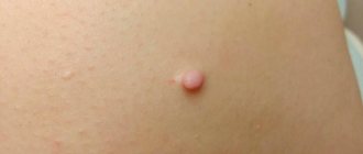
In the photo, the surgeon performs a revision abdominoplasty..
During the operation, it turned out that in the lower abdomen there was no fusion of subcutaneous fat with the muscles of the abdominal wall. Most likely, this is a consequence of seroma not recognized in a timely manner.
As a result, an isolated cavity with a small amount of serous fluid was formed. (See photo)
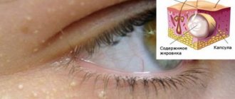
| Using tweezers, the surgeon points to some semblance of a mucous membrane. Such a cavity can exist for a very long time. In some cases (trauma, hypothermia, etc.), the amount of fluid may increase, which is perceived by patients as an enlargement of the abdomen. In addition, the presence of such a cavity with serous fluid, even in a small amount, can lead to suppuration. |
| The only way to deal with such a cavity is to remove the capsule so that the tissues can grow together. The photo shows fragments of the excised capsule. |
The long-term existence of seroma leads to the fact that this cavity never heals, which leads to some mobility of the skin relative to the anterior abdominal wall. In such conditions, seroma can exist for a very long time. Fortunately, this happens extremely rarely.
- Long-existing seroma can lead to deformation of the skin-fat flap, thinning of the subcutaneous fat, which will ultimately worsen the aesthetic result of the operation.
- Seroma may contribute to poorer healing of scars.
Thus, you cannot NOT pay attention to the gray, hoping that it will “resolve on its own,” and it must be treated. Timely treatment guarantees excellent results.
Causes
To better understand what it is - postoperative suture seroma, you need to know why it forms. The main causes do not depend on the competence of the surgeon, but are a consequence of the body’s reaction to surgical intervention. These reasons are:
- Fat deposits. This has already been mentioned, but we will add that in overly obese people whose body fat is 50 mm or more, seroma appears in almost 100% of cases. Therefore, doctors, if the patient has time, recommend liposuction before the main operation.
- Large wound surface area. In such cases, too many lymph vessels are damaged, which, accordingly, release a lot of fluid and take longer to heal.
Diagnostic methods
Postoperative suture gray can be identified by visual inspection of the suture. In a situation where the skin incision area is swollen, a specialist must palpate
If the volume of fluid is large, then during palpation the doctor notes its transfusion under the epidermis. The fluid transfusion process can best be felt in thin patients.
If there is any doubt about the diagnosis, the specialist will refer the patient to undergo an ultrasound examination of soft tissues, which allows one to examine the area of accumulated serous fluid.
Increased tissue trauma
It was mentioned above that seroma of the postoperative suture depends little on the conscientiousness of the surgeon. But this complication directly depends on the skills of the surgeon and on the quality of his surgical instruments. The reason why seroma can occur is very simple: the work with the tissues was carried out too traumatically.
What does it mean? An experienced surgeon, when performing an operation, works with damaged tissues delicately, does not squeeze them unnecessarily with tweezers or clamps, does not grab them, does not twist them, and performs the incision quickly, in one precise movement. Of course, such jewelry work largely depends on the quality of the instrument. An inexperienced surgeon can create a so-called vinaigrette effect on the wound surface, which unnecessarily injures the tissue. In such cases, the ICD 10 code for seroma of the postoperative suture can be assigned as follows: “T 80”. This means “a complication of surgery not noted elsewhere in the classification system.”
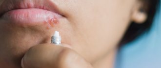
Bubbles have appeared around the seam, liquid is leaking from them, the seam is getting wet, what should I do?
No. 23 760 Surgeon 08/30/2015
A herniated disc was removed 2 weeks ago. The seam was clean, on the 10th day the stitches were removed, but I was allergic to the eraser. Bandages, adhesive plaster. There was redness on the back. On the 5th day after removing the sutures, bubbles appeared around the suture, liquid flowed out of them, and the suture began to get wet. The seams were treated with chlorhexidine and brilliant green. When the seam began to get wet, the doctor prescribed bandages with levomekol, but it didn’t help.
ANSWERED: 08/30/2015
18+ Online consultations are for informational purposes only and do not replace a face-to-face consultation with a doctor. Terms of use
Your personal data is securely protected. Payments and site operation are carried out using the secure SSL protocol.
Do patients who have undergone surgery often develop allergies after surgery ? What is causing this problem? What symptoms does it accompany? Is it possible to get rid of it?
In fact, an allergic reaction is rarely directly related to surgery. After all, by and large, an allergy is a reaction of the nominal system to a particular substance. Therefore, disorders arise already in the postoperative period. But, of course, there are exceptions.
For example, some patients are allergic to latex, which is what surgical gloves are made from. In addition, an allergic reaction may result from tissue contact with certain metals used in surgical instruments. In addition, the immune system often rejects various implants or prostheses, which is accompanied not only by skin manifestations, but also by weakness, the development of an inflammatory process, suppuration and even sepsis.
Excessive electrocoagulation
This is another reason that causes suture gray after surgery and to some extent depends on the competence of the doctor. What is coagulation in medical practice? This is a surgical procedure performed not with a classic scalpel, but with a special coagulator that produces a high-frequency electric current. In essence, this is a targeted cauterization of blood vessels and/or cells by current. Coagulation is most often used in cosmetology. She has also proven herself excellent in surgery. But if it is performed by a physician without experience, he may incorrectly calculate the required amount of current or burn excess tissue. In this case, they undergo necrosis, and neighboring tissues become inflamed with the formation of exudate. In these cases, seroma of the postoperative suture is also assigned the code “T 80” in ICD 10, but in practice such complications are recorded very rarely.
Clinical manifestations of seroma of small sutures
If the surgical intervention was on a small area of skin, and the suture turned out to be small (accordingly, the doctor’s traumatic manipulations affected a small volume of tissue), the seroma, as a rule, does not manifest itself in any way. In medical practice, there are cases where patients did not even suspect it, but such a formation was discovered during instrumental studies. Only in isolated cases does a small seroma cause minor pain.
How to treat it and is it necessary to do it? The decision is made by the attending physician. If he deems it necessary, he may prescribe anti-inflammatory and painkillers. Also, for faster wound healing, the doctor may prescribe a number of physiotherapeutic procedures.
Clinical manifestations of seroma of large sutures
If the surgical intervention affected a large volume of the patient’s tissue or the suture was too large (the wound surface is extensive), the occurrence of seroma in patients is accompanied by a number of unpleasant sensations:
- redness of the skin in the suture area;
- nagging pain that gets worse when standing;
- during operations in the abdominal region, pain in the lower abdomen;
- swelling, bulging of part of the abdomen;
- temperature increase.
In addition, suppuration of both large and small seromas of the postoperative suture may occur. Treatment in such cases is very serious, including surgical intervention.
Symptoms of seroma
The development of seroma can be recognized by specific signs indicating the presence of pathology.
The main symptoms include:
- a feeling of fluid transfusion, which can be especially clearly felt during palpation of the abdomen;
- the appearance of swelling;
- increase in abdominal volume.
As soon as the volume of fluid has reached significant volumes, the following are added to the primary symptoms:
- soreness in the area where serous fluid has accumulated;
- pulling spasms that increase during movement;
- the appearance of redness in the lower abdomen;
- feeling of weakness and fatigue;
- slight increase in temperature.
What seroma looks like, photo
Diagnostics
We have already discussed why seroma of a postoperative suture can occur and what it is. Treatment methods for seroma, which we will consider below, largely depend on the stage of its development. In order not to start the process, this complication must be detected in time, which is especially important if it does not announce itself in any way. Diagnostics is carried out using the following methods:
Examination by the attending physician. After surgery, the doctor is required to examine his patient's wound daily. If undesirable skin reactions are detected (redness, swelling, suppuration of the suture), palpation is performed. If there is a seroma, the doctor should feel fluctuation (flow of liquid substrate) under the fingers.
Ultrasound. This analysis perfectly shows whether or not there is accumulation of liquid in the seam area.
In rare cases, a puncture is taken from the seroma to clarify the qualitative composition of the exudate and decide on further actions.
Prevention and treatment
Prevention of this phenomenon is quite simple. It consists of gentle, light pressure with your hand on the operated area. In this case, you should avoid massaging the scar or pressing on it at an angle, as this can lead to sutures coming apart.
To prevent fluid from accumulating after surgery, plastic surgeons leave drainage tubes in place until the fluid has completely stopped flowing. However, accumulation is possible even after removal of the drainage.
The accumulation can resolve on its own, but the process itself can take several weeks, and its result can be a knot of calcified soft tissue. They are not harmful to the body and are clearly visualized during diagnostic studies.
Conservative treatment
This type of therapy is most often practiced. In this case, patients are prescribed:
- antibiotics (to prevent possible further suppuration);
- anti-inflammatory medications (they relieve inflammation of the skin around the suture and reduce the amount of fluid released into the resulting subcutaneous cavity).
Nonsteroidal drugs such as Naproxen, Ketoprofen, and Meloxicam are more often prescribed.
In some cases, the doctor may prescribe steroidal anti-inflammatory drugs, such as Kenalog, Diprospan, which block inflammation as much as possible and accelerate healing.
Surgery
According to indications, including the size of the seroma and the nature of its manifestation, surgical treatment may be prescribed. It includes:
1. Punctures. In this case, the doctor removes the contents of the resulting cavity with a syringe. The positive aspects of such manipulations are as follows:
- can be performed on an outpatient basis;
- painlessness of the procedure.
The disadvantage is that the puncture will have to be done more than once, and not even twice, but up to 7 times. In some cases, it is necessary to perform up to 15 punctures before the tissue structure is restored.
2. Installation of drainage. This method is used for seromas that are too large in area. When drainage is placed, patients are simultaneously prescribed antibiotics.
Treatment of seroma
Today, several methods of therapy are used, which are described below.
Conservative therapy
Quite often, doctors prefer a conservative method of treatment.
During therapy, patients are prescribed:
- taking antibiotics to prevent re-suppuration;
- the use of anti-inflammatory medications that relieve inflammation of the skin around the suture.
Non-steroidal anti-inflammatory drugs (Naproxen or Ketoprofen) are prescribed. However, in the most severe cases, it is advisable to use steroid medications such as Diprospan or Kenalog.
They are the ones who can block the inflammatory process and speed up the healing process in a short period of time.
Surgery
Significant dimensional characteristics of the liquid become the reason for immediate surgical therapy, including:
- Punctures, during which a specialist using a syringe has the opportunity to remove the contents of the resulting cavity. The procedure can be performed on an outpatient basis. The patient does not feel pain. The main disadvantage of the method is the large number of procedures that must be completed. As a rule, 7-16 visits to the doctor are required for complete recovery. After this, the structural characteristics of the tissue are restored.
- Drainage installations. This method is advisable to use in the presence of too large seroma. In parallel with this, antibiotics should be prescribed.
Folk remedies
Treatment of seroma with traditional methods is impossible.
However, by following the recommendations, you can speed up the healing process of the suture and prevent repeated suppuration:
- Lubricate the seam with antiseptics that do not contain alcohol. Betadine and Fukortsin are perfect for such purposes.
- We apply ointment like Vulnuzan, Levosin.
- We supplement the diet with a vitamin complex.
- In case of severe suppuration of the suture, we treat it with iodine. In this case, the doctor should prescribe antibiotics and anti-inflammatory medications.
If desired, you can systematically make compresses based on larkspur infusion. The pre-washed root system of the plant is crushed in a blender and filled with vodka. For 100 mg of raw material you will need 200 ml of vodka. The composition is infused for 2 weeks.
Before applying the compress, the tincture is diluted 1:1 with water, which will avoid skin burns. The product is applied to a piece of gauze and applied every 2-3 hours to the seam. Using traditional methods really speeds up the healing process.
Folk remedies
It is important to know that regardless of the reasons for the seroma of the postoperative suture, this complication is not treated with folk remedies.
But at home, you can perform a number of actions that promote healing of the suture and prevent suppuration. These include:
- lubricating the seam with antiseptic agents that do not contain alcohol (“Fukorcin”, “Betadine”);
- application of ointments (Levosin, Vulnuzan, Kontraktubeks and others);
- inclusion of vitamins in the diet.
If suppuration appears in the suture area, you need to treat it with antiseptic and alcohol-containing agents, for example, iodine. In addition, in these cases, antibiotics and anti-inflammatory drugs are prescribed.
In order to speed up the healing of stitches, traditional medicine recommends making compresses with an alcohol tincture of larkspur. Only the roots of this herb are suitable for its preparation. They are washed well from the soil, crushed in a meat grinder, put in a jar and filled with vodka. The tincture is ready for use after 15 days. For a compress, you need to dilute it with water 1:1 so that the skin does not get burned.
There are many folk remedies for healing wounds and scars after surgery. Among them are sea buckthorn oil, rosehip oil, mumiyo, beeswax, melted with olive oil. These products should be applied to gauze and applied to the scar or seam.
Postoperative suture seroma after cesarean section
Complications in women whose obstetrics were performed by caesarean section are common. One of the reasons for this phenomenon is the mother’s body, weakened by pregnancy, which is unable to ensure rapid regeneration of damaged tissues. In addition to seroma, a ligature fistula or keloid scar may occur, and in the worst case scenario, suppuration of the suture or sepsis. Seroma in women giving birth after a cesarean section is characterized by the fact that a small dense ball with exudate (lymph) inside appears on the suture. The reason for this is damaged blood vessels at the site of the incision. As a rule, it does not cause concern. Seroma of postoperative suture after cesarean does not require treatment.
The only thing a woman can do at home is to treat the scar with rosehip or sea buckthorn oil to speed up its healing.
Causes of seroma
The appearance of postoperative seroma is most often associated with a large area of surgical intervention, which resulted in detachment of the subcutaneous tissue. Due to the rough impact, tissues begin to bleed and break down under the influence of enzymes. All this provokes the appearance of seroma.
Serous exudate after surgery appears mainly from damaged lymphatic vessels, since they, unlike blood vessels, are not capable of rapid healing. It takes at least a day for the lymphatic vessel to heal. It turns out that the more damage the lymphatic network received, the more serous transudate will be released.
Another reason for the appearance of seroma after surgery is increased bleeding. This occurs in cases where insufficient attention was paid to blood clotting during preoperative preparation.
Complications
Postoperative suture seroma does not always go away on its own and not in everyone. In many cases, without a course of therapy, it can fester. This complication can be provoked by chronic diseases (for example, tonsillitis or sinusitis), in which pathogenic microorganisms penetrate through the lymph vessels into the cavity formed after surgery. And the liquid that collects there is an ideal substrate for their reproduction.
Another unpleasant consequence of seroma, which was not paid attention to, is that the subcutaneous fatty tissue does not fuse with the muscle tissue, that is, the cavity is constantly present. This leads to abnormal skin mobility and tissue deformation. In such cases, repeated surgery must be used.
Treatment of pathology
Serous fluid under the surgical suture may persist for a long time, but in most cases it disappears by 20 days after surgery. The timing of disappearance strongly depends on the nature of the surgical intervention, its complexity and the area of the wound surface. All this time, the doctor must closely monitor the development of seroma.
Treatment of pathology begins if there is too much moisture under the skin and there is a serious risk of developing an inflammatory process or sepsis. The essence of treatment is to remove exudate from under the skin. This is done in various ways.
Vacuum aspiration
This method of treating seroma is used most often. It allows you to get rid of exudate in the early stages of the development of pathology that is not complicated by the inflammatory process.
The doctor makes a small incision in the area where moisture accumulates, into which a suction tube is inserted. After turning on the vacuum device, the moisture accumulated under the skin is mechanically removed.
Using the vacuum aspiration method can significantly speed up the healing of a postoperative wound. In addition, after the procedure, patients note a significant improvement in their well-being.
The main disadvantage of this technique is possible relapses. The fact is that vacuum aspiration only removes exudate, but does not eliminate the cause of its appearance. For this reason, after vacuum aspiration, doctors begin to eliminate the factors influencing the appearance of serous exudate under the postoperative suture.
Subcutaneous drainage
This is a surgical method for treating seroma of a postoperative scar. Its main difference from the vacuum aspiration method is that the doctor does not use special equipment.
Drainage involves removing serous fluid by gravity. To do this, a puncture is made in the area where exudate accumulates, through which a drainage system is inserted under the skin. Its outer part is connected to a collection of withdrawn biological material. After this, the exudate will be drained from under the skin immediately after its appearance.
Prevention
On the part of the medical staff, preventive measures consist of strict adherence to the surgical rules of the operation. Doctors try to perform electrocoagulation more gently and injure less tissue.
On the part of patients, preventive measures should be as follows:
- Do not agree to surgery (unless there is an urgent need) until the thickness of the subcutaneous fat reaches 50 mm or more. This means that you first need to do liposuction, and after 3 months, surgery.
- After surgery, wear high-quality compression stockings.
- Avoid physical activity for at least 3 weeks after surgery.
When does seroma appear?
Lifetime warranty on implants
There is no specific time interval between surgical intervention and the formation of seroma. It is difficult to name the factors that stimulate fluid accumulation. However, the general likelihood of developing this anomaly is as follows: the greater the extent of surgery, the greater the likelihood of developing seroma. Seroma can appear in any part of the body that has been injured or undergone surgery.
It is diagnosed during a mammological examination, but, as a rule, the accumulation is clearly visible to the naked eye and is easily palpable.