Our skin has a complex structure and consists of various cells, and the elements that form them are very diverse. I would like to talk about the most common neoplasms, and also consider an important question - how dangerous they are in terms of cancer and whether it is worth removing them.
All neoplasms are divided into three groups:
- benign,
- precancerous,
- malignant.
Benign is the largest group of neoplasms. There are a lot of elements in this group. Let's look at the most common
Nodes in the veins of the legs: treatment and its features
Treatment of nodes in the veins of the legs begins only after the patient has undergone a series of laboratory and instrumental examinations, the key of which is ultrasound duplex scanning of the vessels of the lower extremities.
Based on the results obtained, the phlebologist can judge the need for minimally invasive surgical intervention and the degree of its safety for the patient. Vein nodules in the leg can be eliminated under local anesthesia in several ways.
- Laser coagulation of a vessel, which involves introducing a special device into the cavity of a blood vessel and “welding” its walls with thermal radiation. Due to this, it stops filling with blood and is completely excluded from the bloodstream.
- Miniphlebectomy, the essence of which is the complete removal of the affected superficial vessels by removing them from the thickness of the tissue through small punctures.
New growths on the skin
The term “neoplasm” implies local excessive growth of any tissue of the body. On the skin they are represented by primary and secondary tumors, nevi and hemoderma. In dermatological practice, tumors are divided into benign and malignant. A detailed photo and detailed description of each of them will be given below.
Why do they occur? The study of skin tumors is still ongoing. The exact reasons for their occurrence have not been established, but scientists put forward several theories about this. Provoking factors may be:
- family history (presence of neoplasms in relatives);
- individual characteristics of a person (light skin and hair, old age);
- exposure to ultraviolet, radiation and x-ray radiation;
- viral infection; long-term trauma to the skin;
- chronic exposure of the skin to chemical carcinogens (nitrosoamines, benzopyrene, aromatic amines, etc.);
- insect bites; metastatic processes in the presence of an oncological process in the body;
- violation of the trophism of the skin, hence chronic skin ulcers; weakened immune system (due to immunosuppressive therapy, HIV infection, etc.).
Types of neoplasms on the skin Neoplasms on the skin, according to their origin, can be divided into primary (those that are formed from the skin tissue itself) and secondary (those that metastasize into the dermis and epidermis from foci of another localization). The latter also include hemoderma. They arise due to pathological proliferation of malignant cells of the hematopoietic system. There is a division of neoplasms into benign, precancerous (precancrosis) and malignant (cancer itself). This classification allows you to determine the treatment method and life prognosis for the patient.
Nevi should be distinguished from skin tumors. These are benign neoplasms that are related to skin developmental defects.
Malignant neoplasms, neoplasms on the skin of a malignant nature in the Russian Federation in the structure of cancer incidence today account for 9.8% and 13.7% in men and women, respectively. People living in areas with high photoinsolation and having fair skin are especially susceptible to the disease. Description of new cases.
Malignant skin tumors include: basal cell carcinoma; Kaposi's sarcoma; liposarcoma; squamous cell carcinoma; melanoma, etc. Basalioma One of the most common epithelial skin tumors. It is formed from atypical cells of the basal layer of the epidermis, which is where it gets its name. The tumor is characterized by long-term progression, peripheral growth, during which the surrounding tissues are destroyed. Basalioma is not prone to metastasis. This pathology develops mainly in elderly people and the elderly, and is localized mainly on the face, neck and head (its scalp). Sometimes basal cell carcinoma is classified as a precancer, because under the influence of certain factors it degenerates into metatypical cancer.
The first manifestation of a developing tumor is a dense, hemispherical nodule that does not rise above the skin. Its color usually matches the skin color or differs slightly (light pink tint).
At the initial stage, the patient does not complain. Over the course of several years, the papule grows, reaching 1 or 2 cm in diameter. Its center gradually collapses, bleeds and becomes crusty.
Under the latter, erosion or an ulcer with a narrow ridge along the edges is found, which over time scars and grows along the periphery.
Basalioma reaches a size of 10 centimeters or more. The once pink papule turns into either a flat plaque with peeling, or a noticeably raised node above the surface of the skin, or a deep ulcer that destroys the underlying tissue (down to the bone).
Liposarcoma This is a neoplasm on the skin from fat cells of mesenchymal origin. In the photos below you can see how big these tumors reach. The description in clinical reference books speaks of liposarcoma as a formation that tends to appear on the buttocks, thighs and retroperitoneal tissue. It is more common in men over 40 years of age.
Initially, a swelling appears, then a node appears. There are no subjective sensations yet. On palpation, the nodule is dense, elastic, and mobile. Subsequently, the tumor grows, turns red, and inflammatory processes begin. Large liposarcoma can compress nerves and blood vessels and even grow into them, causing disturbances in tissue trophism and pain.
Kaposi's sarcoma
This is a systemic multifocal disease of vascular origin with primary damage to the skin, lymphatic system and internal organs. It belongs to tumors of an endothelial nature and develops mainly in individuals with severe immunosuppression.
The morphology of skin lesions of sarcoma is quite diverse. they come in the form of spots, nodules, infiltrative plaques, etc. There are several types of sarcoma: classic (European). endemic (African). Epidemic (for HIV). immunosuppressive (immunodeficiency caused by medications and medical procedures). The first type is observed in the elderly and the elderly and has a favorable course. the elements grow for a long time, tens of years in the proximal direction and do not cause discomfort to the patient. formations are most often localized on the lower extremities, are bluish-red spots up to 5 cm in diameter with smooth edges, reminiscent of hematomas. As they grow, they transform into nodules and merge. large nodes darken and eventually ulcerate. Swelling occurs at the edges of the elements, caused by stagnation of lymph in the lymphatic channel. The African type is severe and affects young people. A fulminant course of the disease is often observed. African Kaposi's sarcoma manifests itself in several types of formations - from nodes to lymphadenopathy. The most malignant type of elements of this type of sarcoma is considered to be “flowery” (growth in the form of vegetation - in appearance it resembles cauliflower). It is characterized by deep lesions of the dermis, subcutaneous tissue and underlying tissues down to the bone. With HIV infection, the tumor can be located literally anywhere in the body, even affecting internal organs. The most typical places are the oral cavity, stomach and duodenum. The current is severe. The immunosuppressive type is similar in its manifestation to HIV-associated.
Squamous cell carcinoma
Malignant tumor of the epithelium. Formed from atypical keratinocytes that proliferate randomly. The process begins in the epidermis, gradually moving to deeper layers. The tumor is characterized by a tendency to metastatic process. Squamous cell carcinoma occurs 10 times less frequently than basal cell carcinoma. It most often affects white-skinned men whose place of residence is a sunny, warm climate.
The localization of spinocellular epithelioma varies. The most favorite place for the formation of squamous cell carcinoma is the border between the mucous membrane and the skin. These areas include the lips and genitals. At the initial stage of cancer development, an infiltrate appears with a raised hyperkeratotic (rough) surface. the color of the formation is usually gray or yellow-brown. At first, as with basal cell carcinoma, there are no complaints. As the tumor grows, it can reach a size of up to 1 cm. At this moment, a dense node that continues to grow begins to be felt. Eventually, the carcinoma approaches the size of a walnut. The tumor grows in two directions - above or deep into the tissue. The latter is usually accompanied by the formation of an ulcer, which affects not only the dermis and epidermis, but also reaches the bone and muscle tissue. An ulcer in squamous cell carcinoma does not heal. The patient suffers from excruciating pain at the site of its formation. Subsequently, the complaints are joined by disturbances in general well-being and infectious complications associated with immunosuppressive processes that accompany any oncological pathology.
Melanoma
This is a tumor of neuroectodermal origin. It consists of malignant melanocytes. UV radiation is considered the main provoking factor. Melanoma develops both from an existing nevus (mole) and on clean skin.
Signs of malignancy include: asymmetry; fuzzy edges; uneven coloring; diameter more than 6 mm; evolution of a pigment spot (any changes in a mole - sudden growth, change in color, etc.) is the most typical sign!
Benign neoplasms
New growths on the skin, classified as benign, as can be seen in the photo and their description, are not prone to rapid growth, metastasis and relapse after removal.
These tumors include:
- atheroma;
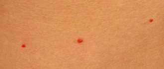
- hemangioma;
- lymphangioma;
- warts;
- moles (nevi);
- fibromas, etc.
Atheroma Damage to the sebaceous gland in the form of a cystic growth. Location: face. The raised formation does not differ from normal skin in color and is tightly fused to it at one point. The contours are clear. Palpation of the atheroma is painless.
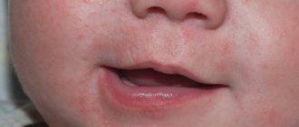
Hemangioma A neoplasm of vascular origin, refers to childhood tumors. It can be either an independent disease or a manifestation of another pathology. The course depends on the patient’s age, location, size and depth to which the hemangioma has grown. Frequent locations are the head, face, neck, but other locations are also possible. The first manifestation is a red nodule (papule) up to 5 mm in diameter. Complaints include bleeding when accidentally touched, and sometimes dysfunction of the organ where the tumor is located.
Lymphangioma There are two possible development options - a congenital defect or consequences of impaired lymph flow. The tumor can be located anywhere, but more often it is the oral cavity, neck, and upper limbs. Capillary lymphangiomas are multiple blisters with a yellowish transparent liquid inside. The same tumors that arose as a result of impaired lymph circulation look like plaques or spots, the diameter of which slowly increases.
Lipoma Tumor of adipose tissue. More often they have a nodular shape. The course is asymptomatic. It is a painless pale pink neoplasm on a stalk. The consistency is doughy. The boundaries are unclear.
Seborrheic warts Also called "senile warts". They arise as a result of impaired differentiation of cells in the basal layer of the epidermis. Appearance: Nodules or plaques raised above the skin. The surface is lumpy. The shape is often round or oval. The color of the wart varies from yellow-brown to black.
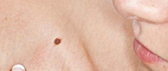
Moles or nevi are a developmental defect and consist of unchanged melanocytes. The characteristic color of the rash is from light brown to black, which is associated with different amounts of melanin (dark pigment) in the cells. More often, moles have a smooth surface and sometimes rise above the skin.
Fibroma Fibroma is a tumor that forms from connective tissue. Dense to the touch. Reaches large sizes. Malignancy (transition to malignancy) of the tumor is possible.
Neoplasms on the skin (photos and descriptions are presented above) of a benign nature, despite their relatively safe nature, can sometimes still develop into precancrosis and even cancer.
Precancerous conditions
Precancrosis is a pathological condition of any tissue of the body, which, with varying degrees of probability, can contribute to the occurrence of a malignant process. The following diseases are considered precancer of the skin: Bowen's disease; Paget's disease, etc. Bowen's disease is an intraepidermal cancer that is prone to transition to squamous cell. It is a chronic inflammatory disease that is associated with excessive proliferation of atypical keratinocytes. Occurs in older people.
The tumor has invasive growth, growing not only into the epidermis, but also into deeper tissues. It can be located on any part of the skin and mucous membranes, but more often on the torso. The elements look like pink spots with fuzzy, rounded edges. Under them there is an infiltrate, due to which the formations are slightly elevated. They are rough to the touch and covered with scales. When the latter are peeled off, an erosive, bleeding surface opens.
Paget's disease is an adenocarcinoma prone to metastasis. The source of growth, as in Bouin's disease, is located intraepidermally. The typical location is the mammary glands, most precisely the area of the nipple and its areola.
Tumor growth is infiltrating (grows into underlying tissues). Clinically manifested as a unilateral itchy plaque with clear contours in women over 40 years of age. The surface is covered with scales and crusts. The element increases in size and begins to metastasize. The outcome is breast cancer.
Diagnostics
New growths on the skin, photos and descriptions of which were presented above, are diagnosed using general principles. These include mandatory medical history collection (patient complaints, clinical manifestations of the disease), examination of the patient, careful visual examination of formations and analysis of data from clinical and instrumental examination methods (MRI, radiography). The main thing in making a final diagnosis is the histological method. It is a microscopic examination of an area of pathologically altered tissue in order to identify atypical cells.
Treatment of skin tumors
Treatment methods for skin tumors include medication, radiation and surgery. The latter is radical (i.e., it allows you to get rid of the disease as completely as possible). New growths on the skin are treated medicinally with antibiotic therapy (photos and detailed descriptions of drugs can be found in clinical reference books), NSAIDs, opioid analgesics and hemostatic drugs. This method is used as a symptomatic treatment, it can alleviate the patient’s condition and somewhat improve the quality of life.
The surgical method is based on the elimination of the tumor. The goal of treatment is final relief from the disease and prevention of relapses. Radiation therapy is more often used for malignant processes, especially in cases where surgical resolution is not possible. The goal is also to prevent tumor regrowth and metastasis. Removal of skin tumors Methods for removing skin tumors are: Cryodestruction (with the help of liquid nitrogen the tumor is frozen, then it dies and disappears). The method is not used on the head, with large formations or when they are located intradermally.
Surgical excision. The scope of the operation can be different - removal of the tumor within healthy tissue (fibroma, nevi), along with the capsule (atheroma, lipoma), etc. up to excision of the surrounding skin, subcutaneous tissue, and nearby lymph nodes (squamous cell carcinoma and other malignant formations). In order not to miss the moment when skin growths become life-threatening, you need to understand what you are dealing with. After studying the photos and descriptions of common pathologies, you can guess the source of the disease and promptly contact a specialist.
Features of the Lakhta Milon laser
Modern science has long appreciated all the advantages of using lasers in medicine. Therefore, today new, more advanced devices are constantly appearing, one of which is the domestically produced Lakhta Milon device.
With its help, you can perform minimally invasive operations in the shortest possible time, differing in:
- high degree of security,
- efficiency,
- speed,
- short recovery period,
- no need for general anesthesia.
Rehabilitation and possible contraindications
After the operation, the patient is recommended to:
- wear compression stockings,
- try to walk a lot,
- avoid strenuous physical activity.
However, in some situations, seals in the veins of the legs are not recommended to be removed using the proposed methods, as this is fraught with great risk for the patient. This is about:
- obesity,
- serious pathologies of the cardiovascular system,
- thrombophilia,
- inflammatory processes of the skin in the area of affected vessels.
A knot in a vein in the leg is not just a cosmetic problem; it must be fought radically and decisively. The phlebologists at the First Family Clinic of St. Petersburg will help you with this. Make an appointment, we are waiting for you at Kolomyazhsky Prospekt, 36/2.
Examples of different types of knots and knots
Subcutaneous
Whiteheads are also a type of nodule on the skin. Also, formations may be evidence of a disease such as basal cell carcinoma or basal cell skin cancer
.
Let us consider in a little more detail the diseases, evidence of which may be the symptom in question. Whiteheads. A fairly common pathology of the dermis. Acne, or milia, is a collection of sebaceous secretions under the skin. Often they do not cause discomfort to a person. Unless it concerns the aesthetic side of the issue.
Whiteheads may also indicate the presence of gastrointestinal diseases. To treat milia, a special diet is used, as well as the use of special-purpose medications.
With skin diseases, the bumps can be not only white, but also red. This may be evidence of acne.
Acne
They begin with a small red tubercle, at the top of which a white purulent head forms over time.
After a short period of time, the pus comes out and a crust forms on top. When the crust heals, a keloid forms in place of the pimple.
Lipomas, hygromas, and atheromas are also distinguished. Let's look at each type in more detail.
Lipoma
Lipoma or wen is a subcutaneous formation, soft to the touch, mobile. Wen can only form in that part of the body where there is adipose tissue.
Hygroma
Unlike lipoma, hygroma is a sedentary formation similar to a tumor. Hygroma is filled with serous fluid. It is formed as a result of herbs received by a person during a fall or blow. The tumor is removed surgically.
Atheroma
Atheroma is partially similar to lipoma; it also forms in the sebaceous duct. Atheroma is a cyst formed as a result of blockage of the sebaceous duct. The size of the formation can be either the size of a pea or the size of a chicken egg.
As mentioned above, nodes can be a manifestation of a disease such as basal cell epithelioma. This is a type of skin cancer. If basal cell tumors appear on the skin, this indicates that the disease is progressing from a mild to a more severe form.
Although not in all cases, nodules on the skin indicate a serious illness, a person in any case should contact people specially trained for this. If something bothers you, let alone causes pain, you need to immediately contact a specialist.
You can find such specialists at the Doctor Nearby clinic. All applicants will be given attention and given a clear and correct diagnosis. Naturally, the treatment will be of the highest level. You can make an appointment by phone without leaving your home.
