Norm
Jaundice occurs due to disruption of bilirubin metabolism, its excretion and accumulation.
Bilirubin tends to form due to cytochromes, red blood cells and myoglobin, which have broken down.
Bilirubin comes in two forms:
- Incoherent. It is toxic, interacts with the protein albumin, and is transported to the liver area through the bloodstream;
- Connected. Cells that are in the liver area combine bilirubin and glucuronic acid, changing it into direct or conjugated bilirubin. Next, the liver produces bile, where bilirubin will be located, and it enters the intestines with it. Next, bilirubin is removed from the feces.
Etiology and pathogenesis of skin pigmentation
The main causes of abnormal skin pigmentation on the face and body:
- chronic exposure to ultraviolet radiation;
- endocrine disorders;
- pregnancy;
- taking medications;
- eating specific foods (for example, rich in carotene);
- environmental pollution;
- professional activity;
- tattoos;
- heredity.
Pathogenetic mechanisms of abnormal pigmentation (according to Fitzpatrick):
- abnormalities in the origin, migration and differentiation of melanoblasts;
- disturbances in melanocyte morphology;
- pathologies of tyrosinase biosynthesis;
- disorders of melanosome genesis;
- pathologies of melanin biosynthesis from tyrosine;
- dynamic anomalies that disrupt the structure of melanosomes and the color of the synthesized pigment;
- disruption of the transfer of melanin granules to keratinocytes.
Types of jaundice
The following types of jaundice are distinguished according to their nature:
- Physiological. It occurs most often in children if their liver tissue is immature. Usually occurs in a large percentage of newborns on the third or fourth days of life. Usually occurs in premature babies. This is due to the fact that the body has not yet had time to adapt to the habitable environment. Jaundice itself can disappear in seven to twenty-one days, and does not harm the child’s body.
- Hemolytic. It occurs when red blood cells are destroyed. Hemoglobin, when released, turns into bilirubin. This effect may occur due to anemia and changes in the composition of hemoglobin protein. It happens that red blood cells are destroyed due to the influence of poisons and various drugs on them. During pregnancy, this type of jaundice occurs due to conflict between the child and the expectant mother.
- Parenchymal (or hepatic). This type of jaundice occurs due to inflammatory processes in the liver tissue due to hypoxia, autoimmune diseases, toxins and hepatitis viruses, which are divided into A, B, C, D, E, F, G.
- Mechanical. This jaundice occurs due to disturbances in the bile flow from the liver to the duodenum. This jaundice can appear due to: stones in the ducts, inflammatory processes, narrowing of the ducts (the disease is dangerous due to complications), neoplasia of the pancreas and liver, roundworms and helminths in the gastrointestinal tract, due to which the bile ducts can be blocked and anemia develops.
Another classification of jaundice is based on its duration:
- Acute jaundice. Rapid simultaneous onset of symptoms;
- Chronic jaundice. Symptoms appear gradually, the speed of their manifestation is associated with the course of the disease that caused jaundice.
Diagnosis of skin diseases with racial pigmentation
Are there skin diseases that more often affect pigmented skin? What does normal skin look like? Do the manifestations of common dermatoses differ on different skin types?
Most dermatoses affect the skin regardless of its color. But on skin with different pigmentation, many types of dermatoses look different. Some diseases occur exclusively or predominantly in dark-skinned people. In addition, some features characteristic of skin with racial pigmentation can in some cases be mistaken for manifestations of the disease.
| Figure 1. Classic presentation of follicular eczema. This variant is common in children with pigmented skin, but is rare in white-skinned children. |
Taking into account all of the above, we can agree that diagnosing skin diseases in dark-skinned patients poses a serious problem for a doctor who deals primarily with white-skinned people. However, one must be aware of the social and psychological consequences of certain diagnoses in certain racial groups and their impact on subsequent patient management.
- Diseases most common in people with racial skin pigmentation
Atopic eczema is very common among children of all races, but research suggests it predominantly affects blacks [1].
| Figure 2. Inflammatory ringworm of the scalp in an Afro-Caribbean child. Infection is more common in urban areas |
The clinical manifestations of this disease in people with different skin colors may also differ.
Lichenification is often more severe, and atopic eczema in blacks often manifests as follicular eczema, which also occurs in white children (Fig. 1). Tinea capitis, as the name suggests, is a fungal infection of the scalp that primarily occurs in children before puberty. Caused by dermatophytes. Most often these are strains of the endothrix infectious agent, Trychophyton tonsurans, which has replaced the ectotrix zoophilic dermatophyte Microsporum canis. The lesions may be inflammatory (swelling, ulcers, and crusts) or appear as multiple patches of baldness that retain short hair fragments or scaling (Figure 2).
| Figure 3. Areas of hypopigmentation on the chin in seborrheic eczema. This condition is more noticeable in blacks |
Over the past few years, outbreaks of tinea capitis among children from Africa or the West Indies have been observed in urban areas of the UK. This racial preference may be due to a number of factors, including the use of hair oils, razor blades, and multiple hair braids [2].
Local remedies are ineffective, so systemic drugs are used for treatment. Griseofulfin is most often prescribed orally daily for 6 to 8 weeks.
Seborrheic eczema is a disease of unknown etiology. Common in children and manifests as areas of hypopigmentation on the face; some of them may peel off (Fig. 3). Topical steroids are ineffective and the disease gradually resolves on its own without treatment.
| Figure 4. Cervical keloid acne. Papules, pustules and plaques are visible on the back of the head |
Seborrheic eczema occurs equally often among representatives of all races, but is more pronounced in blacks, who are correspondingly more likely to seek medical help.
Keloid cervical acne. It is a chronic disease that occurs almost exclusively in blacks and is more common in men than women. Follicular papules develop, no different in color from the skin, most often on the back of the head. Sometimes, as a result of persistent infection, ulcers develop (Fig. 4).
Pustules may coalesce to form keloid plaques. It is believed that the etiology of the disease is due to the curved shape of the hair and hair follicles, as well as the use of razor blades to create very short hairstyles.
| Figure 5: In people with racialized skin pigmentation, keloids can cause significant cosmetic problems. |
Long courses of low-dose antibiotics may be required to prevent recurrent infections. The remaining keloids are flattened by the steroids injected into them.
Pseudofolliculitis of the beard region is a condition similar to the one described above that develops in black men who shave. The hair follicle is crooked and the razor cuts the hair askew, allowing it to re-enter the skin (ingrown hair). This causes the development of a foreign body reaction with the formation of spots and pustules and, subsequently, a keloid.
The patient is advised to press the razor closely to the skin while shaving; The direction of movement of the razor should coincide with the direction of hair growth. In addition, the doctor may recommend growing a beard (which is often unacceptable for the patient) or using chemical depilators. Sometimes topical treatment with retinoic acid or its combination with a topical steroid helps.
| Figure 6. Papular lesions characteristic of papular dermatosis of blacks. There is often a familial predisposition |
The formation of keloids on racially pigmented skin can present a serious cosmetic problem (Figure 5). Typically, treatment does not produce satisfactory results, but sometimes cryotherapy, injecting steroids into the damaged areas and excising them, followed by immediate radiotherapy is effective. Before any surgical procedure, patients should be warned about the risk of keloid scar formation.
Traction alopecia is scar-like baldness that occurs when hair follicles are subject to extreme tension, most often in women who tie or braid their hair tightly. Treatments such as hot combing, hair softening, and chemical straightening, which are more common among dark-skinned women, can also damage hair, leading to breakage and hair loss.
Patient management involves providing patients with detailed explanations of the causes of their condition. It is necessary to try to convince them to change their usual ways of caring for and styling their hair.
| Figure 7. Pigmentation of nails, manifested by longitudinal straight strands, may be a normal variant in dark-skinned people. As a rule, such pigmentation is bilateral |
Oily acne consists of numerous blackheads, papules and pustules on the forehead and temples. It affects both men and women with dark skin who have been using fat-based hair styling products for a long time. Localization of lesions allows them to be distinguished from acne vulgaris.
When the fat-containing product is discontinued, the acne goes away. If the patient insists on using fat, then it may be recommended to replace it with an oil-based gloss. Administration of a keratolytic agent, such as benzoyl peroxide, and topical retinoic acid may help.
Papular dermatosis of blacks manifests as round or thread-like papular lesions with a smooth surface on the face, neck and upper torso (Fig. 6). There is often a family predisposition. Treatment is carried out for purely cosmetic reasons and consists of curettage or cauterization with electrocautery.
Facial Afro-Caribbean childhood rash (LACS) is a condition found only in black children and appears as flesh-colored or lighter-colored patches on the face. The reason is unknown. This condition develops at an earlier age than acne [3]. After a few months, recovery occurs on its own.
Infantile acropustulosis has been described in all racial groups and is predominant among people with racial skin pigmentation. It manifests itself in the form of severely itchy blisters and pustules on the palms and soles. It usually occurs during the first year of life.
| Figure 8. Idiopathic droplet hypomelanosis |
Attacks gradually become less frequent, and complete recovery usually occurs by three years [4]. The etiology is unknown, the differential diagnosis is scabies.
- Variants of the norm in people with racial skin pigmentation
People with dark skin have variants of the norm that are unusual for light-skinned people. You need to be able to distinguish them in order to avoid unnecessary examinations and treatment.
Oral pigmentation is often found on the gums, hard palate, buccal mucosa and tongue. It must be differentiated from secondary oral pigmentation, which occurs as a result of taking medications or ingesting heavy metals. Pigmented dividing lines. Voigt's line is a line of sharp transition from the relatively more colored dorsal surface of the limbs to the ventral surface. Lines of hypopigmentation occur along the midline and in the clavicle region [5].
| Figure 9. Dotted spots on the palmar surface of the hand |
Nail pigmentation. Nail pigmentation, manifested by longitudinal straight strands (Fig. 7), may be a normal variant in dark-skinned people. As a rule, such pigmentation is bilateral and occurs more often on the thumb and index fingers.
This pigmentation is more pronounced in people with darker skin and is more common in older people. It must be differentiated from malignant melanoma.
Patches of hyperpigmentation are very common on the palms and soles of people with racially pigmented skin.
Idiopathic guttate melanosis. This is the name given to small hypopigmented spots that most often appear on sun-exposed areas of the limbs of adults (Fig. 8).
| Figure 10. Example of vitiligo with striking contrast between normal and affected areas |
| Figure 11. Oval pityriasis on black skin is papular, and often after recovery there remains pronounced post-inflammatory pigmentation |
Their edges stand out sharply, but the shape is often irregular.
Such lesions occur on any type of skin, but are more common on pigmented skin. They need to be distinguished from vitiligo. Pinpoint pockmarks and keratoses of the palms and soles are usually located in the palmar and plantar folds (Fig. 9). There is a family predisposition (familial hyperkeratosis of the extremities), but in most cases, lesions develop in people engaged in manual labor [6].
- Unusual manifestations of common keratoses
Common dermatoses may present in unusual ways. Pigmentation may obscure erythema, and dermatoses are often accompanied by post-inflammatory hypo- or hyperpigmentation.
Pigmentation changes are more pronounced and lasting. Vitiligo is much more noticeable on pigmented skin and poses a serious cosmetic problem (Figure 10). Hypopigmentation sometimes develops after the use of topical steroids.
Hyperpigmentation may be primary, such as melasma, but may also accompany other conditions, including acne, drug eruptions, and other inflammatory dermatoses. More often, rashes have papular and follicular components.
Pityriasis oval (Fig. 11) can be papular and follicular, and atopic eczema and pityriasis versicolor appear in the form of follicles.
References 1. Williams HC, Pembroke AC, Forsdyke H., Boodoo G., Hay RJ, Burney PGJ London-born black Caribbean children are at increased risk of atopic dermatitis // J. Am. Acad. Dermatol. 1995; 32: 212-217. 2. Fuller LC, Child FJ, Higgins EM Ninea capitis in south-east London: an outbreak of Trichophyton tonsurans infection (letter) // Br. J. Dermatol. 1997; 136: 139. 3. Williams HC, Ashwirth J., Pembroke AC, Breathnach SM FACE – facial Afro-Carribean childhood eruption // Clin. Exp. Dermatol. 1990; 15: 163-166. 4. Dromy R., Raz A., Metzker A. Infantile acropustulosis // Pediatr. Dermatol. 1991; 8: 284-287. 5. Vollum D. Skin markings in Negro children from the West Indies // Br. J. Dermatol. 1972; 86: 260-263. 6. Henderson AL Skin variations in blacks // Cutis. 1983; 32: 376-377.
Note!
- Clinical manifestations of skin diseases in people with pigmented skin may vary
- Some diseases are found only in blacks
- You need to be able to recognize normal variants in order to avoid unnecessary examinations and treatment.
- Pigmenting diseases in dark-skinned people are common and affect large areas of the skin due to the greater contrast between normal and affected areas of the skin.
- Special hair care practices that are found only in dark-skinned people can cause hair and scalp diseases that are unusual for white-skinned people
What you need to know
Yellow skin color also occurs due to carotene oversaturation. This happens if a person often eats vegetables and fruits that contain it in large quantities.
How to distinguish such yellowing from jaundice? With it, only the skin is stained, but not the mucous membranes.
It happens that the skin turns yellow due to taking medications containing acryquine or picric acid.
Further, the skin turns yellow. If you consume excessive amounts of spices, spicy foods, fast for a long time, or frequently use alcohol or drugs.
Principles of treatment, methods of hardware treatment
Treatment of uneven skin color should begin with identifying the cause of this condition - otherwise, even after solving the problem, you risk relapse and/or worsening of the condition after some time. If a change in tone is caused, for example, by a disease or taking medications, it is necessary to treat the patient and/or discontinue/adjust the prescribed drug. Most likely, this will require interaction between a cosmetologist and dermatologist with doctors of other specialties.
UV filters to exposed areas of your body daily and stop visiting the solarium. To protect against blue light (HEV rays), you should limit the time you use gadgets (monitors, laptops, tablets, smartphones), working with them only when necessary. It is also not recommended to use them several hours before bedtime, and especially not in bed.
Residents of large cities are recommended to use cosmetics with the so-called “pollution protection factor” (PPF). Such cosmetics may contain the following protective complexes: Pollushield, IBR-Pristinizer, 28Extremoin, EXO-P, SymUrban, etc. For example, one of the recent developments includes acids obtained from hibiscus (Hibiscus acids), which due to chelating ( metal-binding) action prevents the appearance of age spots on the skin.
Epidermal Growth Factor (EGF) helps even out complexion - it stimulates cell growth and division, renewing the epidermis. When using products with EGF, there is a gradual increase in the synthesis of DNA, RNA, hyaluronic acid, collagen and elastin - as a result, the appearance of the skin quickly improves. Epidermal growth factor is also called Beauty Factor.
Arbutin evens out the tone well and brightens the skin. In addition, it has an antioxidant effect, improves the protective properties of the skin and promotes its recovery after sunburn. However, the concentration required to achieve a lightening effect and the duration of use of arbutin have not yet been identified. Apparently, the individual sensitivity of the skin, the depth of the pigment and other factors are of great importance.
To correct uneven tone, you can use peeling agents with acids . They reduce the pH in the stratum corneum of the epidermis and at the same time bind calcium ions. Calcium ensures the “gluing” of epidermal proteins, and when it is removed, intercellular connections weaken - keratinocytes begin to peel off, which serves as a signal to activate the division of young cells. As a result, the skin brightens and its tone evens out.
If uneven skin color is associated with uneven distribution of melanin in the epidermis, this problem can be effectively solved using hardware cosmetology methods . There are two approaches used here:
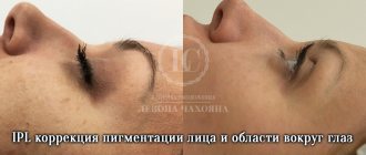
- The first approach is the selective destruction of melanin using heating with visible and near-infrared light, for example, IPL therapy, implemented in the M22-IPL device.
- The second approach is the destruction of the epidermis in order to trigger its renewal. This approach was previously often implemented using laser resurfacing, a method that involves complete vaporization of the epidermis in the treated area. Today, fractional lasers are used to “replace” the epidermis: non-ablative, such as Fraxel, Clear+Brilliant M22-ResurFX, and ablative, such as Acupulse and Ultrapulse.
The effectiveness of fractional lasers in evening out skin color depends on the percentage of the epidermis destroyed, however, these lasers, if used incorrectly, can cause complications in the form of hyperpigmentation. Therefore, when using them, you need to be careful in choosing the impact parameters and procedure techniques.
Questions from our users:
- uneven complexion
- uneven complexion how to deal with it
- uneven skin color in the bikini area
- uneven skin color causes
Diagnostics
To identify jaundice and determine what kind of disease a person has, you need to carry out the following diagnostics:
- CBC, where hemoglobin is determined;
- Blood biochemistry, which shows total bilirubin and its secretions;
- Lipid profile study;
- Test for thyroid hormones;
- Testing for tumor markers;
- Test for roundworms;
- TAM, which determines the level of bilirubin and its secretions;
- Immunological analysis to detect antibodies to viral hepatitis;
- PCR research;
- Antiglobulin test (for newborns);
- Fibrogastroduodenoscopy, which shows inflammation, neoplasia and bile duct stones.
Dyspnea
Diagnostic procedures for each patient are selected individually by the doctor.
