What to consider
When black spots appear on a person’s body, it is important to take into account the following factors:
- causes of dark spots;
- their location;
- the presence of concomitant symptoms and diseases;
- location of black spots in relation to the surface of the skin (entering or merging form of formations);
- associated symptoms (pain, burning, itching).
Depending on the circumstances of their appearance, black spots are classified as a cosmetic defect or a serious pathology, for which you should immediately seek help from a doctor.
It is important to remember that even small black pigment spots on the body can be eliminated medically if they negatively affect a person’s appearance and cause discomfort. For this purpose, various cosmetic techniques are used. In any case, it is important to carefully monitor the dark spots that have formed on the body, since their nature can change from benign to malignant at any time.
Azelik® - a drug to combat comedones
If black spots appear on the body, but there are no papules and pustules, there is a possibility that the condition of the skin can be improved with the help of external medications and cosmetic procedures.
Azelik® is a medicinal product. The main active ingredient in its composition is azelaic acid. It has the following properties5:
- normalizes keratinization processes in follicles;
- fights inflammation by reducing the metabolism of neutrophils and their production of free radical forms of oxygen;
- reduces the concentration of free fatty acids;
- exhibits antibacterial activity against propionibacteria and Staphylococcus epidermidis.
If several open comedones appear on your face, then you need to focus on proper skin care. But when black spots form all over the body, you need to consult a doctor.
The main reasons for the appearance
Why do black spots appear on the body? Clean skin is an indicator of health. Any dark formations on the body or face can not only bring aesthetic discomfort, but also indicate the presence of any problems in the body. Rashes that appear due to the development of the disease most often cause itching and burning and do not give a person peace.
Black spots on the body may appear as a result of an increased pigmentation process. They are also commonly called pigmented. They most often appear in older people, but in some cases they can also be diagnosed in children.
Factors that lead to the appearance of large black spots on the body:
- problems with the functioning of the endocrine system - problems with human physiology (carrying a child, menopause), as well as diseases of the endocrine system (polycystic ovary syndrome and hyperthyroidism) can lead to this condition;
- deficiency or excess of vitamins in the body;
- ultraviolet irradiation;
- bad cosmetic products applied to the body;
- diseases of the biliary tract and liver.
The color of the spots can range from light brown to dark.
How to remove
You can effectively get rid of pigment spots on the skin using various cosmetic procedures. Modern advances in the field of beauty make it possible to quickly and permanently remove defects.
Best practices:
- Chemical bleaching. The cosmetologist applies special acids to the affected area, which affect the upper layers of the skin. Peeling allows you to remove dead cells and excess pigment. It takes about an hour to recover. Immediately after the procedure, redness is observed. In rare cases, the hand may become sore and become covered with a white coating. If the technology is violated, a scar may remain.
- Glycolic peeling, mesopeeling. To perform this, glycolic acid with a concentration of 1% is used. Getting rid of pigmentation occurs due to peeling. The main contraindication is inflammation in the treatment area.
If there are contraindications to the use of cosmetic peelings, then stains can be removed using hardware techniques:
- Cryotherapy. I apply cold – liquid nitrogen – to the pigmented area. As a result, the skin begins to peel off and renewal processes begin.
- Ultrasound. It involves the introduction of therapeutic agents deep into the dermis using waves of a certain length. It is not painful, but it can be unpleasant when a nerve is hit.
- Laser. The beam affects only the pigmented area, without affecting healthy surrounding tissue. The session is painless, but the patient may feel tingling and burning. Treatment of spots can be carried out using local anesthetics. The treated wrist or finger may become red, swollen, and painful, radiating to the nail. After a few days, a crust forms, resembling a bruise, under which new dermis grows.
We recommend reading
- How to get rid of age spots on the neck
- Methods for removing dark spots on the face: cosmetic, pharmacy
- Features of the appearance of black and brown spots on the legs
All of the above methods require repeated visits to a cosmetologist; a course of procedures is expensive, but it allows you to remove any type of stain, including senile stains. If professional whitening is not possible, you can use special medications. They have a soft, gentle effect and require long-term use.
Homemade cream should contain arbutin, vitamin A, iodine, acids and other bleaching agents.
Popular pharmacy products
- Clotrimazole. The medicine is produced in the form of an ointment. It must be applied to stains 3 times a day. Experts recommend taking a course of treatment of 1 month. The first noticeable changes are visible after a couple of weeks.
- Syntomycin ointment. Removal of dark pigment occurs due to the action of active components - castor oil and chloramphenicol. Used under a bandage, which is changed every other day.
- Zinc ointment. Allows you to quickly get rid of various cosmetic defects and pigmentation. Does not cause allergic reactions and has no harmful effects on the skin. When applied up to 5 times a day, noticeable lightening is observed after a week.
- Achromin. The drug has proven itself to be effective and reliable among the entire group of whitening products. An important condition is regular use. Areas with hyperpigmentation should be treated twice daily. After applying the cream, do not go out into the sun for at least 2 hours. If you neglect this recommendation, new stains may appear on your hands. It is better to undergo a course of treatment from autumn to early spring.
It is recommended to treat brown spots on the fingers, palms and the outside of the arms until the best possible result is achieved. For some, the pigment evens out completely, for others, a small dot remains in place of a large spot.
As a result of melanosis
The most common cause of black spots on the surface of the skin is melanosis or melanopathy. Epithelial cells and mucous membranes produce large amounts of melanin. It is he who is responsible for skin tone. The main function of melanin is protective. It helps protect the skin from exposure to ultraviolet radiation.
Melanin production can be severely impaired by pathological factors. As a result of the production of large amounts of melanin, its excess will accumulate in certain parts of the body, leading to the appearance of dark spots of different shades.
Main types of melanosis
The most common types of melanosis:
- Physiological is a completely normal process that is common in certain races.
- Neurodermal is a congenital form of the production of large amounts of melanin. With this condition, dark spots appear not only on parts of the body, but also on the soft membrane of the brain. Most often, against the background of their appearance in the body, muscle atrophy, spina bifida and other serious processes occur.
- Oculodermal - blue-brown spots that form in the area of the eye sclera and on the face along the trigeminal nerve. The lesion most often appears in women with dark skin. It should be noted that oculodermal and neurodermal types of melanosis are congenital. The remaining species can be acquired by a person during his life.
- Dubreuil melanosis is a precancerous form of the lesion. In this case, a single large dark-colored spot with unclear boundaries appears on the skin. At the beginning, its size varies from 2-3 centimeters, and then begins to rapidly increase. Individual parts of the spot can be painted in different colors (from light brown to dark). In most cases, the formation appears on open areas of the body (arms, neck and face). This type of melanosis must be removed, since it, as a rule, degenerates into a malignant form.
- Uremic melanosis. Appears in chronic kidney diseases.
- Cachectic – accumulation of a large number of melanocytes as a result of tuberculosis.
- Endocrine – for diseases of the thyroid gland, pituitary gland and adrenal glands.
- Hepatic – occurs after chronic liver disease (cirrhosis).
- Arsenic - occurs as a result of long-term use of drugs containing arsenic.
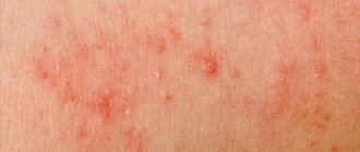
- Toxic reticular melanosis. At risk are people who work for a long time at coal or oil refineries. At first, a person develops red-black spots on the body, which cause burning and itching. Afterwards, lesions with hyperpigmentation (from gray to dark blue) form. They are localized in the lower part of the body (back, legs and shoulders). In sore areas, the skin becomes thinner, peeling occurs, as well as spider veins. Treatment of such a lesion includes avoiding contact with pathological components and strengthening the body's immune system.
If the cause of the appearance of formations on the body is damage to internal organs, then it is important to begin its comprehensive and effective treatment as soon as possible, and only then pay attention to the relief of dark spots.
Skin vasculitis
Skin vasculitis is a group of diseases of a multifactorial nature, in which the leading symptom is inflammation of the blood vessels of the dermis and subcutaneous tissue.
The difficulty in covering this topic is that until now there is no generally accepted classification or even agreed upon terminology of vasculitis. Currently, about 50 different nosological forms have been described, and understanding this diversity is not easy. The diversity of clinical manifestations and insufficiently studied pathogenetic mechanisms have led to the fact that under different names only a variant of the main type of skin lesion may be hidden. Also, in addition to primary vasculitis, which is based on inflammatory damage to the blood vessels of the skin, there are also secondary vasculitis (specific and nonspecific), developing against the background of a certain infectious (syphilis, tuberculosis, etc.), toxic, paraneoplastic or autoimmune (systemic lupus erythematosus, dermatomyositis etc.) process. It is possible to transform skin vasculitis into a systemic process with damage to internal organs and the development of severe, sometimes life-threatening complications.
Skin vasculitis is a polyetiological disease. The most common connection is with a focal infection (streptococci, staphylococci, mycobacterium tuberculosis, yeast, viruses, etc.). Hypersensitivity to a number of drugs, in particular to antibiotics and sulfonamide drugs, is of particular importance. Often, despite a carefully collected anamnesis and examination, the etiological factor remains unclear. Among the risk factors for vasculitis, one should take into account: age (children and the elderly are most vulnerable), hypothermia, excessive insolation, severe physical and mental stress, trauma, surgery, liver disease, diabetes, hypertension. The pathogenetic mechanism for the development of skin vasculitis is currently considered to be the formation of circulating immune complexes with their subsequent fixation in the endothelium, although this has not been definitively proven for all diseases of this group.
Skin vasculitis is a heterogeneous group of diseases, and their clinical manifestations are extremely diverse. However, there are a number of common features that unite these dermatoses:
1) inflammatory nature of skin changes; 2) symmetry of rashes; 3) tendency to edema, hemorrhage and necrosis; 4) primary localization on the lower extremities; 5) evolutionary polymorphism; 6) connection with previous infectious diseases, medication, hypothermia, allergic or autoimmune diseases, and impaired venous outflow; 7) acute or worsening course.
Skin lesions with vasculitis are diverse. These may be spots, purpura, nodules, nodes, necrosis, crusts, erosions, ulcers, etc., but the main clinical differential sign is palpable purpura (hemorrhagic rash that rises above the surface of the skin and is felt on palpation).
There is no generally accepted classification of vasculitis. Vasculitis is systematized according to different principles: etiology and pathogenesis, histological picture, severity of the process, features of clinical manifestations. Most clinicians use predominantly morphological classifications of cutaneous vasculitis, which are usually based on clinical changes in the skin, as well as the depth of location (and, accordingly, the caliber) of the affected vessels. There are superficial (damage to the vessels of the dermis) and deep (damage to the vessels at the border of the skin and subcutaneous tissue) vasculitis. Superficial ones include: hemorrhagic vasculitis (Henoch-Schönlein disease), allergic arteriolitis (polymorphic dermal angiitis), leukoclastic hemorrhagic Miescher-Storck microbiota, as well as chronic capillaritis (hemosiderosis): Majocchi's annular telangiectatic purpura and Schamberg's disease. To the deep: cutaneous form of periarteritis nodosa, acute and chronic erythema nodosum.
Hemorrhagic vasculitis is a systemic disease that affects small vessels of the dermis and manifests itself as palpable purpura, arthralgias, gastrointestinal (GIT) lesions and glomerulonephritis. It occurs at any age, but boys aged 4 to 8 years are at greatest risk. Develops after an infectious disease, after 10–20 days. The acute onset of the disease, with fever and symptoms of intoxication, is most often observed in childhood. The following forms of hemorrhagic vasculitis are distinguished: cutaneous, cutaneous-articular, cutaneous-renal, abdominal-cutaneous and mixed. The current can be lightning fast, sharp and protracted. The duration of the disease varies - from several weeks to several years.
The process begins symmetrically on the lower limbs and buttocks. The rashes are papular-hemorrhagic in nature, often with urticarial elements, and do not disappear with pressure. Their color changes depending on the time of appearance. The rashes occur in waves (once every 6–8 days); the first waves of the rash are the most violent. Articular syndrome appears either simultaneously with skin lesions or after a few hours. Large joints (knees and ankles) are most often affected.
One of the variants of the disease is the so-called necrotic purpura, observed during the rapid course of the process, in which necrotic skin lesions, ulcerations, and hemorrhagic crusts appear.
The greatest difficulties are caused by the diagnosis of the abdominal form of hemorrhagic vasculitis, since skin rashes do not always precede gastrointestinal phenomena (vomiting, cramping pain in the abdomen, tension and pain on palpation, blood in the stool).
The renal form is manifested by impaired renal activity of varying degrees of severity, from short-term unstable hematuria and albuminuria to a pronounced picture of acute glomerulonephritis. This is a late symptom and never occurs before the skin is affected.
The fulminant form of hemorrhagic vasculitis is characterized by an extremely severe course, high fever, widespread rashes on the skin and mucous membranes, viscerapathies, and can result in the death of the patient.
Diagnosis of the disease is based on typical clinical manifestations; in atypical cases, a biopsy is performed. In the abdominal form, surgical supervision is necessary. Observation by a nephrologist is recommended for three months after resolution of purpura.
The term “allergic arteriolitis” Ruiter (1948) proposed to name several related forms of vasculitis, differing in clinical manifestations, but having a number of common etiological, pathogenetic and morphological features.
The pathogenetic factors of the disease are considered to be colds and focal infections. The rashes are usually located symmetrically and are polymorphic in nature (spots, papules, vesicles, pustules, necrosis, ulcerations, telangiectasia, blisters). Depending on the predominant elements, three forms of the disease are distinguished: hemorrhagic type, polymorphic-nodular (corresponds to three-symptomatic Gougerot-Duperre disease) and nodular-necrotic dermatitis (corresponds to Werther-Dümling nodular-necrotic dermatitis). When the rash regresses, cicatricial atrophies and scars may remain. The disease is prone to relapse. Often before the rash, patients complain of malaise, fatigue, headache, and at the height of the disease - pain in the joints (which sometimes swell) and in the abdomen. Diagnosis of all types of the disease is difficult due to the lack of typical, characteristic symptoms. Histological examination reveals fibrinoid lesions of small-caliber vessels with the formation of infiltrative accumulations of neutrophils, eosinophils, lymphocytes, plasma cells and histiocytes.
The clinical course of hemorrhagic leukoclastic microbid Miescher-Storck A sign that makes it possible to distinguish this disease as an independent one is the presence of a phenomenon - leukoclasia (disintegration of the nuclei of granular leukocytes, leading to the formation of nuclear dust) during histological examination. Thus, hemorrhagic leukoclastic microbide can be interpreted as a dermatosis caused by chronic focal infection (intradermal tests with streptococcal antigen are positive), occurring with severe leukoclasia.
Chronic capillaritis (hemosiderosis), in contrast to acute purpura, is characterized by a benign course and is exclusively a skin disease.
Schamberg's disease is a lymphocytic capillaritis characterized by the presence of petechiae and brown purple spots, occurring most often on the lower extremities. Patients are concerned solely as a cosmetic defect.
Majocchi purpura is characterized by the appearance on the lower extremities of pink and liquid-red spots (without previous hyperemia, infiltration), slowly growing to form ring-shaped figures. In the central part of the spot, slight atrophy and achromia develop, and vellus hair falls out. There are no subjective sensations.
Periarteritis nodosa is characterized by necrotizing inflammation of small and medium-sized arteries of the muscular type, followed by the formation of vascular aneurysms and damage to organs and systems. Most common in middle-aged men. Of the etiological factors, the most important are drug intolerance (antibiotics, sulfonamides), vaccination and persistence of HbsAg in the blood serum. The disease begins acutely or gradually with general symptoms - increased body temperature, rapidly increasing weight loss, pain in the joints, muscles, abdomen, skin rashes, signs of damage to the gastrointestinal tract, heart, peripheral nervous system. Over time, polyvisceral symptoms develop. Particularly characteristic of periarteritis nodosa is kidney damage with the development of hypertension, which sometimes becomes malignant with the occurrence of renal failure. There are classic and cutaneous forms of the disease. Skin rashes are represented by nodules - single or in groups, dense, mobile, painful. The formation of nodes along the arteries is typical, sometimes they form strands. Localization on the extensor surfaces of the legs and forearms, on the hands, face (eyebrows, forehead, corners of the jaw) and neck. They are often not visible to the eye and can only be determined by palpation. Necrosis may develop in the center with the formation of long-term non-healing ulcers. Periodically, the ulcers may bleed for several hours (a symptom of a “bleeding subcutaneous node”).
Sometimes the only manifestation of the disease may be reticular or branched livedo (persistent violet-red spots), localized on the distal parts of the extremities, mainly on the extensor surfaces or lower back. It is typical to detect nodules along the course of livedo.
Diagnosis of the disease is based on a combination of damage to a number of organs and systems with signs of significant inflammation, fever, changes primarily in the kidneys, heart, and the presence of polyneuritis. There are no laboratory parameters specific for this disease. Dynamic clinical observation of the patient is crucial for diagnosis.
Acute erythema nodosum is a panniculitis that is characterized by the presence of painful pink nodules on the extensor surface of the lower extremities. Accompanied by fever, malaise, diarrhea, headache, conjunctivitis and cough. Among adults, erythema nodosum is 5–6 times more common in women, with a peak age of 20–30 years. The disease is based on hypersensitivity to various antigens (bacteria, viruses, fungi, neoplasms and connective tissue diseases). Half of the cases are idiopathic. Diagnosis is based on history and physical examination. A complete blood count, chest X-ray (detects bilateral adenopathy in the hilar region), a throat swab, or a rapid streptococcal test should be performed.
Chronic erythema nodosum is a group of different types of dermohypodermatitis nodosum. Women aged 30–40 years are most often affected. Nodes of various sizes appear on the legs with reddened skin over them, without a tendency to necrosis and ulceration. Inflammatory phenomena in the area of the rash and subjective sensations (arthralgia, myalgia) are mild. Clinical variants of chronic erythema nodosum have their own characteristics, for example, the tendency of the nodes to migrate (Beferstedt's erythema migrans) or the asymmetry of the process (Vilanova-Pinol hypodermitis).
Tactics for managing a patient with skin vasculitis
- Classify the disease (characteristic clinical picture, anamnesis, histological examination).
- Search for an etiological factor, but in 30% of cases it cannot be established (search for foci of chronic infection, microbiological, immunological, allergological and other studies).
- Assessment of the general condition and determination of the degree of disease activity: general blood and urine analysis, biochemical blood test, coagulogram, immunogram. Degree of vasculitis activity: I. Rashes are not abundant, body temperature is not higher than 37.5, general symptoms are insignificant, ESR is not higher than 25, C-reactive protein is not more than ++, complement is more than 30 units. II. The rash is abundant (extends beyond the lower leg), body temperature is above 37.5, general symptoms are headache, weakness, symptoms of intoxication, arthralgia; ESR is higher than 25, C-reactive protein is more than ++, complement is less than 30 units, proteinuria.
- Assessment of signs of systemicity (research according to indications).
- Determination of the type and regimen of treatment depending on the degree of activity: Art. I. — treatment on an outpatient basis is possible; II Art. - in the hospital. In all cases of exacerbations of skin vasculitis, bed rest is necessary, since such patients usually have pronounced orthostasis, which should be observed until the transition to the regressive stage. A diet excluding irritating foods (alcoholic drinks, spicy, smoked, salty and fried foods, canned food, chocolate, strong tea and coffee, citrus fruits) is recommended.
- Etiological treatment. If it is possible to eliminate the causative agent (drug, chemicals, infection), then resolution of the skin lesions follows quickly and no other treatment is required. But we must remember that when sanitizing foci of infection, an increase in the vascular process may be observed.
- Pathogenetic treatment.
- Preventive measures: medical examination, prevention of provoking factors (infections, hypothermia, insolation, stress, etc.), rational use of medicines, employment, physical therapy, sanatorium treatment.
Treatment of hemorrhagic vasculitis
- Glucocorticosteroids (prednisolone up to 1.5 mg/kg) alleviate the manifestation of skin-articular syndrome, but do not shorten the disease or prevent kidney damage. Prescribed in severe cases and under the cover of heparin, because they increase blood clotting.
- Nonsteroidal anti-inflammatory drugs (NSAIDs) in usual therapeutic dosages. The choice of a specific drug is not of fundamental importance (indomethacin, diclofenac, acetylsalicylic acid).
- Anticoagulants and antiplatelet agents. Heparin for a common process: 300–400 units/kg/day. The duration of the course should be at least 3–5 weeks. Under coagulogram control.
- Therapeutic plasmapheresis, when the manifestations of the disease are not eliminated by the listed means.
- Nicotinic acid in tolerable doses intravenously.
- You should not use: antihistamines (possibly only at the very beginning of the disease), calcium supplements, all vitamins.
Treatment of skin vasculitis
1) NSAIDs (naproxen, diclofenac, Reopirin, indomethacin, etc.); 2) salicylates; 3) Ca preparations; 4) vitamins P, C, antioxidant complex; 5) vasodilators (xanthinol nicotinate, pentoxifylline); 6) 2% solution of potassium iodide, 1 tbsp. l. 3 times a day (erythema nodosum); 7) anticoagulants and antiplatelet agents; detoxification methods IV drip; 9) glucocorticosteroids (GCS) 30–35 mg/day for 8–10 days; 10) cytostatics; 11) ultra-high frequency therapy, diathermy, inductothermy, ultrasound with hydrocortisone, ultraviolet irradiation.
External treatment. For erosive and ulcerative rashes
1) 1–2% solutions of aniline dyes; 2) epithelializing ointments (solcoseryl); 3) ointments containing glucocorticoids, etc.; 4) lotions or ointments with proteolytic enzymes (Chymopsin, Iruksol); 5) Dimexide applications;
For knots - dry heat.
Treatment should not end with the disappearance of clinical manifestations of the disease. It continues until laboratory parameters are completely normalized, and in the next six months to a year, patients are given maintenance treatment
Literature
- Adaskevich V.P., Kozin V.M. Skin and venereal diseases. M.: Med. lit., 2006, p. 237–245.
- Kulaga V.V., Romanenko I.M., Afonin S.L. Allergic diseases of the blood vessels of the skin. Lugansk: “Etalon-2”, 2006. 168 p.
- Berenbein B. A., Studnitsin A. A. et al. Differential diagnosis of skin diseases. M. Medicine, 1989. 672 p.
I. B. Mertsalova, Candidate of Medical Sciences RMAPO, Moscow
Contact information about the author for correspondence
Skin mastocytosis
With such a lesion, spots with black dots appear on the person’s body. The disease occurs due to the pathological proliferation of mast cells (they are responsible for the state of human health) and their accumulation in large numbers in the epithelium. Doctors divide the disease into a skin form, in which dark spots, nodules and spider veins appear on the human body, and a systemic form (the spots spread to internal organs).
Mastocytosis in a child occurs in the first years of his life. The most common form of mastocytosis is cutaneous. The disease goes away on its own as you grow older.
In elderly and adult people, the disease spreads not only to the skin, but also to internal organs (heart, kidneys, liver, spleen).
There are the following types of disease:
- Maculopapular - multiple dark rashes appear on the skin, which, when scratched, turn into blisters and hives. This type of mastocytosis is also called urticaria pigmentosa.
- Nodal form. With this condition, a person is diagnosed with small blisters ranging in size from 7 to 10 millimeters. They can be pink or light brown, often merge and form large plaques.
- Solitary form. In this case, one large dark spot with a size of 5 to 6 centimeters appears on the body. It is usually localized on the shoulders, abdomen, back and neck. If you accidentally touch the lesion, it will turn into a blister and cause severe itching.
- Erythroderma - yellow-brown dense spots. They have no boundaries, are easily deformed and provoke the formation of cracks and ulcers. Most often, the spots spread to the buttock folds and hollows.
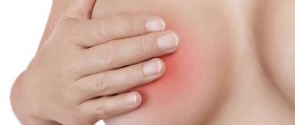
- Telangiectasia is a large number of dark spider veins in the neck and chest. In most cases, such formations are formed in women.
Treatment of any type of mastocytosis should be comprehensive and include the use of hormonal agents, cytostatics, antioxidants and antiallergic medications. If black spots appear on the body in a single quantity, then it can be eliminated surgically.
Treatment of pigmentation
There are several ways to help get rid of brown spots on the skin. An integrated approach and a combination of different types of treatment are possible. Freckles, juvenile or age-related lentigo, chloasma resulting from pregnancy or taking medications - these spots can be corrected with medication, through cosmetic procedures, as well as using masks at home.
Medicines
Pharmacies sell preparations for external use that have a good lightening effect. The most popular antifungal agents are: Clotrimazole, zinc and sulfur ointments. Antibacterial compounds noticeably whiten brown spots: syntomycin ointment based on chloramphenicol, as well as salicylic ointment. Pharmacists can offer salicylic-zinc paste. It provides a good exfoliating effect, reduces the production of melanocytes and brightens dark areas of the skin.
If hyperpigmentation appears as the main symptom or consequence of a serious illness, it requires the attention of a doctor. After a proper examination, he will explain how to treat the pathology. If the spots appear due to endocrine diseases, you will need to take hormonal medications. If there is a disorder in the gastrointestinal tract, medications may be prescribed to treat gastritis or peptic ulcers, as well as probiotics.
Cosmetic procedures
Pigment bleaching is carried out in several ways. Cosmetologists can offer:
- Laser resurfacing . The procedure is painful, but highly effective. It is performed under anesthesia and is usually used on the face.
- Chemical peeling is a radical measure in which brown spots are removed using a special acid composition. The result will appear after 3-10 sessions.
- Phototherapy , which gives a good effect after the first procedure. The skin is treated with waves of pulsed light. This radiation is safe for humans. It destroys melanin without damaging the epithelium.
- Mesotherapy is a method in which a medicinal cocktail is injected subcutaneously into the patient. The composition is selected individually. In addition to eliminating brown spots, mesotherapy also solves other problems, for example, rejuvenating the face. Experts consider this procedure less traumatic and more effective than laser resurfacing.
Details of each procedure, as well as possible side effects, can be found in consultation with a cosmetologist.
Folk remedies
At home, you can use whitening masks prepared with your own hands. The following recipes are popular:
- Half a lemon is grated on a fine grater, mixed with sour cream in a 1:1 ratio, applied in a thick layer to brown spots and left to act for 30 minutes. Wash off the mixture with warm water. This mask is made 2 times a day until the desired result is achieved.
- Gauze wipes are moistened with hydrogen peroxide and applied to pigmented areas. As the gauze dries, it is moistened again. The duration of the procedure is 15 minutes, and it is performed once a day, no longer than two weeks. The course can be repeated after a week break.
- Chop fresh parsley and pour in 5 tbsp. l. raw materials 100 g of vodka. Parsley is infused in the refrigerator for 7 days, then 100 g of cold water is added and the mixture is shaken. Use this tincture to wipe pigment spots 5-6 times a day. The course of treatment is at least 1 month.
- For mild spots and sensitive skin, gentle products are used. You can soak a gauze pad in yogurt and apply it to the affected area of skin for 15 minutes. The procedure is repeated 1-2 times a day. The minimum course is 2 weeks.
- Fresh cucumber is grated and left in the refrigerator for 1 hour. Then the paste is applied to the brown spots, left for 15 minutes, and then washed off with warm water. This procedure is best done before bedtime.
In addition, cosmetics are used at home to whiten pigmented areas - special creams, lotions, serums. Preference should be given to expensive professional cosmetics. Cheap drugs do not contain natural substances and can harm the skin.
Acanthosis nigricans
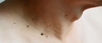
Acanthosis nigricans also leads to the appearance of dark pigmentation on the human body. This is a rare type of skin dermatosis, which manifests itself in thickening of the stratum corneum of the skin, age spots and papillomas.
In most cases, pigmentation spreads in the folds of the skin: in the armpits, under the knees, near the neck, under the breasts, in the groin area and thighs.
Why do black spots appear on the body? The main factors leading to the onset of the disease are not precisely known. Doctors believe that acanthosis may indicate diseases of the endocrine system or the presence of malignant and benign formations in the body.
Pigmentation in acanthosis nigricans can be light brown or dark in color, do not have a clear boundary, and also spread over large areas of the body. The skin in the affected areas becomes thicker and is often covered with a large number of small papillomas. This form of rash does not affect the patient’s condition in any way, but only brings cosmetic discomfort.
To get rid of acanthosis nigricans, it is first important to eliminate the root cause of the lesion. To do this, the doctor prescribes the use of immunostimulants, vitamin complexes and cosmetic gels. You can see the black spots on the body in detail in the photo.
Freckles on the skin
Almost every person has a certain number of freckles on their body. Sometimes they spread to the hands, back, and chest. Freckled rash appears in humans as a result of heredity. It is formed due to the fact that melanin in the skin is distributed unevenly.
The peak prevalence of freckles on the human body occurs in the spring and summer. Freckles are numerous small rashes (size 2 to 3 millimeters). Their color ranges from light yellow to dark brown.
The freckled rash may become darker when exposed to sunlight. Experts believe that the skin of people who are susceptible to the formation of hemp is particularly sensitive.
Freckles do not need treatment. Many people even like such rashes on the body. If a person wants to eliminate them, then he can use a specialized whitening cream. To prevent the appearance of rashes, it is important to have the right diet, start taking a complex of vitamins, and also protect your skin from the rays of the sun.
Preventive actions
To prevent the development of spots on the body, the following recommendations should be followed:
- in sunny weather, it is mandatory to wear a hat;
- eat foods with plenty of vitamin C, and also limit your intake of vitamin A;
- Before going outside, you should use sunscreen;
- wash your face with fermented milk products (kefir, yogurt).
Burning and itching during pigmentation indicates liver disease. When scratching the affected area of the body for a long time, the skin becomes yellow. Eliminating the root cause of dark spots will help get rid of the problem and prevent metastasis.
Prevention of occurrence
To preserve the beauty of your hands for a long time, it is necessary to begin treatment of pigmentation in a timely manner and carry out proper care:
- use lightening creams and ointments;
- exfoliate regularly;
- protect from frost;
- apply protective cream when going out into the sun;
- eat foods high in vitamin C.
It is possible to get rid of age spots on your hands, but sometimes it requires a lot of money or time. The best way to track your results is by using a photo, where all the changes are clearly visible. If you managed to remove all the dark areas, do not forget to prevent their appearance, otherwise they may pop up again in the same place and cause problems.
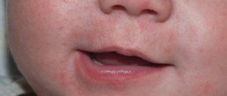
Spots on a child's body
Skin is a barrier between the outside world and the internal organs and systems of a person. She takes on negative influence from the outside. Black spots on a child’s body are skin developmental defects. They can be of different nature, color and shape. If they occur after the birth of a child, then they are classified as congenital; if after the child grows up, then they are classified as acquired.
Melanin directly affects human skin color. When certain parts of the body are exposed to certain external factors, increased production of melanin begins, which leads to skin pigmentation.
A child may develop dark spots for the following reasons:
- As a result of heredity. The presence of a large number of moles and other pigmented formations in relatives often leads to their appearance in the child.
- A disturbance in the production of hormones in a pregnant woman while carrying a child.
- Negative effects of external factors on a woman’s body (strong radiation, exposure to chemical components, harmful work, sudden climate change).
- Infection of the genitourinary system.
- Genetic diseases, one of which is the pigmentation process.
Treatment in a child
It is important to remember that the skin of a newborn baby is very sensitive and delicate, so it is quickly exposed to any negative processes. Anatomical and physiological features (thin epithelium, humoral and immature immunity) provoke a direct impact on the body of external factors. Pigmentation in a child is a serious reason to visit a treating specialist. Only an experienced doctor can determine the danger and shape of a dark spot after carrying out diagnostic measures.
Treatment of dark formations on the child’s body, if they do not cause any discomfort, involves dynamic monitoring of the general condition. Lightening and whitening preparations are almost never used to eliminate dark spots on a child’s body. Many products contain hormones or chemical components that are dangerous for the baby. After consulting a doctor, pigmentation in a child over 6-7 years old can be eliminated using parsley juice, lemon, cucumber and other folk remedies. If the size of the formation increases or new ones appear, it is important to seek help from a doctor.
How to properly care for your face and body with blackheads?
To reduce the risk of the formation of open comedones, it is necessary to properly care for your skin - clean it regularly. It is necessary to remove excess sebum and dead skin particles. For washing, you need to select mild cosmetics designed for your skin type, for example, problematic or oily. This product may contain glycolic or salicylic acid. Homemade peels and scrubs can be suitable for cleaning, but they must be used carefully so as not to injure the skin.