The structure of the nose and its sinuses
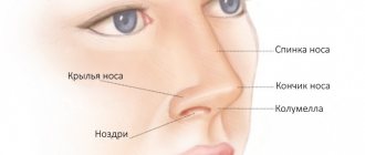
The anatomical structure of the nose consists of a cavity and paranasal sinuses. The nasal cavity is limited by five boundaries:
- posterior border - choana;
- anterior border - the plane separating the nose from the vestibule;
- the upper wall is the vault, separated by a perforated plate, bone and the body of the sphenoid bone;
- bottom wall – bottom;
- outer wall – medial side of the maxillary sinuses.
An inseparable part of the nose is its sinuses:
- maxillary (maxillary) - a paired air-bearing organ localized on the body of the maxillary bone;
- sphenoid (main, sphenoidal) - located in the body of the sphenoid bone at the point of transition of the anterior cranial fossa to the anterior one;
- ethmoid labyrinth (cells of the ethmoid bone) - a paired organ located in the center between the orbits on the side of the palatine, lacrimal, frontal sphenoid, maxillary bones;
- frontal - a paired organ located behind the superciliary arches on the frontal bone.
The tumor mainly develops in the maxillary sinuses, gradually covering the ethmoid labyrinth and the entire nasal cavity. Much less frequently, the lesion is diagnosed in the frontal and sphenoid sinuses.
Osteoma
Most often it is localized in the frontal and ethmoid sinuses, less often in the maxillary sinuses and very rarely in the basal sinus. The tumor has a wide base or a narrow stalk.
On radiographs and SCT scans (at ACMD-Medox we use it most often for diagnosing osteomas), osteoma has the appearance of an additional bone formation of a compact or spongy structure, with a clear contour, and a homogeneous structure. If the tumor grows, it can occupy the entire sinus, and then begin to deform it, growing even more.
Classification
Depending on the location of the pathological process, oncological processes are divided into several groups:
| Damage to the epithelial layer | Squamous cell carcinoma |
| Verrucous cancer | |
| Spinocellular carcinoma | |
| Adenocystic cancer | |
| Mucoepidermoid carcinoma | |
| Mucous adenocarcinoma | |
| Adenocarcinoma | |
| Transitional cell carcinoma | |
| Undifferentiated cancer | |
| Other | |
| Soft tissue damage | Fibrosarcoma |
| Malignant fibroxanthoma | |
| Rhabdomyosarcoma | |
| Malignant hemangiopericytoma | |
| Neurogenic sarcoma | |
| Other | |
| Damage to osteochondral tissue | Osteoma |
| Chondroma | |
| Osteogenic sarcoma | |
| Chondrosarcoma | |
| Other | |
| Damage to hematopoietic and lymphoid tissue | Extranodal β - cell marginal zone lymphoma MALT - type |
| Hodgkin's disease | |
| Diffuse β-cell lymphoma | |
| Anaplastic large cell lymphoma, peripheral T-cell lymphoma | |
| Mixed tumors | Malignant melanoma |
| Teratoma | |
| Esthesioneuroblastoma | |
| Malignant melanoma | |
| Other | |
| Secondary tumors | |
| Unclassified tumors |
Treatment of malignant neoplasms of the nose and paranasal sinuses
The treatment plan is drawn up only after histological examination of the tumor. Combination treatment is recommended (surgical in combination with radiation therapy, chemotherapy). For inoperable nasal tumors, radiation treatment, chemotherapy, and palliative measures in the form of ligation and transection of large afferent blood vessels (external carotid arteries) are performed. However, even despite earlier treatment, timely treatment, and surgical intervention, quite often malignant tumors recur within a few months.
Causes
Oncological processes develop against the background of chronic hyperplasticity. Based on practice, cancer of the nose and sinuses becomes a consequence of complications:
- cysts of radicular or follicular type;
- purulent glandular-fibrous polypous rhinosinusitis;
- purulent polypous rhinosinusitis, complications of which are caused by dysplasia and metaplasia of the epithelium;
- hyperplastic sinusitis with dysplasia;
- chronic sinusitis;
- pigmented nevus;
- perforation of the nasal septum;
- leukoplakia.
The development of a malignant tumor is influenced by such diagnoses as squamous cell papilloma, hemangioma, fibromatosis, osteoblastoclastoma, pleomorphic adenoma of the minor salivary gland. Nasal cancer is diagnosed at any age, but a greater prevalence of the disease is recorded in old age after 60 years. A malignant tumor ranks 35th among all oncological pathologies. The risk group includes:
- patients who had nasal polyps removed or cauterized;
- people working in hazardous industries. Inhalation of carcinogens and chemicals affects the condition of the mucous membrane;
- unfavorable ecology and other environmental factors.
Types of growths in the nose
Have a growth appeared in your nostril or on your nose? It is necessary to determine whether these are papillomas, warts or a polyp.
| Types of growths | ||
| Name | Description | Localization |
| Papillomas | Epithelial tumors caused by HPV | At the entrance to the cavity |
| Along the walls | ||
| On top of the partition | ||
| Polyps | Round formations from overgrown nasal mucosa, non-painful | Lattice labyrinth (typical for adults); |
| Maxillary sinuses (more often in children). | ||
| Warts | Growths caused by HPV and prone to self-destruction | Any location, both inside and outside, most often at the tip |
The papilloma virus can provoke the growth of tumors in the nose.
Return to contents
Papillomas
A seal growing on the nasal septum can be of the following types:
- Flat, in the form of a speck in the surface layer of the mucous membrane or skin. Characteristic of a chronic process.
- Pointed, medium-sized, flesh-colored. Characteristic of an acute course.
- Thread-like with an elongated shape, requires removal.
According to another classification, there are:
- Inverted seal. It grows on the cartilage of the nose or on the sides of the walls. More often diagnosed in old age. It bleeds and makes breathing difficult.
- Exophytic growth on the bridge of the nose or at the entrance to the nostril. It has a dense structure with a lumpy shell, red in color. Makes breathing difficult.
You can become infected with papilloma through sexual or domestic contact.
Return to contents
Causes
The main provocateur of the appearance is HPV during primary infection or when activated against the background of reduced immunity. You can catch a pathogen:
- How to get rid of nasal polyps at home - 5 effective methods of treatment with folk remedies
- during unprotected sexual intercourse;
- when visiting public places without due attention to personal hygiene.
Return to contentReturn to content
Symptoms
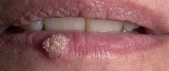
Often the growth that has grown in front of the nostril does not hurt. Possible itching and tingling in the growth area. Due to its epithelial origin, when papilloma is damaged, nosebleeds often develop. The appearance of the formation is a flesh-colored bulge, less often with a brownish or reddish tint. As the tumor grows, it takes on the appearance of a cauliflower.
Treatment of papilloma in the nose is aimed at eliminating the virus and getting rid of the growth.
Return to contents
Therapeutic measures
A growth on/in the nose should only be treated under the supervision of a doctor. Self-treatment can lead to curvature of the bridge of the nose and other complications.
| An approach | Facilities | Description | Notes |
| Medication | For necrotization of unhealthy tissues | “Verrukatsid”, “Kondilin”, “Solcoderm”, “Vartek” | Doctor's prescription required |
| Keratolytic | "Lyapis", "Kolomak", solcoseryl ointment, "Salipod" patch | ||
| Antiviral and immunomodulatory | Oxolinic ointment | ||
| People's | Pulp of golden mustache leaves | Apply within 7 days | |
| Rowan and cabbage juices, as well as squeezes from celandine, leaves and stems of nettle, calendula flowers | Use until no longer available | ||
| Chalk powder | Apply for 4 weeks | ||
| Mixture of honey and onion juice | Use until disappearing | ||
| Removal | Cryodestruction | Treating the build-up with liquid nitrogen | Not applicable to genital papillomas |
| Laser | Burnout | Without pain and scars | |
| Electrocoagulation | Exposure to high frequency current pulses | For large tumors | |
| Radio knife | Excision with a special scalpel | May leave scars, but papillomas go away forever |
Nasal polyps usually do not cause pain.
Return to contents
Nasal polyps
These formations grow from the nasal mucosa and do not hurt. They are able to grow both inward and outward from the maxillary sinuses or the ethmoid labyrinth of the nose. Polyps develop on the mucous membrane in humans in 3 stages:
- the tumor is located on the upper cartilaginous part;
- gradually spreads along the septum;
- occupies the entire cavity.
Return to contents
Etiology
The causative factors for the appearance of nasal polyps are:
- frequent inflammation and infections, which lead to detachment of the nasal mucosa;
- allergies;
- getting stuck and injuring the nasal cavity with foreign bodies;
- cystic fibrosis;
- asthma;
- congenital or acquired deformation of the nasal septum;
- sinusitis.
Symptoms
A growing nasal polyp causes snoring, sneezing, and decreased sense of smell and breathing.
The early stage is asymptomatic. Subsequently, the following are observed:
- breathing problems;
- problems with smell;
- sneezing;
- snore;
- nasal discharge.
As the patient develops, he feels:
- Dry nose: mechanism and factors of occurrence, connection with diseases, how to treat
- headache;
- ringing, noise, congestion in the ears;
- burning and soreness in the nasal cavity;
- weakening of hearing.
Return to contents
Treatment of polyps
| An approach | Facilities | Description | Notes |
| Medication | Corticosteroids | Nasal sprays based on beclomethasone, fluticasone, budesonide, mometasone | Allows you to reduce polyps and prevent relapse |
| Antiallergic | "Loratadine", "Cetirizine" | ||
| Antibacterial | "Augmentin", "Sumamed", "Amoxicillin" | ||
| Surgery | Endoscopic excision | Recommended if medications are ineffective |
Corticosteroids are prescribed only by a doctor with precise dosages and frequency of administration, so that mucosal atrophy does not occur and the nasal cartilage does not become deformed.
Return to contents
Warts
The peculiarity of warts on the nose is the ability to self-destruct. Since the provocateur of the problem is HPV, the appearance and disappearance of the growth depends on the activity of the virus. But if you accidentally tear it off, it forms again and often more than one due to infection of healthy tissues. There are filiform, flat, and simple warts. These are dense white formations with a hard crust of the stratum corneum of the skin. They often grow outside/under the nose or in the nostril. Growths are often found on the skin and other parts of the body.
Return to contents
Why do they appear?
The provocateur is HPV, or more precisely, the activity of the virus due to weakening of the immune system. Provoking factors:
The reason for the growth of warts in the nose can be poor hygiene, weak immunity, or a malfunction in the body’s hormonal processes.
- lack of hygiene;
- stress;
- disruptions at the hormonal level;
- metabolic disorders;
- pregnancy;
- chronic and viral pathologies.
Return to contents
How to get rid of it?
| An approach | Facilities | Description | Notes |
| Drug | Antivirus | "Viru-Merz", oxolinic ointment | Dosages, frequency of use, course of treatment are determined by the doctor |
| Immunomodulatory | Vitamins, preparations with barrier-protective properties | ||
| Drugs with necrotizing properties | "Kondilin", "Vartek", "Verrukatsid" | ||
| Keratolytics for melting growths | "Salipod", "Kolomak", salicylic ointment, "Lapis" | ||
| Cryotherapeutic | "Cryopharma" | ||
| Surgery | Cryodestruction | Liquid nitrogen freezing | Requires a professional approach, because with incorrect excision there is a risk of relapse |
| Laser | Burnout | ||
| Folk recipes | Lubricate with salted sour cream | With doctor's permission | |
| Apply onion leaves | |||
| Tomato pulp compress |
Return to contents
Symptoms
In the first stages of development, nasal cancer develops asymptomatically. Doctors misdiagnose the tumor, mistaking it for chronic inflammation.
The first signs of a tumor of the nose and paranasal sinuses:
- discharge. First, the patient notices congestion in one nasal passage, breathing is impaired on one side, which is explained by the tumor growing into the cavity. Over time, the face swells, the eyeballs shift, and mucus production increases. When the maxillary sinuses are affected, the secretion is mucopurulent mixed with blood;
- pain syndrome. At first there is a headache. With a tumor of the ethmoid labyrinth, the head hurts already at the beginning of the development of the disease. Pain in the nose appears already at a late stage of cancer. It spreads to the temples, eyes and ears, so the patient sometimes cannot determine the exact location of the lesion. With cancer of the maxillary sinuses, toothache increases. Neurological signs of cancer of the nose and sinuses, for example, numbness of the cheeks and upper lip, increase in the later stages, when the tumor grows and extends into the pterygopalatine fossa.
Stages of development of neoplasms
The severity of signs and symptoms directly depends on the stage of the disease. When the tumor is small, a sign of its appearance is a feeling of congestion. This symptom looks like the beginning of a cold, so often at this stage a person does not diagnose and treat growths. Treatment boils down to the use of vasoconstrictor drops, which ultimately do not help. During this period, the body is more vulnerable to all kinds of infections, and otitis media, tonsillitis and other inflammatory diseases of the ENT organs can occur.
At the next stage, symptoms such as loss of smell and the appearance of a nasal voice appear. If the tumor blocks the opening of the auditory tube, signs of hearing loss appear.
In the third stage, the symptoms of the disease appear most intensely. Nasal discharge, headaches, constant congestion, breathing through the mouth - all these are signs that the polyp has reached an impressive size.
The symptoms of the disease are actually not as harmless as they look. To get rid of them, you must definitely contact an ENT doctor to prescribe competent treatment.
Unfortunately, few patients come to an otolaryngologist at the first signs of illness, when conservative treatment is still possible. Most of them turn to an ENT doctor when only surgical treatment can really help.
Specific signs
In the later stages of oncology development, characteristic clinical features appear that help identify the location of the tumor.
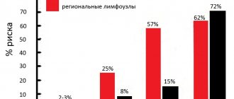
Thus, cancer of the maxillary sinuses causes forward displacement of the eyeballs, swelling of the cheeks, and decreased visual acuity. When the posterolateral and posterior wall is damaged, the temporal fossa swells and exophthalmos develops. Complete obstruction of the nasal passages indicates cancer of the medial wall. A tumor of the ethmoidal labyrinth is recognized by lacrimation, deformation of the inner corner of the eye, and changes in the shape of the lower eyelid.
Already in the later stages of development, nosebleeds appear, protrusion of the eyeballs worsens, the hard palate is deformed, and the retropharyngeal, cervical and parotid lymph nodes enlarge.
Treatment of nose cancer
Initially, it is important to tell your doctor about the symptoms of nasal cancer. The cavity, as well as the sinuses, have a complex anatomy. Therefore, treatment of the disease must be carried out with special care. There are blood vessels and nerves nearby, which are very important. Also located nearby are the carotid artery, oral cavity, eyes, and brain. Therefore, surgery for a diagnosis of nasal cancer is considered quite complicated. If possible, specialists prescribe conservative treatment.
When diagnosing paranasal sinus cancer, a team of doctors such as a speech therapist, dentist, neurosurgeon, reconstructive surgeon, plastic surgeon, and oncologist should work.
Thanks to such an integrated approach, it is possible to ensure effective treatment of nasal cancer, as well as complete rehabilitation of the patient (social, aesthetic, functional), and a return to normal life. Modern methods of treating cancer of the nose and paranasal sinuses include the following options:
- open surgery. It is carried out in the presence of large tumors with a diagnosis of nasal cancer, and resection of the upper jaw is performed. They also restore those structures that have been lost, using microsurgical techniques and implants made in 3D;
- invasive endoscopic interventions for nasal cancer; during this process, no incisions are made on the face. With the help of such operations, you can get excellent results in terms of cosmetology and minimize hospital stay. And this will not worsen the results of treatment of nasal oncology. If large processes are expected, then before the start of surgery, embolization of the vessels that provide nutrition to the tumor is performed, this minimizes blood loss;
- radiation therapy. This type of treatment for nasal cavity cancer can be independent or used simultaneously with chemotherapy. In some situations, IMRT is used, a special technique for radiation treatment for nasal cancer. It reduces damage to healthy tissues that are around;
- chemotherapy. Standard regimens are offered to treat symptoms of nasal cancer, but experimental protocols are also used in which specialists use targeted drugs and administer intra-arterial chemotherapy. Antitumor drugs are injected directly into the vessels when treating nasal cancer, so that there is minimal toxic effect on internal tissues and organs.
Clinical features of some types of nasal tumors
Esthesioneuroblastoma is formed from neuroepithelial cells. The tumor is located in the upper part of the nasal passage, gradually covering the sinuses, brain, base of the skull and orbit. In the photographs it looks like a soft tissue polyp, which gives metastases to the mediastinum, cervical lymph nodes, bones, lungs, and pleura. This type of nasal cancer occurs in older adults, middle-aged men, and children. The disease progresses in three ways - rhinological symptoms worsen when the ethmoidal labyrinth, maxillary sinuses, or orbit are affected, nasopharyngeal symptoms worsen when the tumor spreads to the nasopharynx, choana or ethmoid labyrinth, or neurological symptoms worsen when cancer cells grow into the base of the skull.
Risk factors
All conditions that cause chronic inflammation of the nasal passages or sinuses (infections, allergies) increase the risk of nasal polyps. The following conditions are most often associated with polyps:
- asthma;
- sensitivity to aspirin;
- allergic fungal sinusitis, allergy to fungal spores;
- cystic fibrosis (or cystic fibrosis);
- Churg-Strauss syndrome (a rare disease that causes inflammation of blood vessels);
- vitamin D deficiency;
- Some genetic variations related to the functioning of the immune system may increase the likelihood of developing nasal polyps.
Diagnostics
During the initial examination, the doctor conducts an external examination and palpates the tissues. The specialist notes asymmetry of facial features due to a soft tissue tumor. Rhinoscopy reveals a narrowing of the lumen of the nasal cavity and/or nasopharynx, and oropharyngoscopy reveals a spasm of the masticatory muscles, due to which the mouth does not open completely. If, upon palpation of the lymph nodes in the neck on both sides, compactions are felt, this indicates metastases.
After the consultation, the doctor issues a referral for tests. A cytological examination of the lymph nodes and maxillary sinuses is carried out. A histological examination is prescribed - a biopsy of the lymph nodes and tissues of the nasal cavity.
The next stage is an instrumental examination:
- CT/MRI of the sinuses, upper jaw, base of the jaw and eyes. Tomography indicates the exact location of the tumor focus and its spread to surrounding tissues. If there are dark areas in the images, a more detailed examination is prescribed;
- fibrolaryngoscopy for in-depth examination of the nasopharynx;
- Ultrasound of the abdominal cavity and neck to look for metastases;
- chest x-ray to look for distant metastases;
- fibrobronchoscopy to search for metastases in the mediastinum;
- FGDS to search for metastases in the gastrointestinal tract;
- angiography to search for metastases in the great vessels;
- CT/MRI, PET of the chest and abdominal organs to search for metastases;
- puncture of the maxillary sinuses and nasal cavity to determine the cellular composition of the tumor and its structure.
After clarifying the diagnosis, the doctor determines the stage of development of the disease:
Prognosis of benign tumors of the nasal cavity
As practice shows, a large number of benign tumors of the nasal cavity are not prone to malignancy and are characterized by slow, non-invasive growth. Thanks to this, doctors give a positive prognosis for the patient’s further full recovery. Moreover, modern medicine has various tools for conducting successful and effective operations.
Postoperative relapses are often accompanied by bleeding nasal polyp and papilloma. Of all benign tumors of the nasal cavity, the most unfavorable are chondromas and osteomas. This also applies to chondrosarcoma and osteosarcoma, which are prone to malignancy and cause destruction of surrounding tissue.
Removal of osteomas and chondromas is accompanied by extensive tissue defects, the development of choanal atresia, the formation of synechiae in the nasal cavity, and the like. Under the influence of these factors, persistent disruption of nasal breathing and complete loss of smell occurs.
Doctors recommend that at the first manifestations of benign tumors in the nasal cavity, immediately consult a specialist in order to prevent their development and begin treatment in a timely manner.
Treatment
Treatment tactics are determined by the doctor based on the clinical characteristics and stage of the disease.
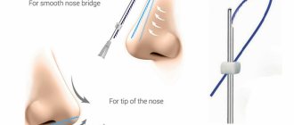
Surgery
Most tumors are treated with surgery. An open or endonasal operation is performed. For example, a malignant tumor of the mucous membrane and sinuses is operated on through an external approach. The doctor exposes the anatomical structures of the cavity, maxilla, ethmoid sinus and orbit. The operation is preceded by radiation exposure, the total focal dose of which does not exceed 50 g.
Operations depending on severity:
| Stage 1 | The tumor is removed manually through access to the nasal cavity. If the lesion is small, then removal using cryodestruction is possible |
| Stage 2 | An open operation is performed. The outer wall and nasal turbinates are excised, cells of the ethmoid sinus and parts of the upper jaw are resected in the presence of cancer cells in the maxillary sinus |
| Stage 3 and 4 | The upper jaw is resected. If the orbit is affected, it is completely excised. Only the bone walls remain |
In case of metastasis, the primary focus is removed, and the cervical tissue is excised. Kreill's operation is rarely performed for massive lesions of the cervical lymph nodes.
Chemotherapy
Patients with poorly differentiated types of cancer are exposed to radiation and chemotherapy. In addition, drugs are prescribed for the following indications:
- verified tumors based on histology and cytology results;
- distant metastases;
- metastases in regional lymph nodes;
- relapse;
- unimpaired functionality of the cardiovascular system, kidneys, liver and respiratory organs;
- blood counts are satisfactory - platelet count is more than 100,000, granulocytes - more than 200, and hemoglobin and hematocrit are normal;
- the patient’s personal refusal to undergo surgery;
- correction of an inoperable tumor into an operable one.
Platinum, methotrexate, bleomycin, fluorouracil, and doxorubicin are prescribed.
Targeted therapy is prescribed for the treatment of squamous cell carcinoma of the head and neck with metastases after a course of chemotherapy, locally advanced squamous cell carcinoma of the head and neck coupled with radiation. Targeted drugs are also used when chemotherapy is ineffective in treating recurrent squamous cell carcinoma.
Diagnosis of nasal polyps
A doctor can usually make a diagnosis based on symptoms and a visual inspection of the nasal passages using a special tool with a flashlight.
In addition, you may need:
- Nasal endoscopy . A narrow tube with a lighted lens or tiny camera (nasal endoscope) allows the doctor to view the inside of the nasal passages and sinuses in detail. The doctor places a tube in the nostril and guides it into the nasal cavity.
- Computed tomography helps to accurately determine the size and location of polyps deep in the sinuses and evaluate the area of inflammation. The test can also detect other possible problems, such as structural abnormalities or other types of benign or malignant tumors.
- Allergy tests (skin tests or blood tests). Usually prescribed to determine the cause of chronic inflammation.
- If we are talking about a child, the doctor may order a test for cystic fibrosis . Because of it, the function of the external secretion glands (sweat, intestinal glands, pancreas), as well as the lungs and liver is disrupted. Cystic fibrosis can affect all organs, but the main symptoms are in the respiratory and digestive systems.
- Blood test for vitamin D.
Prevention
To prevent malignant tumors of the nose and sinuses, it is important to promptly treat inflammation of the nasal structures, prevent the development of hyperplastic processes and immediately remove polyps.
Subsequently, after completing the course of treatment, patients are observed by an oncologist. The first two years should be examined monthly, the next five years - every six months. If there is an increased risk of relapse, routine examinations are prescribed individually.
List of references on the topic:
- Gantsev Sh.H. Oncology – M, 2012 – P.204-205.
- Golovin D.I. Errors and difficulties in diagnosing tumors, D.: Medicine. Leningr. department, 2015 305 pp.
- Golovin D. Im Dvorakovskag I. V. Tumors of the nose and paranasal sinuses. L.: Medicine. Leningr. department 2014. 94 p.
- Selected lectures on clinical oncology/Ed. IN AND. Chissova, S.L. Daryalova. – M., 2010
- Matyakin E.G., Alferov V.S. Chemotherapy of head and neck tumors // Mat. 2nd Ros. oncol. conf. “Current trends in the development of drug therapy for tumors” December 8–10, 2016 – M., 256 p.
- Podvyaznikov S.O., A.I. Paches, T.D. Tabolinovskaya Diagnosis and treatment of malignant tumors of the nasal cavity and paranasal sinuses // SIBERIAN JOURNAL OF ONCOLOGY. . — 2011.
- Tumors of the head and neck: hands/ A.I. Paches. - 5th ed., add. And revised - M.: Practical Medicine, 2013. -478 p.
- Khasanov A.I. Comparative assessment of the effectiveness of treatment of locally advanced malignant tumors of the upper jaw of the nasal cavity and paranasal sinuses Vestn RORC im. N.N. Blokhin RAMS. 2006. T. 17. No. 1 P. 45-49
- Shine A.A. Oncology. M – 2014 365 pp.
- Encyclopedia of Clinical Oncology/Ed. M.I. Davydova. – M., 2014 –P.140-179.
Treatment of benign tumors of the nasal cavity
Benign tumors of the nasal cavity, due to the danger of proliferation and malignancy, and disruption of normal respiratory function, must be removed surgically. Surgery can stop the presence of chronic decompensated diseases (coronary artery disease, heart failure, severe hypertension, bronchial asthma, respiratory failure, diabetes mellitus, liver cirrhosis, renal failure) and the patient’s advanced age.
The method of removing a benign tumor of the nasal cavity directly depends on its growth pattern, size and type. The structure of the tumor and the tissue it affects also play a role.
Small adenomas, papillomas and fibromas are removed under local anesthesia by the endoscopic method using an electrocoagulating loop. In the case of a bleeding nasal polyp, the tumor must be removed along with the part of the nasal septum to which it is attached. The base of the tumor is cauterized by electrocoagulation or cryotreatment. In this way, relapses can be prevented.
Large benign tumors of the nasal cavity are removed with a radio wave knife, laser or scalpel. During the operation, a surgical microscope is used to clearly differentiate tumor cells from surrounding tissues.
If we talk about small vascular benign tumors of the nasal cavity, they are removed by cryodestruction, electrocoagulation or using a laser. Removing large angiomas can cause severe bleeding. In this regard, the carotid arteries are pre-ligated before the operation. If the tumor grows deep into the surrounding tissue, sclerosis of the tumor is performed or occlusion of the vessels feeding it is applied.
Chondromas and osteomas, which grow into structures adjacent to the nose and into the bone walls, quite often doctors have to remove them in parts. For this purpose, not only endonasal, but also external surgical approaches are used. In some cases, surgery may be triggered by resection of bone structures. The appearance of facial defects is also not excluded, since the volume of tissue will increase. In such situations, reconstruction must be carried out using plastic surgery techniques.
Possible complications
When we talk about cancer, complications almost always arise. But their character depends on the process itself, its distribution, and the anatomical structures that are nearby and involved in this process.
The most common complications of nasal cancer include the following:
- vision is impaired. When nasal cancer is in its final stage, the disease spreads to the eyes, brain, and skull. Due to this pathology, the functioning of organs, as well as vision, is disrupted;
- problems related to food arise. Since the tumor grows with nasal cancer, it becomes difficult for a person to open his mouth and chew food. These moments contribute to general exhaustion, weight loss;
- there is a risk that the disease will relapse;
- metastasis. If cancer of the paranasal sinuses is in the last stages, then metastases may occur, during which the lymph nodes will be affected.