Causes of pityriasis rosea
Currently, the exact cause of the pathology is not clear. Many representatives of the medical community believe that the development of pityriasis rosea occurs under the influence of the herpes virus, but this opinion has not yet been confirmed. It is believed that the disease can be triggered by taking certain medications.
Risk factors also include:
- Decreased immunity due to acute respiratory viral infections, sore throat, colds;
- Presence of chronic diseases;
- Stressful situations;
- Disruptions in the metabolic process;
- Illiterate skin care;
- Allergic manifestations;
- Hypovitaminosis;
- Injuries and damage to the skin, including insect bites.
Pityriasis rosea develops due to hypothermia or as a consequence of vaccination. Sometimes the cause is pathologies of the digestive system.
Make an appointment with a dermatologist by phone or by filling out the online form
| Select a clinic | Skin rash | Dermatologist at the clinic | Dermatologist at home |
What symptoms does it manifest?
The severity of erectile dysfunction symptoms can range from minor symptoms to serious disorders. Characteristic features:
- decrease or disappearance of spontaneous erections;
- decreased ability to have repeated sexual intercourse during the day;
- reduction in the number of full sexual acts with ejaculation;
- inability to cause an erection during masturbation;
- the need for additional erotic stimulation;
- insufficient erection intensity.
Due to problems in the sexual sphere, a man becomes irritable and nervous. He may develop depression and worsen his mood.
The main symptom of impotence in men is the complete absence of an erection. Other signs include premature ejaculation, prolonged ejaculation and weakened erection.
Symptoms of pityriasis rosea
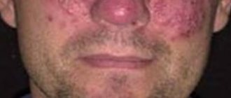
The beginning of pityriasis rosea is the formation of a maternal plaque - a single spot of a fairly large size and a round shape. Its diameter is from 2 cm, the color is pinkish, but gradually the central part of the spot turns yellow. Over time, peeling begins here.
After 7-10 days, multiple oval-shaped spots form on the surface of the skin. They are smaller than the maternal plaque - about 0.5-1 cm in diameter. For the most part, the rash spreads along the back and shoulders, covers the sides of the body, and runs along the lines of skin tension. In the center of all spots, the skin peels off, and a red border appears along the edges of the rash.
The spots that appear do not merge with each other. The pathology may be accompanied by itching. Sometimes the disease manifests itself in an atypical form - without the appearance of a maternal plaque. Pityriasis rosea lasts for several weeks, after which the rash goes away on its own. However, there is also a longer course – up to six months. Pityriasis rosea may be accompanied by weakness and malaise, drowsiness and fever.
Useful information on the topic:
- Calling a dermatologist to your home
- Fungal infections
- Rash on the body
- Sexually transmitted diseases
- How to get tested
- Wart removal
- Removal of condylomas
- Tests for STIs
- Removal of papillomas
- Skin rash
- Pyoderma
- Skin structure
- Mushroom tests
- Diagnosis of sexually transmitted infections
Pityriasis rosea in children
In childhood, pityriasis rosea manifests itself mainly after acute respiratory viral infection. The scales on the rash are usually sparse or absent altogether. The spots most often form in the inguinal folds and armpits. With this disease, the child can continue to live a normal life, the only limitation concerns water procedures. Do not use washcloths or rub the skin with hard towels. During the period of exacerbation of the disease, excessive sweating should be prevented, so physical activity will have to be limited.
It is important to follow all doctor's recommendations. As a rule, drug treatment is not required, but special attention should be paid to restoring the body's immune defenses. In this regard, a balanced diet, room ventilation, and adherence to general hygiene rules play an important role. If there is itching or an atypical course of the pathology, the doctor prescribes medications. But it is not recommended to use folk remedies.
Pityriasis rosea in pregnant women
In pregnant women, pityriasis rosea can develop due to hormonal changes in the body (deficiency of female sex hormones) or a weakening of the immune system due to toxicosis. The disease does not affect the condition of the fetus and does not cause pathologies. However, it is necessary to identify the causes of the disease and eliminate them, since the health of the mother depends on the health of her baby.
The main treatment in such circumstances will be restorative methods. Doctors categorically prohibit the ingestion of decoctions of medicinal herbs, especially St. John's wort, sage, and oregano. Interferons are used to strengthen the immune system, but they should be taken with caution so that your own protective functions do not decrease even further. Multivitamin complexes are often prescribed.
When the disease develops, pregnant women should pay attention to their diet and do special gymnastic exercises. For those who are concerned about itching, doctors recommend treating the rash with vegetable oil, chamomile infusion, and calendula decoction. It is very important to avoid stress, which has an extremely negative effect on the body, including causing pityriasis rosea.
General information
An ointment based on sulfur is widely used for various diseases of the dermis. The simple composition makes the ointment accessible to different segments of the population, because prices per jar start at only 28 rubles.
The drug is used not only to relieve unpleasant symptoms, but also as the main medicine that can eliminate the cause of the disease. The main component, which is sulfur in its pure form, better known as a chemical element, is supplemented by the following auxiliary substances:
- petrolatum;
- water;
- emulsifier.
In the pharmacy you can find sulfur ointment packaged in glass jars with a plastic lid or in aluminum tubes.
Prevention of pityriasis rosea
Simple preventive measures can prevent the development of the disease. To protect your body from pityriasis rosea, you need:
- Observe the rules of personal hygiene;
- Avoid synthetic and woolen clothing;
- Carefully select cosmetics, paying attention to their composition, quality, expiration dates;
- Adjust your diet so that your diet contains all the necessary substances;
- Avoid products with artificial colors and additives;
- Forget about bad habits;
- Temper yourself;
- Take vitamins as prescribed by your doctor.
It is advisable not to indulge in strong tea and coffee, fatty and fried foods. At the first manifestations of pityriasis rosea, you need to protect the areas of skin with rashes from direct sunlight and consult a doctor.
Contraindications and negative reactions
A contraindication to the use of the product is hypersensitivity to any components in the ointment. Treatment with the drug should not be carried out during pregnancy, lactation and under the age of 6 years.
When using ointment, it is important to avoid getting it into the eyes, mucous membranes of the nose and mouth. If this happens urgently, it is necessary to wash off the ointment with plenty of water to prevent burns. If negative reactions occur, you should seek medical help.
Side effects include allergic reactions. Most often they manifest themselves as redness on the skin and the occurrence of itching and burning. In severe cases, hives may occur. In case of any negative reactions of the body, you should stop using the product. Overdose causes similar symptoms. Against this background, severe burns may occur.
When treating dermatological problems, the product is applied slowly to damaged areas with precise movements. It is important to observe the rules of personal hygiene. After completing the procedure, hands should be washed thoroughly.
Yam ointment can be purchased without a prescription. It is stored in a dry, dark place at a temperature in the range of 0°C - 30°C. When the drug is frozen, its medicinal properties are lost. Shelf life – 2 years. After the expiration date, the product must not be used. In pharmacies you can buy various analogues of yam ointment. But they must be prescribed by a doctor after diagnosis.
Diagnosis of pityriasis rosea
Diagnosis of the disease is not difficult, since the manifestations of the pathology are quite typical. But the doctor must make sure that the rash is caused by pityriasis rosea, and not by eczema, syphilis, psoriasis or fungal infection of the skin - mycosis.
In addition, the disease can occur in an atypical form, so special studies and tests are carried out to confirm the diagnosis.
| Appointment with a dermatologist at the clinic. Call a dermatologist at home. | Reception is strictly by appointment, make an appointment by phone: +7 | Prices for services | Reviews about the clinic |
Methods of infection
To date, there is no consensus on the methods of infection.
There is an assumption that the source of the disease is the herpes virus and that people with low immunity, those who have recently suffered viral infections, colds, and who are often hypothermic are primarily at risk of contracting Zhiber's disease. Women and children have a significantly higher risk of infection than men. Observations show that people over 40 years of age and infants practically do not become infected with this infection. Is this disease contagious? There is no clear answer to this question. There is an opinion that it can be transmitted through tactile contact or airborne droplets. A prerequisite for a person to become infected is the presence of a provoking factor. In practice, it has been noted that transmission of this virus from one person to another occurs infrequently.
Treatment methods for pityriasis rosea
In the typical course of the disease, treatment may not be required, but only a doctor can make such conclusions based on the examination results. In such situations, the patient is recommended to:
- Hypoallergenic diet;
- Limiting water treatments (shower instead of bath), using a soft washcloth;
- Wearing clothes made from natural fabrics;
- Avoiding activities that cause excessive sweating.
In case of protracted course of pityriasis rosea, antiviral medications, treatment of rashes with salicylic alcohol, and topical antibacterial agents - ointments, can be prescribed. Antihistamine medications are used to reduce itching and can also prevent the spread of rashes. In some cases, treatment with antibiotics is required.
Ointments used for pityriasis rosea
For severe, disturbing itching, anti-inflammatory and antiallergic creams and formulations are prescribed. Ointments are used to treat the disease:
- Hydrocortisone – has an anti-inflammatory effect, relieves itching;
- Olettrinovaya is an antibacterial drug;
- Prednisolone - a composition with healing properties that can relieve inflammation;
- Loriden A - ointment with antipruritic, anti-parotid, anti-edematous effects;
- Sinalar – eliminates inflammation due to antibacterial properties;
- Lassara paste is an antiseptic that discolors stains;
- Rioloxol - antibacterial and anti-inflammatory;
- Flucinar – effectively relieves itching, eliminates flaking and inflammation;
- Sulfur – inhibits the inflammatory process.
Tsindol suspension also helps dry the skin, eliminate inflammation and itching. Ointments are applied to the pink plaques in a thin layer in accordance with the instructions and directions of the doctor. The treatment course is 2-3 weeks, the frequency of use of each drug is determined individually. At the same time as using medications, you should be careful with the affected skin.
However, any ointments, like all other medications, should be used exclusively as prescribed by a doctor, so as not to aggravate the situation. In addition, the effectiveness of each of the listed external remedies greatly depends on the patient’s age, the state of his immunity, the degree of spread of lichen, the presence of chronic pathologies and other characteristics of the body.
Calling a dermatologist to your home for pityriasis rosea
When the first signs of pityriasis rosea appear, you should consult a doctor. Today there is a convenient service for calling a doctor to your home, which provides many advantages:
- Saving time - no need to go to a medical facility or wait for your turn at the office door. You can simply arrange a doctor’s visit at a time convenient for you;
- Comfort – communicating with a doctor at home is much easier, because the doctor is not distracted by other patients and clinic staff, he can spend as much time as necessary on examination and consultation;
- Calmness – for children of all ages, a visit to a medical facility is often stressful, which plays a negative role in the development of many diseases, including pityriasis rosea. A doctor’s home visit will save the child from unnecessary worries;
- No risk – the development of pityriasis rosea often occurs against the background of weakened immunity. In such circumstances, contact with other patients is undesirable, but it is almost impossible to avoid it in the corridors of the clinic, as well as in public transport on the way to the medical facility. At home, such a risk is eliminated;
- If the disease is accompanied by fever, calling a doctor at home will be the best solution;
- Examination at home is the most convenient option for older people, mothers with small children, and patients with disabilities.
The quality of medical care at home is no different from that provided in a medical facility. The doctor gives the necessary recommendations, provides full consultation, diagnoses the disease and prescribes adequate treatment.
Common symptoms and manipulations in dermatology:
- Skin rashes
- Calling a dermatologist to your home
- Itching in the urethra
- Itchy skin
- Skin rash
- Prevention of casual sex
- Skin neoplasms
- Pyoderma
- Pityriasis rosea
- Streptoderma
- Scabies
- Peeling skin
- Fungal infections
- Skin infection
- Pus on the skin
- Blisters on the skin
- Papillomas on the foreskin
- Sexually transmitted diseases
- Skin structure
Types of erectile dysfunction
Depending on the cause of occurrence, the following types of impotence are distinguished:
- Endocrine. Associated with a lack of the hormone testosterone.
- Anatomically determined. It occurs due to curvature of the penis, its partial or complete removal, pain due to a hernia in the scrotum.
- Neurogenic. It develops against the background of diseases of the nervous system, for example, neuropathy or sclerosis.
- Vascular. It is explained by problems with blood vessels, which lead to impaired blood supply to the penis and the inability to achieve an erection.
Where to go with pityriasis rosea
Diagnosis and treatment of pityriasis rosea is carried out by a dermatologist. There are at least two important reasons to see a doctor rather than self-medicate. The first is the need to define the disease. Often, pityriasis rosea has some similarities with other skin pathologies such as fungal infections, psoriasis, mycosis of smooth skin or syphilis.
The second reason is to prevent complications. Despite the fact that the disease usually goes away on its own, sometimes lack of treatment or incorrect selection of drugs creates a serious risk of complications. They consist in the spread of rashes, the appearance of prolonged severe itching, and skin pigmentation after recovery.
With a strong decrease in immunity, a complicated course of the disease with an increase in temperature is possible. In people with blood pathologies or cancer who have undergone chemotherapy, the disease can develop again. The use of folk recipes and all kinds of folk remedies can cause contact dermatitis and even chemical burns. When using dyes, for example, celandine, diagnosing the disease is difficult, and a bacterial infection enters the body.
Lichen planus
Lichen ruber planus, described by Wilson in 1869, is a chronic inflammatory disease characterized by monomorphic papules on the skin and visible mucous membranes, most often on the oral mucosa and red border of the lips, accompanied by itching of varying severity. Today, this disease remains an urgent problem associated with the constant frequency of its detection, the lack of a unified pathogenetic concept, as well as the presence of severe forms and a chronic course, often resistant to therapy [1, 4, 6, 13]. In the general structure of dermatological morbidity, lichen planus (LP) accounts for 0.78–2.5%, among diseases of the oral mucosa - 35%. This disease occurs in all races, in all age groups and in both sexes, although the mucous membrane is more often affected in women from 40 to 60 years of age [1, 19].
Etiology and pathogenesis. In modern literature, various theories of the development of LP can be traced, such as viral, neurogenic, hereditary, intoxication and immunoallergic [1, 10, 15, 17].
The immunoallergic theory of the development of this pathology, based, according to various authors, on a decrease in the number of T cells in the patient’s blood and their functional activity, deserves the most attention at present. Some authors have shown a decrease in T-helper cells and an increase in the T-helper/T-suppressor ratio [11–14].
On the oral mucosa, manifestations of LP are associated with the presence of pathologies in patients with the gastrointestinal tract (gastritis, colitis, etc.), liver, and pancreas. Also, in a number of patients there is an undoubted connection between the development of the disease and vascular (hypertension) and endocrine (diabetes mellitus) pathology. Trauma to the oral mucosa, including those caused by dental pathology, has a certain significance in the development of the disease on the oral mucosa: sharp edges of teeth, poorly fitted removable plastic dentures, missing teeth, etc. [1, 6, 7].
Recently, there have been increasing reports of the development of lichen planus of the skin and oral mucosa in response to the action of certain chemicals on the body, including drugs. The so-called lichenoid reactions have been described in people whose work is related to the development of color film, who have contact with paraffinylenediamine, who have taken tetracycline (tetracycline lichen), para-aminosalicylic acid (PAS), gold preparations, etc. Thus, the disease in some cases may present is an allergic reaction to certain medicinal and chemical irritants.
Clinical manifestations of LP are characterized by the formation of a monomorphic rash, consisting of flat, polygonal, with a shiny surface and centrally recessed papules of pinkish-violet or crimson-reddish color, with a diameter of 2–3 mm. On the surface of the papules there is a peculiar shine with a waxy tint, which is especially noticeable in side lighting. Papules, merging, form small plaques, on the surface of which there are small scales. When the surface of papules and especially plaques is lubricated with vegetable oil, small whitish dots and intertwined web-like stripes appear through the stratum corneum (Wickham's symptom), this is due to uneven thickening of the granular layer of the epidermis. When pathological lesions resolve, persistent hyperpigmentation often remains. Dermatosis is accompanied by itching, often very intense, depriving patients of rest and sleep [1, 7–10, 15, 19].
LLP is localized on the flexor surfaces of the forearms, in the area of the wrist joints, on the inner surface of the thighs and extensor surfaces of the legs, as well as in the inguinal and axillary areas, and the oral mucosa. The pathological process usually does not involve the skin of the face, scalp, palms and soles. In the area of the extremities, the rash may have a linear (zoniform) arrangement.
About 25% of patients with LP have only lesions of the mucous membranes (mouth, glans penis, vestibule of the vagina) and are not accompanied by manifestations on the skin. On the mucous membrane of the cheeks, grayish-opaline dotted papules are formed, grouped in the form of rings, networks, laces, on the surface of the tongue - flat, whitish opal plaques with clear jagged edges, reminiscent of foci of leukoplakia, on the red border of the lips (usually the lower) - small purple ones plaques, slightly flaky, with a grayish-white mesh on the surface [1, 7–10, 15, 19].
Changes in nails with pronounced longitudinal striations, sometimes in the form of ridges, hyperemia of the nail bed with focal clouding of the nail plates of the hands and feet are observed in some patients with LP [1, 15].
LCP is characterized by an isomorphic reaction to irritation. Often, typical elements of dermatosis are located linearly at the sites of excoriation (Koebner phenomenon). The disease lasts a long time, often for many months. There have been cases of generalization of dermatosis with the development of secondary erythroderma (lichen ruber planus generalisata) [1, 7–10].
Several atypical forms of lichen planus are identified [2, 3, 17]:
- hypertrophic, warty form (lichen planus hypertrophicus, seu verrucosus);
- atrophic and sclerotic forms (lichen planus atrophicus, lichen planus sclerosus);
- pemphigoid, or vesicular, form (lichen ruber pemphi goides, seu bullosus);
- lichen ruber moniliformis (lichen ruber moniliformis);
- pointed, perifollicular form (lichen planus acuminatus, sen planopilaris);
- erosive-ulcerative form (mucous membrane).
Diagnosis . With classic manifestations of LP, diagnosis is not difficult and it is established clinically. In doubtful cases, histological examination can help, where a typical mononuclear infiltrate will be present, disrupting the basal line of keratinocytes [1, 3].
In some patients, differential diagnosis of LP with psoriasis and syphilis is carried out, since lichen planus rashes on the skin may resemble psoriatic elements and syphilitic papules. However, papules in LLP have the characteristic color of the rash, a polygonal shape, an umbilical depression in the center of the papules, a Wickham grid, and the absence of the phenomena of stearin stain, varnish film and pinpoint bleeding allows us to differentiate lichen planus with psoriasis. Papules are round and hemispherical in shape, their “ham” color, density, positive serological reactions to syphilis make it possible to distinguish syphilitic papules. It may be difficult to make a diagnosis if the rash is localized on the oral mucosa.
The clinical picture of LP of the oral mucosa is differentiated from leukoplakia, lupus erythematosus, syphilitic papules and other diseases.
Leukoplakia, unlike LP, has keratinization in the form of a solid grayish-white plaque and does not have a patterned lesion.
In lupus erythematosus, the lesion is hyperemic, infiltrated, hyperkeratosis is present only within the focus of inflammation in the form of tender points, short stripes, sometimes merging along the edge of the lesion in the form of stripes and arcs; in the center of the lesion there is atrophy, which will not occur with LP.
Syphilis papules are usually larger, round or oval in shape, their surface is covered with a grayish-white coating, which is usually removed when injured, and pale treponemas are found on their surface. Positive serological reactions to syphilis [1, 3, 15, 19].
Treatment approaches
LLP is often a chronic but benign disease, sometimes asymptomatic, and does not require systemic treatment. However, taking into account the frequent chronicity of the process and the many severe and atypical forms, the complexity of its pathogenesis, successful treatment is possible only with complex and individualized treatment using modern tools and methods [4, 11, 16, 18].
It is especially important to consider the factors that contributed to the onset of the disease. It is necessary to eliminate risk factors - household and professional hazards, concomitant diseases, foci of focal infection. They carry out sanitation of the oral cavity and prosthetics. Food products should not cause irritation to the mucous membranes of the mouth. Attention is paid to previous treatment and drug tolerance [11].
Due to the fact that the material basis of the disease is immune inflammation (delayed-type hypersensitivity reaction - DTH), correction of immunity is of particular importance and the disease itself responds well to immunosuppressants. To interrupt the cooperative connection of immunocompetent cells, glucocorticosteroids in combination with 4-aminoquinoline derivatives (hydroxychloroquine, chloroquine) are prescribed as basic therapy, especially for common and resistant forms. However, drugs that suppress immune processes should be used only in the presence of documented visceral damage or if the erosive process interferes with eating or speaking.
In the presence of intense itching, in the acute period of the disease, histamine H1 blockers and antiserotonin drugs and catecholamine blockers are indicated. Along with this, sedatives and antidepressants are prescribed to help normalize sleep and reduce itching.
Vitamin therapy has a beneficial effect on metabolic processes. The proliferation and differentiation of keratinocytes is influenced by vitamin A (daily dose for adults - 100,000 IU). Retinoids - derivatives of vitamin A (Tigazon, Neotigazon, Etretinate) reduce the intensity of the inflammatory reaction, affect the condition of cell membranes and normalize proliferation processes. Retinoids are effective against lesions of the oral mucosa and red border of the lips. In recent years, analogues of vitamin A - carotenoids - have been successfully used, especially in atypical forms, in particular erosive-ulcerative, as well as in cases of damage to the oral mucosa and genital organs.
Vitamin E (alpha-tocopherol acetate), used as an antioxidant and inhibitor of the cytochrome P450 system, allows for complex treatment with corticosteroids to reduce the daily dose and shorten the duration of steroid therapy. The multivitamin preparation Aevit is indicated for patients with a long-term chronic course of the disease, with verrucous forms and damage to the mucous membranes.
In chronic relapsing dermatosis, agents that improve oxygen supply to tissues are indicated.
External treatment with applications of corticosteroid ointments, solutions and shaken mixtures with menthol, anesthesin, citric acid, and antihistamines is prescribed for intense itching. Hypertrophic foci are destroyed by cryodestruction or electrocoagulation. Erosive-ulcerative lesions are subject to local treatment with epithelizing agents, including Solcoseryl, sea buckthorn oil, and rosehip oil.
Currently, the method of combined phototherapy (UVAB, ultraviolet irradiation) is successfully used. This therapy affects immune responses by damaging immune cells in the skin. At the same time, the superficial lymphocytic infiltrate in the dermis disappears, and the cellular composition in the epidermis is normalized.
Of the listed means and methods, only their rational choice - sequential stage-by-stage (course) application, taking into account the individual characteristics of the patient and the nature of the course of the disease - allows one to achieve positive results.
Recently, immunotropic therapy for lichen planus has been increasingly used, including the use of exogenous interferons (Reaferon, Interlock) and interferonogens (Neovir, Ridostin).
If the disease is suspected to be caused by any drug or chemical, its use should be discontinued. A thorough examination of patients is necessary to identify internal diseases. First of all, you need to examine the gastrointestinal tract, blood sugar levels, and neuropsychic state [1, 11, 15, 19].
In the absence of symptoms, treatment is not required. To reduce itching, antiallergic drugs are used. The doctor may also prescribe vitamins, sedatives, and physiotherapy.
Physiotherapeutic procedures include electrosleep, diadynamic paravertebral currents, and inductothermy of the lumbar region. Antipruritic shaken suspensions, corticosteroid ointments, 2–5% tar-naphthalan ointments are prescribed externally. The prognosis for life is favorable.
As with any chronic process, LP requires local and systemic therapy [15, 19].
Local drugs of choice are corticosteroids. Class I and II corticosteroids are prescribed. For verrucous process, occlusive dressings with class II corticosteroids are recommended. Intralesional administration of drugs is used, but this method should only be used for very persistent verrucous plaques.
Corticosteroids are effective for lichen planopilaris of the scalp, but they should be applied to the periphery of the plaque and not to its center. Also, betamethasone preparations in the form of foam are very suitable for this localization of the lesion.
For mucosal lesions, especially if they are erosive or ulcerated, triamcinolone pastes or gels are prescribed, which improves the condition in 65% of such patients after 2 weeks. The use of 0.025% fluocinolone based on 4% hydroxycellulose gel in combination with chlorhexidine and miconazole gel for an antifungal effect improved the condition in 50% of patients and served as a prevention of oral candidiasis.
You can rinse the mouth for 5 minutes with one soluble tablet (500 mcg) of betamethasone, originally intended for systemic use. However, this rinsing method is only useful for lesions that have eroded de novo and not for lesions that have eroded due to trauma.
Fluocinonide 0.025% 6 times a day for 2 months is active compared to placebo and without side effects. Betamethasone valerate in aerosol form was used for 2 months with good results in 8 of 11 patients. Clobetasol propionate 0.05% was recently found to be more effective than fluocinonide 0.05%.
Intralesional administration of corticosteroids has been used for lichen planus of the oral mucosa. The preferred drug is triamcinolone acetonide, which is prescribed at a dose of 5–10 mg/ml weekly or twice a week for 3–4 weeks [4, 18, 19].
Topical retinoids are used for lesions in the oral cavity. Fenretinide, for example, gave excellent results when used twice daily, with no local or long-term side effects. Retinoic acid 0.1%, tretinoin 0.025%, and isotretinoin gel 1% were all effective, but less effective than topical triamcinolone or flucinonide [19].
Systemic treatment
Cyclosporine A (CyA) specifically targets cell-mediated hypersensitivity reactions and is the drug of choice for LP. The initial dose is usually 5 mg/kg/day. It is reduced to 2 mg/kg/day as soon as possible: regimens with a treatment duration of more than 6 months should be avoided. Blood pressure should be monitored weekly and kidney function monthly. The best indication for the use of CyA is severe erosive LP.
Systemic corticosteroids may be used instead of CyA. Prednisone can be prescribed at a dose of 1 mg/kg/day (or a lower dose). The dose is reduced over 1 month. When the drug is discontinued, a rebound effect may occur. Common side effects of corticosteroids are common.
For erosive LP with chronic active hepatitis, azathioprine (50–100 mg/day) is recommended. Normalization of transaminase levels is usually accompanied by improvement of lesions in the oral cavity. However, if a patient has antibodies to the hepatitis C virus, all immunosuppressive treatments should be avoided, since immunosuppressive drugs may contribute to the development of liver cancer.
Other drugs that have been shown to be beneficial in reversing the clinical process of LP include thalidomide, hydroxychloroquine, retinoids, and levamisole.
Hydroxychloroquine is used at a dose of 200–400 mg/day for several months in patients who have lesions in the oral cavity. When analyzing the risk-benefit ratio, retinal side effects must be taken into account and carefully monitored.
For systemic treatment of LP, acitretin (0.25–0.75 mg/kg/day) and isotretinoin (0.25–0.50 mg/kg/day) are used. However, these drugs should not be prescribed to women of childbearing age due to their well-known teratogenicity [19].
In severe forms of erosive oral lichen planus that do not respond to traditional treatment, PUVA therapy (PUVA = Psoralens + UltraViolet A) can have a positive effect. A dose of 0.6 mg/kg 8-methoxypsoralen is administered orally 2 hours before irradiation with long-wave ultraviolet light. In one study, irradiation was administered 12 times at 2–3 day intervals for a total dose of 16.5 J/cm2. In another study, 20 sessions were performed, 3 per week, with a total cumulative dose of 35.9 J/cm2. After treatment, clinical symptoms and erosive lesions disappeared. Side effects are similar to those observed after whole body PUVA therapy [16, 19].
Evaluation of treatment effectiveness. Provided that the therapy is correctly selected, regression of the pathological process occurs within 1–2 weeks, the rash disappears after 1–1.5 months. Clinical cure (recovery) is characterized by the complete disappearance of papules, in the place of which hyperpigmented or depigmented spots remain. The latter can remain for an indefinite period of time (from several weeks to several months). Erosive-ulcerative, hypertrophic and atrophic forms of lichen planus are usually resistant to therapy, and the rashes persist for several months or even years.
Primary prevention consists of sanitation of foci of chronic infection, treatment of psychoneurological disorders; Overwork and stress should also be avoided. Prevention of exacerbations: prescribing water treatments - baths, showers (not recommended in the acute period of the disease), hydrogen sulfide and radon baths; diet (exclusion from the diet of coffee, spices, chocolate, honey, alcohol) [1, 10, 17, 19].
Literature
- Bazyka D. A., Bazyka A. D. Etiology, pathogenesis and therapy of lichen planus // Vestn. derm. and veins 1977, No. 11, p. 58.
- Gadzhimuradov M.N., Gunaeva A.A. Atypical forms of lichen planus: clinical manifestations, differential diagnosis and treatment // Clinical dermatology and venereology. 2009, No. 3, p. 85–80.
- Differential diagnosis of skin diseases. Ed. B. A. Berenbein and A. A. Studnitsyn. M., 1983. P. 269.
- Korsunskaya I.M., Nevozinskaya Z.I., Zakharova A.B., Konstantinov E.M., Andryushkova Yu.A. Experience in the treatment of lichen planus with the drug Glutoxim // Russian Journal of Skin and Venereal Diseases. 2008, No. 1, p. 44–46.
- Maksimovskaya A. N., Tsarev V. N., Guseinova S. S. Bacteriological basis for the treatment of lichen planus of the oral mucosa using laser radiation. In the book: Proceedings of the VI Congress of the Russian Dental Association. M., 2000, p. 275–277.
- Manukhina O. N. Clinical course of lichen planus of the oral mucosa against the background of decreased functional activity of the thyroid gland. M.: Science to Practice, 1998. pp. 145–147.
- Mashkilleyson A. L. Diseases of the mucous membrane of the oral cavity and lips. M.: Medicine 1963. P. 188.
- Pashkov B. M. Lesions of the oral mucosa in skin and venereal diseases. M.: Medicine 1963. P. 182.
- Petrova L.V. Features of the clinical course of lichen planus of the oral mucosa // Ros. magazine cutaneous veins bol. 2002; 3:28–31.
- Pototsky I.I. Directory of dermatovenerologist. Kyiv, 1985. P. 88.
- Rabinovich O. F. Immunological aspects of the pathogenesis of lichen planus of the oral mucosa (clinic, diagnosis, treatment). Diss. Dr. med. Sci. M., 2001, 190 p.
- Raikhlin A. N. Subcellular mechanisms of development of lichen planus of the oral mucosa and its treatment: Abstract of thesis. dis. ...cand. honey. Sci. M., 1986.
- Slesarenko N. A. Lichen planus (modern immunological and biochemical aspects) and methods of pathogenetic therapy: Abstract of thesis. dis. ...Dr. med. Sci. M., 1995.
- Spitsyna V.I. Pathogenesis of immunodeficiency in patients with lichen planus of the oral mucosa // Russian Dental Journal. 2002, No. 3, p. 30–34.
- Thomas P. Habiff. Skin diseases. Diagnosis and treatment. M.: “MEDpress-inform”, 2008. 672 p.: ill.
- Faizulin R. A. The use of dimocyphon and selective phototherapy of patients with lichen planus, taking into account immunological blood parameters: Abstract of thesis. dis. ...cand. honey. Sci. M., 1992. 21 p.
- Fedorov S. M., Selissky G. D., Timoshin G. G. Lichen planus // Skin diseases. M.: GEOTAR-Medicine, 1997. pp. 67–69.
- Khamaganova I.V. Advantan (methylprednisolone aceponate) in the complex treatment of lichen planus // Bulletin of Dermatology and Venereology. 2004, No. 3.
- Rebora A. Lichen planus. European guidelines for the treatment of dermatological diseases. Ed. A. D. Katsambasa, T. M. Lotti. M.: “MEDpress-inform”, 2008. pp. 371–374.
A. S. Bisharova, Candidate of Medical Sciences
GBOU DPO RMAPO Ministry of Health and Social Development of Russia, Moscow
Contact information about the author for correspondence
What tests need to be taken for pityriasis rosea?
The diagnostic method and set of necessary studies are determined by a dermatologist. Typically, the patient takes a blood test to study general indicators. Tests are often ordered to detect antibodies to viral agents, including syphilis and herpes. To exclude fungal damage to the skin, microscopic examination of scrapings from the site of the lesion is carried out.
Additionally, the doctor may prescribe an immunogram if there is a suspicion of a significant decrease in the body's defenses. The cause of pityriasis rosea is an allergic reaction, so testing for allergens is possible. If necessary, the doctor prescribes additional examinations and consultations with specialists. Typically, such measures are taken in case of protracted or atypical course of the pathology.
Why erectile dysfunction may occur
All existing causes of impotence in men are divided into the following groups:
- Organic. Associated with the state of the body when there is some physiological obstacle to erection. The risk of impotence is increased by smoking and alcohol abuse.
- Psychogenic. Erection problems are caused by a man’s psychological state: strong feelings, stress, depression, problems in his relationship with his sexual partner.
- Mixed. Here, a man is influenced by both organic and psychogenic factors.
In many cases, the cause of erectile dysfunction is problems with blood vessels associated with age-related changes in their walls. For example, when blood vessels are damaged by cholesterol plaques, obstacles appear to the normal flow of blood, so it cannot fill the corpora cavernosa of the penis. Therefore, it is so important to visit a doctor on time, since problems with blood vessels increase the risk of stroke and heart attack.
Among vascular problems, erectile dysfunction and impotence can result from:
- atherosclerosis;
- injuries to the abdomen and pelvis that damage the vessels supplying the penis;
- diabetes;
- hypertension.