RITTER DERMATITIS EXFOLIATIVE
(G. Ritter, German doctor, 1820-1883; Greek derma, dermat [os] skin + -itis; Latin exfoliare tear off leaves, peel off; synonym:
Ritter’s disease, exfoliative dermatitis of newborns, keratolysis neonatorum
) - one of forms of staphylococcal and streptococcal infections of newborns, characterized, as a rule, by a severe course and characterized by severe intoxication and local changes in the skin in the form of edema, hyperemia and detachment of the epidermis. Described in 1870 by Ritter. Etiologically close to other forms of neonatal pyoderma (see Pyoderma). R.d.e. in a child is one of the indicators of unfavorable sanitary hygiene. the state of the maternity hospital, newborn wards and the poor quality of care for newborns, women in labor and postpartum women.
Anatomical-physiol. skin properties and immunobiol. The characteristics of the body of newborns contribute to easier penetration of coccal flora into the body (see Newborn). It is believed that a violent reaction on the part of the skin with characteristic detachment of the epidermis is a consequence of rapid intoxication as a result of the introduction of a coccal infection.
Causes of the disease
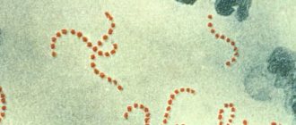
Exfoliative dermatitis (or Ritter's exfoliative dermatitis
) is a childhood skin disease that is caused by staphylococcus (there is an assumption that streptococci can also influence the development of dermatitis).
Exfoliative dermatitis is a complicated form of another skin disease - pemphigus of newborns
and is considered one of the most severe forms of dermatitis. The cause of infection lies in the imperfect immune system of newborns and the undeveloped barrier function of the epidermis.
Diagnosis
The diagnosis is made on the basis of the wedge, pictures. R.d.e. differentiated from pemphigus of newborns (see Pyoderma), which is characterized by blisters filled with serous-purulent fluid and a more benign course; with phlegmon (see) of newborns, characterized by hyperemia and thickening of the skin, necrosis of the subcutaneous tissue; with epidermolysis bullosa, in which the blisters are often located on the extremities, usually in places exposed to trauma (see Epidermolysis bullosa); with pemphigus in early congenital syphilis, in which blisters appear on the skin of the soles and palms on an infiltrated base - papules with positive serols. reactions (see Syphilis, congenital). R.d.e. should be distinguished from congenital ichthyosis, or congenital ichthyosis-form Broca's erythroderma, which is detected from the moment of birth and is characterized by the absence of erosive surfaces and blisters, the presence on the skin of dense, collodion-like films and other signs of ichthyosis (see), as well as from desquamative erythroderma Leiner, starting with diaper rash and the formation of the so-called. milky crust on the head in the first weeks of a child’s life and subsequently manifested by redness, infiltration and pityriasis-like peeling of the skin (see Erythroderma).
Signs of Ritter's exfoliative dermatitis
The disease includes a number of typical symptoms:
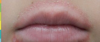
- Redness and swelling of the skin
- The appearance of so-called “flaccid” bubbles (there is little liquid in such bubbles, they are not elastic)
- After some time, erosions form in place of the bubbles
Typically, skin lesions begin near the mouth or near the navel and gradually spread to larger areas, covering the body and/or limbs of the child. All symptoms are accompanied by a serious condition and high body temperature.
Symptoms
Primary erythroderma begins spontaneously and acutely, while secondary erythroderma can be a manifestation of psoriasis, eczema or long-term dermatosis. During the pathological process, several stages can be distinguished:
- erythroderma begins with rashes, with primary bullae, papules, pustules appearing, hyperemia occurs with a predominance of exudative processes in some areas and swelling of the skin, which is accompanied by thinning of the epidermis;
- during the involution of primary rashes, there is a parallel addition of new ones, peripheral growth, a tendency to merge and the formation of extensive foci of inflammation;
- when pustules and vesicles are opened, areas of erosion are formed, covered with hemorrhagic or purulent crusts, the process very quickly covers more and more new areas of the skin and can become total, however, in some cases spontaneous resolution of erythroderma may occur;
- areas of skin folds are characterized by weeping with the rapid addition of a secondary infection;
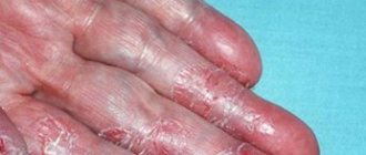
- peeling in large plates occurs on the entire surface of the affected skin;
- subjective sensations in the areas of rashes are reduced to pain, burning, severe itching, general weakness, weakness, arthralgia , while the increase in body temperature reaches subfebrile values, concomitant diseases worsen;
- at later stages, thickening of the epidermis occurs, involvement of mucous membranes, nails and hair in the pathological process, which causes their loss; in many, alopecia , nails exfoliate, break and have dystrophic changes;
- an increase in symptoms due to intoxication causes tachycardia , chills , muscle tremors, high (up to 40°C) temperature, enlarged lymph nodes, hepato- and splenomegaly, indigestion, and the mammary glands become hardened.
During the prodormal process, severe itching occurs, cracks form, which is fraught with the addition of a secondary infection. Complications of the process occur in the form of eczematization and furunculosis .
Stages of exfoliative dermatitis
- Initial (erythematous) stage of Ritter's exfoliative dermatitis. The skin turns red and swelling appears.
- The exfoliative stage of dermatitis is a dangerous period of the disease, which occurs a few days after the onset of the disease. The skin begins to peel off, leaving behind erosions, and the lesions begin to merge with each other. Every day larger and larger segments of the skin are affected.
- Regenerative. The final stage of the disease, in which symptoms subside, but large-lamellar disorders appear.
If the disease is mild, small areas of skin are affected, there are no papples, recovery occurs in two weeks.
Diet for erythroderma
Diet for skin diseases
- Efficacy: therapeutic effect after a month
- Time frame: three months or more
- Cost of products: 1400-1500 rubles per week
In the treatment of erythroderma, diet, work and rest schedules play an important role. In this case, the diet should be gentle, hypoallergenic, balanced and rich in vitamins. Recommended Products:
- berries, fruits and vegetables, preferably fresh and rich in vitamins, especially A;
- light broths and soups;
- porridge;
- boiled river fish;
- low-fat dairy products.
Patients are usually prohibited from eating fatty and spicy foods, sausages, fatty fish, cocoa-containing products, synthetic sweets, and limiting the consumption of salt, vegetable oils and meat.
Treatment methods for exfoliative dermatitis
Treatment of exfoliative dermatitis primarily includes drug therapy:
- Cephalosporins (antibiotics) effectively act on staphylococci, destroy them and prevent further reproduction.
- Solutions of Poliglyukin and Hemodez are used infusionally. These agents neutralize dehydration during infection.
- The introduction of gamma globulin provokes increased body resistance to infections.
- Compresses with silver nitrates and external treatment of lesions with potassium permanganate will speed up the skin healing process.
Any treatment of exfoliative dermatitis must be carried out after preliminary consultation with a dermatologist. All procedures must be performed by specially trained personnel.
Therapeutic therapy includes the use of appropriate antibiotics, parenteral (intravenous) drugs (to strengthen anti-staphylococcal immunity), and special skin care.
Home remedies
Folk remedies for Ritter's exfoliative dermatitis will be an excellent additional therapy and an excellent preventive measure. The main thing is to find suitable folk recipes that will help soothe the skin and make it smooth and silky.
Here are the most effective recipes used for Ritter's exfoliative dermatitis.
- Birch tar . Birch tar will help get rid of dermatitis. You need to add a teaspoon of this product to any hypoallergenic cream, heat the mixture in a water bath to 70 degrees, and then lubricate the lesions with it. Birch tar works best on the legs and knees.
- Potatoes and sour cream. To create this miracle remedy, you need to add 50 grams of sour cream to freshly prepared mashed potatoes, and then wrap the mixture in a bandage or gauze and apply a similar compress to the affected areas.
- Yarrow . Gauze compresses soaked in yarrow decoction are an excellent way to cope with the manifestations of dermatitis.
Introduction
This publication analyzes the results of the use of biological therapy and the position of international clinical guidelines for severe forms of psoriasis, information about which is very limited for practical use, which is especially important in the context of the growing number of severe forms of psoriasis, which is encountered not only by dermatologists.
Psoriatic erythroderma (PE)
PE is a rare and severe variant of psoriasis vulgaris with an incidence of 1 to 2.25% among patients with psoriasis [1].
PE usually presents with symptoms such as erythema, swelling, itching, poorly defined psoriatic plaques, superficial peeling, hair loss, and keratoderma-type lesions of the palms and soles [2–7].
In addition, nail changes are very common in PE, and the severity of the disease can range from mild changes to severe onychodystrophy, often affecting the fingernails rather than the toenails [3, 5, 6].
Some authors note that PE is characterized by generalized inflammatory erythema with or without exfoliation involving at least 75% of the body surface area. Other authors argue that at least 90% of the body surface area should be affected [2, 3, 7].
In addition, patients with PE may have systemic symptoms: fever, tachycardia, fatigue, malaise, chills, dehydration, lymphadenopathy, arthralgias, myalgia, insomnia, sweating, diarrhea, constipation, weight loss, rarely severe heart failure at the end of the day (from -due to excessive water loss and edema) and cachexia [3, 7, 8]. Laboratory results may reveal massive protein and fluid loss, leukocytosis, anemia, elevated levels of C-reactive protein and erythrocyte sedimentation rate, blood electrolyte abnormalities, temperature abnormalities (hypo- or hyperthermia), and in rare cases, liver pathology with functional test changes [ 5, 9, 10].
The diagnosis of pancreas can be confirmed histologically by the characteristic pattern of perivascular infiltrates, predominantly consisting of lymphocytes and histiocytes, with capillary dilatation and hyperkeratosis. Additional histologic evidence of PE includes several features of classic psoriasis, including parakeratosis, acanthosis, spongiosis, Munro microabscesses, and occasionally keratinocyte apoptosis [9, 11, 12]. However, due to peeling and loss of the epidermal stratum corneum, Munro's microabscesses and parakeratosis may not be noticeable on histological examination [11]. In addition, to confirm the diagnosis of PE, especially if it develops without a previous history of psoriasis vulgaris (VP), the physician must exclude other possible causes, such as atopic dermatitis, lichen pilaris, drug rash, connective tissue diseases, Sézary syndrome, and others. malignant neoplasms [3, 9, 13].
PE can be divided into two main clinical subtypes. The first is characterized by the presence of psoriatic plaques with the gradual development of generalized erythroderma, in which psoriatic plaques remain differentiated from erythroderma. This form has a relatively stable and favorable prognosis. The second subtype, which is more often observed in patients with psoriatic arthritis, is often characterized by rapidly developing erythema of the entire body and the absence of clear boundaries of psoriatic plaques. The course of the disease in this type of patient is relatively unstable and may be associated with abnormal vital and laboratory parameters. With the second subtype, the prognosis is unfavorable and death is possible. The first clinical subtype is often associated with a chronic and long-lasting course, while the second subtype is more likely to be acute, rapidly progressive and relapsing [7, 14].
An important aspect to consider in patients with psoriasis is that the use of certain drugs, such as oral corticosteroids and methotrexate, can also lead to the development of erythroderma [15, 16]. Finally, there is literature data on the development of PE when using acitretin, etretinate, infliximab, lithium, and antimalarial drugs. The development of PE has been described in several systemic diseases, such as human immunodeficiency virus, leukemia, T-cell lymphoma and gout [7, 17, 18].
Pathogenesis of PE
The pathogenesis of the development of CAP has been studied quite well recently. It has been shown that class I antigens HLA-Cw6, HLA-B57, HLA-B13 and HLA-B17 are associated with the development of CAP, and mutations in IL36RN are associated with the development of pustular psoriasis, while very little is known about the genetic basis of the development of PE [19, 20 ].
The paucity of data on HE forces us to rely on information obtained from small studies, case reports, case series, and a small number of comparative studies involving patients with HE, patients with CAP, and healthy controls. In 2005, scientists demonstrated a statistically significant increase in serum immunoglobulin E in PE compared with patients with CAP. This discrepancy has been explained by a Th1/Th2 imbalance favoring Th2 differentiation [21].
A recent study of 16 patients further developed the idea of Th2 involvement. It made 3 important discoveries regarding the pathogenesis of PE: first, the Th1/Th2 ratio was significantly lower in patients with PE than in patients with CAP; secondly, the levels of interleukins IL-4 and -10 were significantly higher in patients with PE than in healthy individuals and patients with CAP; third, the interferon IFN-γ/IL-4 ratio in patients with PE was <1.0 and was the exact opposite of the other two groups [22].
Given the important contribution of Th17 to the pathogenesis of CAP, the role of Th17 in the pathogenesis of PE has recently been investigated. Th17 cells secrete IL-17, -22 and IFN-γ, enhancing the production of inflammatory chemokines by T cells, dendritic cells and neutrophils [23].
Using immunohistochemical analysis, Moy et al. found that in PE, Th17 are the most predominant pool of T cells after Th2. The authors also noted the similarity of the increase in the content of Th17 cells in both PE and the erythrodermic variant of atopic dermatitis [24].
At the molecular level, excessive levels of tumor necrosis factor (TNF-α) in psoriatic plaque contents have also been demonstrated, suggesting that the rapid systemic release of TNF-α in PE may contribute to the rapid onset and severity of the disease [25].
Treatment of PE
First of all, treatment of PE should include compensation of hemostasis: water, proteins and electrolytes; prevention of hypothermia, as well as treatment of any secondary infections to prevent septic complications. According to some authors, sepsis, most often caused by Staphylococcus aureus, is a particularly severe and potentially fatal complication that most often develops in these patients [26].
In 2010, the American Medical Board National Psoriasis Foundation published consensus recommendations for the use of cyclosporine or infliximab as first-line treatment for patients with unstable erythroderma. At the same time, acitretin and methotrexate were recommended for patients with a more stable clinical picture [3].
Since the number of clinical studies on the treatment of generalized forms of psoriasis is limited, the effectiveness of a particular therapy is difficult to reliably assess.
Many years have passed since the 2010 recommendations were developed. During this time, new classes of biological agents, such as IL-12/23, -17 inhibitors, were created and well studied. Updated data on the use of new drug groups are very useful for the treatment of such patients due to the increasing incidence of psoriasis, as well as the formation of severe generalized forms. Based on the analysis of practical and scientific data, updated AAD-NPF clinical guidelines were published in 2022, which analyzed the level of evidence for approved and widely used genetically engineered biological drugs (see table) [27].
Conclusion
Although the evidence for the effectiveness of genetically engineered biologics in PE is certainly limited, biologic therapy appears to be an effective approach to treating this form of psoriasis. There is evidence of beneficial use of biologics in these serious conditions.
Personal positive experience with the use of IL-17 inhibitors in various forms of psoriasis, as well as in PE, obviously once again confirms the unified hypothesis of the pathogenesis of PE, where Th17 type and IL-17 play an important role. The presence of a well-studied universal remedy allows you to obtain predictable results of therapy and save valuable time, which is especially important for patients with generalized forms of psoriasis. However, larger controlled clinical trials are needed to evaluate the comparative effectiveness and safety of different biologic agents in patients with PE.
Pharmacy products
In parallel with drug therapy, it is recommended to use La-Cri cream, which strengthens the natural protective barrier of the epidermis and has an antimicrobial and anti-inflammatory effect. The active components of the cream are extracts of licorice, violet, string, walnut, as well as bisabolol and panthenol. In addition to eliminating the symptoms of exfoliative dermatitis, they have a regenerating, healing and soothing effect.
La-Cri cream is suitable for babies from the first days of life, does not contain fragrances and is not addictive.
Prevention
Simple preventive measures will help prevent the sudden onset of Ritter's exfoliative dermatitis:
- Monitor your skin frequency regularly. Be sure to follow daily rules of personal hygiene; compliance with them will help maintain the health of epithelial tissues.
- Balance your diet. Try to saturate your diet with enough meat, fish, cereals, fresh vegetables and fruits. Fermented milk products are also important. And, of course, the best prevention of dermatitis is to stop drinking alcohol, smoking and other bad habits.
- Look after yourself. Skin health is only in our hands. Treat chronic skin diseases, rashes, acne, boils and other rashes in a timely manner. And at least once a year, try to be examined by a dermatologist. Avoid scratching the skin, pustular abscesses, and, if possible, avoid contact of the epidermis with toxic changes.
Men and women whose relatives suffer from skin cancer, seborrhea, psoriasis and other unpleasant diseases need to be especially careful. Patients with signs of allergies must take antihistamines regularly.
Pathogenesis
The pathogenesis is based on a total inflammatory process of the epidermis, although it has not been fully studied, but it is not clear why such different factors provoke a single skin response. Presumably the epidermis reacts to the skin as a “graft that is an enemy of the host.” When foreign agents enter the body and encounter histological compatibility antigens on unique HLA protein molecules that regulate the response and react to “foreign agents”, they trigger the process of destruction of cells - keratinocytes, in which mutations have occurred under the influence of pathogens. At the same time, the production of anti-inflammatory cytokines, T-killers and T-suppressors begins, proliferation increases, the process of antibody synthesis begins and the development of an allergic reaction. Against the background of an increase in mitotic activity in the basal cells of the epidermis, they are transported to the surface of the skin, which is clinically expressed by large-plate peeling and impaired microcirculation, increased blood perfusion through the skin and the occurrence of disimmunoglobulinemia.
At the systemic level, changes are observed in the cardiovascular and central nervous systems, serious disturbances in water-salt and protein metabolism, temperature dysregulation, heat loss and the development of hypothermia .
The main theories of the development of pathogenesis include toxic-allergic, but exogenous infections such as fungi, staphylococci and scabies mites, as well as malignant proliferation and paraneoplastic process .
Many toxic erythroderma are characterized by impaired blood circulation and kidney or liver function in response to provoking factors with a time limit.
Clinical researches
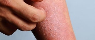
Clinical studies conducted by Vertex have proven the effectiveness, safety and tolerability of the products. The products are suitable for daily care of children's skin with mild and moderate forms of atopic dermatitis during remission. As a result of using the products, a decrease in the activity of inflammation, dryness, itching and flaking was noted.
It has been proven that La Cree cream eliminates dryness and flaking of the skin, retains its own moisture and, importantly, protects the skin from cold and wind. Cream for sensitive skin significantly reduces itching and irritation, relieves redness of the skin, moisturizes and gently cares for it. The creams are recommended by the St. Petersburg branch of the Union of Pediatricians of Russia.
Sources:
- Fundamentals of health development in children. Textbook. — Moscow: Mir, 2016. — 384 p. Tulchinskaya, V.D. Nursing assistance to children / V.D. Tulchinskaya. - Moscow: Russian State University for the Humanities, 2016. - 368 p.
- Andropova T.V., Gudina M.V., Odintsova I.N., Hygiene of children and adolescents, publishing house: Siberian State Medical University Publishing House, 2022 - 101 p.
- Cohen Bernard A. Pediatric dermatology, publishing house: MEDpress-inform, 2015
- Boniface Ernesto, Differential diagnosis in pediatric dermatology, publishing house: Panfilov Publishing House, Binom. Knowledge Laboratory, 2014
Photos of dermatitis
Photo album on the diseaseTests and diagnostics
To make a diagnosis of erythroderma, clinical and laboratory confirmation is necessary. The most important task of a dermatologist is to establish the cause of the disease.
A series of skin biopsies, immunohistochemical, virological tests, bacterial cultures, PCR tests, HIV testing, determination of lipid, zinc and amino acid , as well as other studies to exclude malignancy are mandatory.