We are all designed in such a way that in case of illness we wait until the last minute, hoping that it will pass without effort on our part. Alas, in most cases a miracle does not happen. Nail fungus is one of the diseases that must be treated!
However, it can be treated in different ways. You can go to traditional healers, you can waste money on expensive medicines, going through everything one by one, or you can face the truth, admit the existence of a problem and turn to specialists.
Treatment of nail fungus
Dermatologists
- Ayvazyan Linda Volodyevna
Experience: 5 yearsDermatovenerologist, mycologist, trichologist
Rating: 0/5 — 0 votes
Make an appointment
- Osipova Daria Sergeevna
Experience: 14 years
Deputy chief physician for medical work. Dermatovenerologist
Rating: 0/5 — 0 votes
Make an appointment
At the Kutuzovsky Children's Center in Moscow, they perform tests for nail fungus with highly accurate results and at reasonable prices. The research is carried out using modern methods. The results will be ready soon. The price of nail scraping for fungus is indicated on the website.
Causes of false negative fungal tests
No one is immune from incorrect results of laboratory tests for fungus. What if the patient takes antifungal medications on his own during the delivery of the material? Then the study will show a negative result, but the fungus will be in the body. On the eve of blood sampling or scraping, the person steamed his nails and used hygiene products. In this case, the result will be false negative.
Remember!
The material must be taken from a specific area where there is a sufficient amount of the pathogen.
It is difficult to take a sample with deep mycosis, when the pathogen is absent in the superficial areas of the nail. Sometimes sampling occurs from an area where the concentration of the pathogen is low. Then the conclusion will be negative for the presence of fungus. Conducting laboratory diagnostics using low-quality reagents also leads to unreliable results.
Indications for analysis
Diagnosing nail fungus by visual signs is difficult. The clinical picture is similar to other lesions, and treatment must be different in all cases.
The doctor recommends getting tested for nail fungus if you have the following symptoms:
- Unnatural color of the nail plate. Diseased tissues lose transparency. The nail becomes yellow or turns brown. At the very beginning of the disease, the lesion may look like a small point, and as the fungus grows, the process spreads to the entire plate, to the skin, to neighboring fingers.
- Changes in nail density and mobility. Some types of fungus cause softening and delamination of the plate. Loose growths form. It is necessary to do a nail fungus test if part of the plate has moved away from the finger or has become noticeably thicker.
- Changing the shape of the plate. Fungi do not always lead to visible tissue changes. Patients often turn to a dermatologist with complaints of discomfort while wearing shoes. This occurs due to the deformation of the diseased nail - it becomes convex.
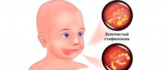
Types of fungal diseases
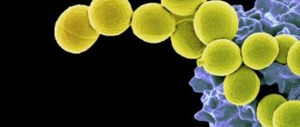
Microscopic pathogenic fungi in medical practice have a common name - mycoses (Greek mycosis). Today, more than 100 types of parasitic and pathogenic microfungi are classified; we will highlight the main infections that affect adults and children.
Dermatomycoses
Common fungal diseases that affect the skin, nails and hair. The source of infection can be a person or an animal. They manifest themselves in various symptoms; we will indicate only the most common diseases in our geographical area:
- Rubromycosis
is a disease caused by the anthropophilic fungus Trichophyton rubrum. It is characterized by a variety of clinical manifestations and localization of lesions on any part of the body; it can affect smooth skin, hair follicles and nails; - mycosis of the foot
(athlete's foot), which also affects the interdigital folds. Very similar to candidiasis, and sometimes polymycosis infection occurs; - favus
is a rare form accompanied by severe baldness of the head. Can be transmitted through combs, underwear and shaving and haircutting tools; - microsporia
– trichomycosis, the causative agent of which is the microsporum fungus. On smooth skin it appears as red spots of a clear shape with a peripheral ridge, and on the scalp – as small flaky lesions; - trichophytosis
(synonymous with ringworm). Externally manifested by pink-red focal skin lesions on any part of the body; - epidermophytosis of skin folds.
Accompanied by itching, redness and peeling.
Keratomycosis
Mycoses are predominantly of the stratum corneum. Among them, the most famous are piedra, erythrasma and pityriasis versicolor. Superficial mycoses are often ignored by many ordinary people, because if lichen versicolor can be primarily identified by yellowish-brown spots covered with pityriasis scales, then erythrasma is often perceived as age-related darkening of the skin. This is due to the fact that the disease progresses slowly and is localized on the inner thighs, in the groin folds and under the mammary glands in women. Faint darkening of the skin affected by erythrasma is covered with small pityriasis-like scales, and although the disease causes almost no itching, it sharply reduces the protective properties of the skin and spoils the appearance of the infected person.
Candidiasis
Diseases caused by yeast-like fungi of the genus Candidosis, which includes more than 150 species. They are considered the most dangerous and most common, as they are found throughout the world and can be transmitted from people, birds and pets. Moreover, in addition to skin manifestations, candidiasis can:
- affect mucous membranes;
- penetrate deeply into tissues and organs;
- cause septic diseases and allergic changes in the body.
Candidiasis can manifest itself as a localized and widespread rash on the hands and feet, lesions of the nail folds and scalp, in the form of stomatitis, cheilitis and gingivitis. With internal infection, they cause vulvovaginitis, urethritis, pleuropneumonia, endocritis, meningitis and other diseases.
Visceral and systemic mycoses
Fungal infections of internal organs caused by infection with taxonomic microfungi and accompanied by severe damage to the skin, visceral (internal) organs, subcutaneous tissue, nervous system and even the musculoskeletal system. This group includes more than two dozen fungi, among which there are pathogenic and conditionally pathogenic. The most common are actinomycosis, aspergillosis, histoplasmosis, coccidiosis, sporotrichosis, chromycosis and others. As a rule, deep mycoses are transmitted indirectly and are difficult to diagnose. Some pathogens demonstrate amazing survivability and resistance to drug therapy, high pathogenicity and significant contagiousness and can lead to disability and death. Most systemic mycoses act as opportunistic infections in patients with hepatitis and AIDS.
Preparation for scraping
The doctor can take a test directly at the appointment if the basic requirements are met:
- Nail plates without decorative cosmetics.
- During the last month, the affected area was not treated with antifungal ointments and other drugs. Otherwise, scraping for nail fungus will give a false negative result. The study will have to be repeated.
- The nail was not trimmed for 6-10 days.
- There was no contact with water or detergents for 3 days.
Causes of false positive fungal tests
Errors in analysis results do occur. There are a number of factors for the appearance of a false positive conclusion for the fungus. For example, a person did not properly prepare for collecting material. The result of the analysis will be inaccurate, quite possibly a false positive. A lot depends on the laboratory. Collection of biomaterial should be carried out only with a sterile instrument. The test tube must be original and the packaging must be intact. If one of these items is contaminated with fungi, the test result will certainly be a false positive. The reliability of the research procedure depends on the laboratory assistant. Hands must be wearing sterile gloves. This will prevent contamination of biological material. If you do not violate the procedure for preparing for analysis, there should be no erroneous results. It is better to contact a trusted specialist or a specialized laboratory.
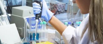
How the study is performed
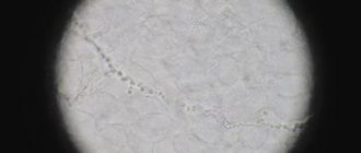
To test for nail fungus, laboratory staff first dissolve the biological material in alkali. The keratin from the plate should go into the liquid phase. Then a preparation is prepared, which is examined through a microscope. Modern equipment allows us to examine the spores and mycelium of a fungal infection and make an expert opinion on the presence of the disease.
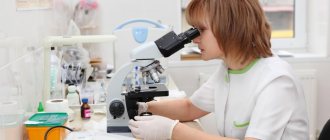
The advent of light microscopes has significantly accelerated the diagnosis of mycoses. Previously, a patient could take a scraping for nail fungus and wait for the culture result only after 3 weeks.
The price of a nail fungus test includes only an unambiguous conclusion about the presence or absence of infection. If more accurate information is needed, the doctor may prescribe PCR tests or other studies.
Methods for diagnosing fungal infections
The Northwestern Center for Evidence-Based Medicine offers traditional and innovative research methods. We carry out the following analyses:
Microbiological methods
Microscopy
– the most accessible and simplest diagnostic method. The study is aimed at confirming infection with superficial mycosis, for which biological material is taken from the patient, which is potentially considered infected: scraping of a pathologically changed nail or skin, eyelash, hair. Refers to qualitative analyzes and allows only to establish or refute the fact of infection. The study takes several days: the resulting material is processed (stained) in a certain way and submitted for microscopic examination to identify the elements of the fungus (spores, hyphae). Microscopy allows you to quickly confirm mycosis, but the type of pathogen and its quantitative concentration are established only for yeast-like and mold species. Therefore, it is often supplemented with cultural research (bacterial culture). The results of microbiological studies must be interpreted by the treating specialist.
Enzyme-linked immunosorbent assay (ELISA)
Linked immunosorbent assay
– a modern and highly reliable method for identifying fungi in a patient’s venous blood. It is a qualitative and quantitative method and can be used as a primary diagnosis and act as a confirmatory analysis of superficial and visceral mycoses.
The method is based on the detection and identification of immunoglobulin protein to a specific pathogen. Antibodies and antigens provide reliable information about infection with aspargillosis, candidiasis, cryptococci and dimorphic microfungi. The test result is interpreted as “positive” (there is an infection) or “negative” (there is no fungal infection). In some cases, the study may give a questionable result; as a rule, this happens if the patient has recently taken antibacterial drugs.
The study takes from 1 to 5 days. If it is necessary to identify the dynamics of the disease, ELISA is carried out every 14 days.
PCR
A highly accurate research method based on the polymerase chain reaction and taking no more than 3 days. It can be used to register any fungal pathogens, but has one drawback - targeted research. This means that the laboratory must obtain information about the specific species of microfungus whose spores and hyphae need to be identified. Blood, sputum, prostate secretion or urine can be provided for analysis, but in the last three options it is necessary to ensure maximum purity of the biological material. The most effective and appropriate in the complex diagnosis of systemic and visceral forms of fungal diseases. The study provides qualitative and quantitative results that are interpreted only by the attending physician.
Serological method
Classic studies in which IgG prepitins, enolase, proteinase and mannoprotein antigens are more often detected. The study is variable and can be based on the agglutination reaction, titration and RSC. They allow you to obtain information only about the fact of carriage of a mycotic infection or indicate a previously suffered fungal disease. The analysis may be based on blood serum testing. With extensive serodiagnosis, microfungus can be detected in other physiological fluids of the patient.
The results are interpreted by the treating specialist. In this case, the serological method is often used as a control study for subsequent adjustment of therapy and determining the effectiveness of treatment.
How often should you get tested?
A scraping is taken at the initial visit to the doctor to make a diagnosis. The doctor tells you how much a nail fungus test costs and what information he wants to get. Based on the results of the study, treatment is prescribed. If the lesion is small, repeat testing may not be necessary.
If the fungus has already led to deformation of the nail, a change in its structure, treatment control will be required. To do this, scraping is done several times at the discretion of the doctor. Based on the test results, the doctor will be able to adjust the treatment for the fastest and most sustainable results.
Risk groups and prevention of fungal infections
Fungal pathogens are found in minimal quantities on the skin of any person. But uncontrolled use of medications, especially hormones and antibiotics, can provoke their active growth and subsequent lesions. It should also be taken into account that deep mycotic infections can enter the body through open wounds. Compliance with sanitary rules for treating any injuries associated with a violation of the skin minimizes the risk of disease with systemic and visceral microfungi.
High humidity and constant above-zero temperatures are an ideal environment for the life and reproduction of microscopic fungi. Accordingly, the risk group a priori includes employees and visitors of swimming pools, fitness clubs, bath complexes, spa salons, as well as workers in laundries and catering establishments.
People with weakened immune systems, a depressed nervous system, a tendency to allergies, critical weight loss and metabolic disorders are also prone to fungal infections. Therefore, the most effective prevention is strengthening the immune system, impeccable adherence to personal hygiene rules and systematic examinations by a therapist and dermatologist.
References
- Russian Society of Dermatovenereologists and Cosmetologists. Federal clinical guidelines for the management of patients with trichophytosis. M.: 2015.
- Federal clinical guidelines. Dermatovenereology 2015: Skin diseases. Sexually transmitted infections. — 5th ed., revised. and additional - M.: Business Express, 2016. - 768 p.
- Verma S, Madhu R. The great Indian epidemic of superficial dermatophytosis: An appraisal. Indian J Dermatol 2017;62:227-36.
- Shen JJ, Jemec GBE, Arendrup MC, Saunte DML. Photodynamic therapy treatment of superficial fungal infections: a systematic review. PhotodiagnosisPhotodynTher. 2020;101774. doi:10.1016/j.pdpdt.2020.101774