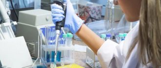
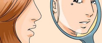
Scraping for pathogenic skin fungi is a microscopic analysis that is performed to identify fungal infections of the epidermis (mycosis). Using this technique, it is possible to determine the fact of infection with a fungus (preliminary analysis) and determine the type of pathogen (final analysis). The causative agents of mycoses can be pathogenic and opportunistic fungi. In order for the dermatologist to prescribe an effective course of therapy, the patient is sent for mycological examination.
general characteristics
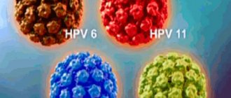
Fungal skin diseases (dermatomycosis), caused by pathogenic dermatomycete fungi (dermatophytes), are divided into 4 groups. The first group is keratomycosis. Pathogens parasitize in the most superficial parts of the stratum corneum of the skin or on the cuticle of the hair. This group includes pityriasis versicolor, erythrasma, nodular trichosporia and axillary trichomycosis. The second group is epidermomycosis. The pathogens also parasitize in the stratum corneum, often affecting the nails, but, unlike the pathogens of keratomycosis, they cause an inflammatory reaction from the underlying layers of the skin. This group includes: athlete's foot, athlete's foot and superficial yeast lesions of the skin and mucous membranes (candidiasis). The third group is trichomycosis. They differ from the causative agents of epidermomycosis in their ability to infect not only nails, but also hair, growing into their cuticle and penetrating into the cortex of the hair. This group consists of: trichophytosis, microsporia and scab. The fourth group is deep dermatomycosis: actinomycosis, deep blastomycosis, chromomycosis, sporotrichosis, as well as visceral forms of candidiasis and mycosis caused by certain molds. This study determines only the presence of a dermatophyte, without determining the species.
Methods for diagnosing fungal diseases
Scrapings are taken exclusively from smooth skin.
A dermatologist most often prescribes this test to patients with lichen that affects the shoulders, back, chest and neck. The doctor scrapes off the top layer of dead skin and obtains a sample containing mycelium and fungal spores.
Tests for heterotrophic microorganisms include:
- scraping for pathogenic skin fungi;
- microscopic diagnostics;
- cultural research.
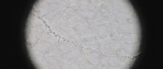
Using microscopic diagnostics, clear contours of the microorganism are revealed. To do this, the biomaterial is clarified in an alkaline environment, which dissolves organic matter and only fungi remain for further diagnosis.
A culture test will help identify the causative agent of the infection. The analysis is performed within 7-10 days, and the doctor can make the final diagnosis based on the results of cultural diagnostics on the 13th day, since primary fungi grow over a long period of time. Other methods for diagnosing skin diseases caused by microorganisms include:
- enzyme immunoassay blood test;
- PCR research;
- sowing.
Patient preparation rules
scraping the epidermis
Standard preparation conditions (unless otherwise determined by the doctor):
3 days in advance Stop taking any cosmetic products for facial skin care (creams, soaps, lotions, etc.), local medications.
Do not wash your face on the day of delivery. hair Standard preparation conditions (unless otherwise determined by the doctor):
3 days in advance Stop taking any hair care products, topical medications.
Notes:
To examine hair from the affected part of the head, cut it into a clean container at a distance of 0.1 mm from the roots or pull it out with hair follicles; do not wash your hair on the day of submitting the material.
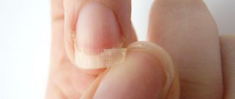
nails Standard preparation conditions (unless otherwise determined by the doctor):
3 days in advance Stop taking any cosmetics (varnishes, nail strengtheners, etc.), local medications.
Notes:
Nails must be clean and dry. You should also not treat your nails with a metal file before the procedure. Trim the nails of both hands (the length of the cut nails should be at least 2 mm). BM is delivered in a sterile plastic container with a screw cap or in a paper envelope.
You can add this study to your cart on this page
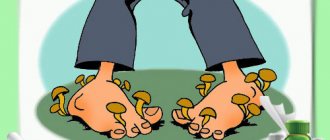
Symptoms of fungal skin diseases
It is difficult to determine the causative agent of infection in the early stages, since there are no external manifestations of the disease. With a large number of pathogenic microorganisms, the first symptoms appear: the skin becomes pale and looks unhealthy; peeling of the skin is observed; hair becomes brittle and begins to fall out; spots form on the skin with an unpleasant odor; the structure of the nails is destroyed; discomfort, itching and burning are felt.
Pathogenic fungi reproduce in favorable conditions (humid environment) and can live outside the human body for a long period of time. It is possible to become infected through direct contact, when the pathogen comes into contact with healthy areas of the body. You can “get” an infection through someone else’s shoes or clothes; this often happens on the beach, in a bathhouse or in a swimming pool.
The incubation period lasts from 2-3 days to 3-4 months. The first symptoms of the disease are cracks in the skin, itching, rashes, and exfoliation of the upper layer of the epidermis.
Indications and contraindications for taking scrapings for pathogenic skin fungi
The procedure is indicated for the purpose of preventive diagnostics for visiting a swimming pool, bathhouse, sauna and other public places. The analysis begins when mycoses are suspected, in particular due to the patient’s complaints of cracks, rashes, itching and peeling of the epidermis. Using the study, you can detect microorganisms, determine their type and make an accurate diagnosis.
There are no contraindications to the manipulation, since it is painless and is carried out within a few minutes. Taking a scraping does not cause discomfort or other unpleasant sensations.
Decoding the research results
The result is considered positive when mycelium, pseudomycelium and fungal spores are detected in the scrapings, as well as in the presence of symptoms of mycotic skin lesions. It should be understood that mushrooms are a component of normal human microflora. The absence of symptoms with a positive fungal test result indicates a healthy carrier state. In addition, this diagnostic method does not allow identifying the type of pathogen. This requires additional research, including inoculation of skin scrapings on nutrient media.
Do you need to do a skin scraping test for fungi?
The Med-Yutas Medical Center conducts microscopic examinations for fungi in Moscow clinics located at st.
Grimau, 10A, building 2 (metro station Akademicheskaya) and Michurinsky Avenue, 21B (metro station Ramenki). To obtain a consultation and make an appointment, use the online registration form or contact the clinic reception. The procedure is performed by a laboratory microbiologist, duration – 10 minutes. The cost of examining skin and nails for fungus is from 790 rubles.
INITIAL CONSULTATION
from 2,500 rub.
What is the danger?
Without treatment, a fungal infection can serve as an “entry gate” for infectious diseases, such as erysipelas. The worst thing is that in many patients this process sooner or later moves from the feet to the hands. This is a real tragedy for women. If your toenails can be hidden in shoes or socks, then your hands are always visible. Extending nails, using polish, etc. only worsens the process. Such tricks form the so-called “greenhouse effect” and the fungus begins to feel just great, multiplying and destroying your nails, health and peace of mind.
Unfortunately, once the fungus has taken root, it will never leave its host without treatment. The process can only progress and spread to other parts of the body. So, itchy, scaly spots may appear in the groin area, head, thighs and other parts of the body.
We should not forget about the constant allergization of the body, weakening of the immune system, exacerbation and worsening of chronic diseases such as diabetes and bronchial asthma.
Microscopic examination for micromycete fungi
general information
A disease caused by fungal infection of the skin and its appendages (nails, hair (eyelashes)) is called superficial mycosis. When infected, fungi invade and form colonies in the outer layer of skin (epidermis), hair and hair follicles (“bulbs”), and nail apparatus.
Diagnosis of superficial mycoses is carried out by a dermatologist. The research algorithm is as follows:
- a general examination of the patient is carried out;
- the affected area is examined using a Wood's ultraviolet lamp;
- A scraping is taken from the skin, the affected hair is removed with tweezers along with the bulbs and examined under a microscope for the presence of fungi.
The most common cause of the development of superficial mycoses is fungi of the genus Dermatophytes, the infection of which is called dermatophytosis. In addition, the pathology can be caused by:
- fungi of the genus Candida (candida) - an opportunistic fungus that is part of the normal microflora of the body, and in large quantities is the causative agent of candidiasis;
- pathogenic fungi of the genus Trichosporon (trichosporon - in the scalp causes the disease white piedra (the formation of soft nodules of yeast cells around the hair shafts), and in people with weakened immunity it can cause the development of trichosporonosis (systemic damage to the body by fungi of this type));
- parasitic fungi Microsporum, which is the causative agent of microsporia - a disease of the hair and head
- pathogenic fungi Epidermophyton, which cause dermatomycosis and should not normally be found on human skin and mucous membranes;
- Hortaea mushrooms - their representative Hortaea werneckii is the causative agent of shingles in humans;
- Malassezia (Malassezia) is an opportunistic fungus that is part of the normal microflora, but with an increase in the number it causes malassezia of the skin: dandruff, seborrhea (an inflammatory skin disease that occurs due to an increase in the amount of sebum) and atopic dermatitis (an inflammatory skin lesion of an allergic nature); Malassezia furfur is the causative agent of pityriasis versicolor, etc.
Transmission of superficial mycoses from one person to another in several ways:
- in personal close contact with a person affected by the infection;
- when wearing someone else's shoes, using a comb, hats, scissors, hygiene and household items;
- when visiting public places, for example, baths, swimming pools, showers, hairdressers, etc., as well as children's and educational institutions.
The infection enters the body through wounds, cracks, diaper rash, and ulcers. A superficial examination of the affected areas allows us to make only an assumption about the presence of a disease, since fungal damage is visually similar to other diseases that affect the skin and its appendages (for example, psoriasis). The presence of fungi is determined by microscopic examination of samples of skin, hair, and nails taken from the affected areas. This test does not indicate the type of mushrooms detected, but only states the fact of their presence. For a more in-depth study to determine the type of fungi that caused mycosis, the biomaterial under study is inoculated on nutrient media.
Skin mycoses
Depending on the symptoms of the disease, the type of fungus, and the degree of its penetration into the skin, mycoses are divided into two categories:
- superficial mycoses - those that affect
- deep mycoses - affecting the dermis (the connective tissue part of human skin lying under the outer layer of skin, the epidermis) and deeper soft tissues lying under the skin, and with a significant deepening - bone tissue.
In addition, mycoses are divided according to the area affected, in particular:
- mycoses of smooth skin, in which fungi are localized in the face, torso, and limbs);
- keratomycosis - the formation of fungal colonies on the surface parts of the skin (stratum corneum of the epidermis) and fungal follicles;
- mycoses of the scalp;
- Onychomycosis is fungal infection of the nail apparatus.
The most common mycoses of the skin and its derivatives are:
1. Superficial mycoses
a) dermatophytosis:
- Rubrophytia is the most common fungal disease of the feet, affecting the skin and nails, as well as any areas of smooth skin and vellus hair, including skin folds, nails and skin of the hands;
- trichophytosis is a highly contagious fungal disease of the skin, hair, and nails caused by fungi of the genus Trichophyton (in common parlance lichen);
- microsporia is a fungal infection of the skin involving long or vellus hair, caused by fungi of the genus Microsporum (in common parlance - “ringworm”);
- favus - mycosis, which is caused by the parasitic fungus Trichophyton schonleinii, which affects mainly smooth skin (in very rare cases, the effect of the fungus can spread to hair, nails, and internal organs);
- epidermatophytosis inguinalis is a fungal infection accompanied by itching and rash, localized in the folds of the skin of the groin area, and sometimes in other large folds of the body;
- Athlete's foot is a common fungal infection that affects the skin and nails of the feet.
b) candidiasis – damage to the mucous membranes and skin by opportunistic candida yeast fungi;
c) keratomycosis – infection of the stratum corneum of the epidermis by a fungus, one of the representatives of which is lichen versicolor.
2. Deep mycoses:
- chromomycosis is a deep mycosis, more often found in the subtropics and tropics, which affects the skin, subcutaneous tissue, and in some cases, internal organs;
- keloid sporotrichosis is an infectious disease caused by the dimorphic fungus Sporotrix, shencki, and less commonly, s. Shenki var. luriei;
- North American blastomycosis is a fungal infection of animals and humans, most often affecting the lungs, common in North America, the causative agent of which is the dimorphic fungus Blastomyces dermatitidis.
The causative agents of skin mycoses are quite often the yeast fungi Candida and Malassezia, as well as dermatophyte fungi - Trichophyton, Epidermophyton, Microsporum.
Representatives of yeast fungi are part of the normal microflora, being, in fact, conditionally pathogenic fungi. Exposure to provoking factors on the body (high temperatures, humidity, decreased defense response, failure to comply with personal hygiene rules, etc.) can cause their excessive growth. Signs of skin candidiasis are red or pink rashes in the folds of the skin. Tinea versicolor, caused by Malassezia furfur, is manifested by multiple foci of peeling on the skin. The resulting spots can be of different colors - pink, white, brown, which is why the disease received such a “bright” name.
Dermatophyte fungi - Trichophyton, Microsporum, Epidermophyton, are pathogenic. Normally, they should not be found on human mucous membranes and skin. The nutrient medium for these fungi is the protein keratin. It is contained in the upper layers of skin and hair, as a result of which the location of parasite colonies can be observed in different parts of the body. Hair affected by fungi breaks and falls out, the skin itches and peels, and ring-shaped rashes typical of infection appear on it.
Infection with agents of superficial mycoses occurs through personal contact with an infected person, when sharing personal items (combs, scissors, hats, etc.), when visiting public places (baths, saunas, swimming pools). Mushroom particles are resistant to external influences. They can remain on surfaces for a long time.
Infection can also occur through animals, among which the most common carriers of infection are dogs and cats. Factors that increase the risk of contracting mycosis are decreased immune defense, the presence of damaged skin areas, and diaper rash.
Microscopic examination of skin scrapings is a preliminary test and is carried out to confirm/refute the presence of lesions by opportunistic or pathogenic fungi without specifying the specific causative agent of the infection.
Hair mycoses
Fungal infections of the scalp are characterized by certain characteristics. The most common causative agents of infection include the dermatophyte fungi Trichophyton, Epidermophyton and Microsporum. The infection is caused by fungal spores landing on the head. This occurs when there is contact with a carrier of the infection, which can be a person or animal, or violation of personal hygiene rules. Fungal spores are resistant to environmental factors and remain viable for a long time on contaminated surfaces.
After entering the scalp, fungi cause the formation of one/several lesions, characterized by fragility and hair loss, itching and redness of the skin.
One type of mycosis of the skin and hair is called microsporia. Its causative agents are the fungus Microsporum ferrugineum, which is pathogenic for humans (anthropophilic), and the fungus microsporum lanosum, which is zoophilic (a threat to humans and animals). When affected by fungi, the hair becomes whitish and breaks off, leaving stubs up to 6 millimeters.
Another hair disease, trichophytosis, develops when infected with the fungi Trichophyton tonsurans (more often) and Trichophyton violaceum (less often). The cause of infection is contact with a person suffering from mycosis, the use of his personal belongings - scissors, comb, hats. The most common places of infection are hairdressers, preschools and educational institutions.
Before conducting a microscopic examination of the hair, it is removed from the affected area with tweezers. Examining the affected hair under a microscope allows you to determine the structure of the hair and detect fungal particles in it.
Mycoses of the nail apparatus
Fungal infection of the nail plate is called onychomycosis. This disease is observed only in humans. Infection can occur through personal contact or through objects used by the patient, at home or in public places. There are a number of factors, both external and internal, that create favorable conditions for the development of fungus. Thus, the likelihood of infection increases:
- when receiving minor injuries;
- when wearing rubber shoes;
- with increased sweating;
- when the body's resistance decreases;
- in case of dysfunction of the thyroid gland;
- in the presence of chronic and somatic diseases;
- with immune deficiency.
Nails may be affected:
- dermatophyte fungi (E.floccosum, T.rubrum, T.violaceum, T.tonsurans, M.gypseum, T.schonleinii, T.mentagrophytes v.interdigitale);
- yeast-like fungi Candida;
- mold fungi (Penicillium. spp., Scopulariopsis brevicaulis, etc.).
Every second patient with onychomycosis is affected by several types of the listed fungi, and the disease spreads to the nails of both the upper and lower extremities.
The most informative methods for confirming infection are microscopy of fragments of the nail and scrapings taken under it, as well as culture of the material and isolation of the culture.
To conduct a bacteriological study, material is taken from the visible affected area (optimally at the border between the affected and healthy area). The types and clinical form of fungal nail infection differ, which influences the choice of sampling site. The fungus can be located:
- under and inside the nail (distal and proximal forms);
- in the outer layers (surface form);
- in the posterior nail fold (paronychia) - the area located behind the cuticle.
Material for research is taken from the affected area of the fingernails or toenails. Depending on the clinical form of nail damage, the sampling technique changes, which is reflected in the table:
| Clinical form onychomycosis | Sampling area | Methodology used |
| superficial | nail plate | surface scraping |
| distal-lateral subungual | nail bed | section of the nail plate |
| nail plate | scraping of the nail bed, closer to the proximal edge | |
| proximal subungual | nail bed | section of the nail plate |
| periungual fold | nail dust obtained using a drill | |
| nail plate | biopsy (removal) of the nail | |
| scraping from under the removed part of the nail |
How to take a scraping
The procedure can be carried out directly at the doctor’s appointment. The doctor unseals a sterile scalpel, glass slides or a special container with a lid. To test for fungus, scrape the top layer of epithelium from the scalp, limb or other part of the body where the lesion is found, and take a few hairs, if any. It is important that particles of biomaterial do not fall on healthy areas, otherwise pathogenic spores will quickly spread. The test tube is carefully packaged and signed.
After taking a scraping for a fungal infection, no traces remain on the skin. The doctor removes only the upper layers, consisting of dead cells, next to which there are no longer any vessels. If blood appears during scraping, it is enough to apply a sterile napkin to the wound.
Recovery time
In the most severe cases of long-term, advanced mycosis, the average duration of therapy will take 6-7 months. This period of time is determined by the rate of growth of toenails - the nail plate completely changes just during this time. This period does not mean that the patient will need to regularly visit the doctor for the entire six months. Scheduled follow-up visits to the doctor are necessary only 2-3 times during therapy, including the initial appointment (now we are not talking about visits for a hardware pedicure procedure).
Case study
Patient A. came for an initial appointment in November 2009 with complaints of changes in the structure and color of nails, itching in the groin area. He has been ill, according to him, for about six years. Treatment with Orungal, Lamisil and Batrafen Lac did not bring relief. The patient experienced great anxiety and even despair when the process moved to his fingernails, which became the decisive impetus for visiting the mycologist. Upon examination, the nails of A.'s big toes are completely affected: thickened, crumbling, yellow in color. Detachment of the epidermis, itching, and cracks in the skin between the fingers were also observed. On the first and fifth fingers of the right hand there are yellow spots, thickening and detachment from the nail bed. Inguinal region: large, clearly defined scaly plaques, excoriations.
At the beginning of December, the results of A.'s tests were completely ready. Using an accurate history, physical examination and laboratory methods, eczema, onychodystrophy, psoriasis of skin folds, erythrasma and candidiasis of skin folds - severe skin diseases with identical external signs - were excluded. To confirm the fungal etiology of this process, material was taken from the nail plates and the skin of the groin area. In both cases, microscopy revealed fungal mycelium, and the exact pathogen was identified in culture - Trichophyton rubrum.
Already at the first examination, it became obvious that for a successful recovery the patient needed a therapeutic spectrum, including broad external therapy, therapy with systemic antimycotics, and therapy for dystrophic processes in the nail bed that arose as a result of the duration of the disease. In addition, recommendations were given aimed at improving microcirculation and improving blood flow to the nail plates, which should ensure the delivery of the necessary nutrients, also prescribed to accelerate nail growth and reduce treatment time.
The third visit was scheduled for A. at the end of February. Upon examination, a significant improvement in condition was observed. So, in particular, in the groin area the skin is absolutely healthy, blood biochemistry tests are normal. The treatment regimen has been adjusted (in the process this must be done once every two months).
The last visit took place at the end of April 2010. Based on the results of the examination and repeated tests (blood biochemistry and microscopy), patient A. is absolutely healthy.