Skin rashes are a sign of syphilis infection.
Syphilis is a severe infectious disease of an inflammatory nature caused by the pathogenic bacterium Treponema pallidum. First of all, it affects the skin and mucous membranes of the body, manifesting itself in various rashes, erosions and ulcers.
Syphilis is divided into three stages, each of which has its own manifestations, therefore, what a syphilis rash looks like will depend on the stage of development of the disease.
Infection and incubation period
Treponema pallidum affects the body through the slightest damage to the skin and mucous membranes, and it does not have to be an open wound, just a thinned area of the skin in a certain area.
After penetration into the body, it is concentrated in the lymphatic system and gradually spreads throughout all fluid environments of the body, accordingly, infection can occur through:
- blood;
- mother's milk;
- saliva;
- vaginal fluid;
- sperm;
- lubricant that is released during sexual intercourse and when aroused.
Treponema pallidum is concentrated in all body fluids.
Routes of infection:
- In 95% of cases, infection with Treponema pallidum occurs through sexual contact and it does not matter whether it is protected sex or not. A condom reduces the risk of infection, but does not eliminate it completely. The type of sex also does not matter. The risk of infection is the same during vaginal, anal and oral intercourse. In all cases, an exchange of body fluids occurs, in one case it is sperm and vaginal fluid, in the other saliva. Anal sex is even much more dangerous than usual, since the pathogen penetrates through damage to the mucous membranes, and the rectum constantly suffers from microcracks. And during vaginal sex, the acidic environment inhibits the spread of infection, but the rectum is deprived of such protection.
- Infection in the home occurs less frequently, but is possible through close contact with an infected person. The pathogen remains active in the open air until the liquid in which it is concentrated dries; therefore, infection can occur from a cup with infected saliva on it or from bed linen with discharge from a patient’s skin rash.
- Infection of a child from the mother during pregnancy, childbirth or breastfeeding is another route of infection. Cases of such infection are minimal, since pregnant women undergo mandatory testing for syphilis.
- Infection through blood leads to disease in 100% of cases. The patient’s blood, especially at the second stage of the disease, contains the maximum concentration of treponema in the body. This method of infection can occur through donor blood transfusion, when using one syringe (especially in drug addicts).
- The professional route of infection occurs only among doctors, since they have direct contact with infected people.
Important. Infection during unprotected sex is 95%.
Incubation period and its manifestations
After entering the body of a healthy person, the pathogen is concentrated in the lymphatic system, since this is the only body system with suitable conditions for it. During this period, the pathogen adapts to the conditions of the body and multiplies for further spread throughout the body.
The period of adaptation of Treponema pallidum is called the incubation period. This time is characterized as asymptomatic, since the disease does not manifest itself in any way.
Incubation can last from 10 to 90 days, the average time is 20-45 days. When the first symptoms appear depends on the state of the body. Incubation will decrease if the infected person has a severely weakened immune system, and a healthy body will slow down the spread of infection.
After adaptation of treponema pallidum in the body through the walls of small blood vessels, it spreads through the bloodstream and the first symptoms of the disease begin to appear. The entire period of the disease, from the manifestation of the first symptom to the most severe damage to the body, is divided into stages of development, each of which is distinguished by certain clinical manifestations.
A distinctive feature of the disease is damage to the skin, which manifests itself as a rash, but depending on the stage it looks different, but has similar characteristics:
- cyclical appearance;
- disappearance, even without treatment;
- does not leave scars after disappearance (except for the tertiary stage);
- despite the strong inflammatory process, there is no increase in temperature and symptoms of intoxication of the body.
Important. Treponema pallidum is very sensitive to acidic and alkaline environments, therefore, for prevention purposes, you can use 0.01% Chlorhexidine bigluconate and Gebitan solution.
How is it transmitted?
Syphilis is caused by Treponema pallidum, which lives in the external environment for only 3 minutes. Therefore, the main route of transmission of the disease is sexual. Infection of the fetus is possible in utero (vertical route) or intrapartum, when the child passes through the mother’s birth canal.
The household route of transmission is uncommon; infection is possible from persons with the tertiary stage of syphilis, when treponema pallidum gets on dishes, linen, towels, etc. from decaying gums. Hematogenous transmission of syphilis through blood transfusion cannot be ruled out.
Cases of infection of medical workers through contact with the blood of a patient are not that rare. It is possible to become infected through “bloody” objects: a shared toothbrush, razor, manicure set, etc.
Skin manifestations of primary syphilis
The beginning of the primary stage of syphilis is marked by the appearance of a skin lesion at the site of infection, which looks like a red spot; this is the primary manifestation of the disease - chancre. It occurs as a result of the cells of the immune system limiting pathogenic microorganisms at the site of their penetration. Chancre is a kind of protective reaction of the body to the invasion of pathogenic microorganisms.
Its localization will depend on the route of infection; in case of sexual infection, it is located on the genitals, in the area of the anus or in the oral cavity; in case of domestic infection, it is located in any place on the skin. If the infection occurs in a rare way, primary syphiloma is absent through the blood and the disease immediately manifests itself as symptoms of secondary syphilis.
Distinctive characteristics of chancre:
- at the beginning of development it looks like a red spot with a dense structure;
- after two weeks it increases in size, acquiring an increasingly dense structure and the appearance of a nodule;
- in the process of maturation it turns into erosion or ulcer with scanty secretion;
- remains unchanged for about 30 days and begins to heal;
- does not cause discomfort or pain, since the toxins of the pathogen have an analgesic effect;
- chancre is a single formation;
- round regular shape with clear outlines;
- size – up to 10 mm, in rare cases up to 15 mm;
- surface is smooth.
In the photo, erosion is the primary manifestation of the disease.
Chancre is a typical manifestation of syphilis at the first stage, but in rare cases the disease can occur in an atypical form and the skin manifestations will look different.
Rash with primary syphilis of atypical form:
- Chancre panaritium is an atypical syphiloma localized on the phalanx of the finger. Its development begins with a small ulcer with dense inflamed tissue around its circumference. It has an irregular shape and unclear outlines. After ulceration, the discharge of purulent contents begins. The inflammatory process involves a large part of the finger, which becomes inflamed and takes on a swollen appearance and a purple tint. Chancroid felon is accompanied by inflammation of the lymph nodes in the area of the elbow joint. Atypical chancre causes pain and may be accompanied by an increase in temperature and a clinical picture of intoxication. Syphiloma can become chronic and not heal for several months.
- Indurative edema is an atypical manifestation of chancroid. It is most often found on the external genitalia, in men it is the foreskin or scrotum, in women it is the labia minora and the clitoris. It looks like an inflamed area of the mucous membrane, which increases in size several times and acquires a red tint. The fabric is characterized by increased density and does not leave dents when pressed. When subjected to mechanical action, it does not cause pain; it persists for several weeks, in rare cases until the onset of secondary syphilis.
- Chancroid amygdalitis is an atypical form of chancre that affects the larynx and tonsils. Manifests itself as an ulcer or erosion on the oral mucosa or tonsils. It has a round shape with clear edges and swollen tissue around the circumference. The surface of the chancre is smooth, size up to 10 mm. Chancroid amygdalitis is very similar to a regular sore throat, but there are still fundamental differences. It affects only one side, is accompanied by enlargement of the submandibular lymph nodes and is not accompanied by an increase in temperature, like ordinary sore throat. When swallowing it causes pain.
The initial stage of development of chancre felon.
Lesions of the mucous membranes and rashes on the skin with syphilis of the first stage end here and the disease does not manifest itself in any way until the onset of secondary syphilis.
In a very rare case (only 5% of those infected), symptoms of intoxication of the body may appear:
- temperature increase;
- fever;
- general deterioration of health.
Important. Atypical chancre is difficult to diagnose and very often other diseases are treated instead. They can be distinguished by concomitant inflammation of the lymph nodes.
Syphilis of the nose
Nasal syphilis occurs during different periods of the disease.
Nasal involvement in early congenital syphilis
With early congenital syphilis, the nose is affected in 75% of cases. The disease manifests itself either immediately after birth or during the first 4 to 5 weeks of the child’s life. A dry runny nose, accompanied by noisy exhalation and inhalation, is quickly replaced by severe rhinitis. The child begins to sniffle, snort and sneeze, and refuses to breastfeed. The mucous membrane at the entrance to the nose becomes hyperemic, thickens and becomes crusty. A viscous secretion is released from the nasal passages. Due to the development of the ulcerative process, blood appears in the discharge over time. From an epidemiological perspective, children during this period become extremely dangerous to others.
Sometimes newborns develop gummous infiltration in the nose. The disintegration of gummas is accompanied by damage to the bone structures, as a result of which a perforation of the nasal septum develops, and various types of retraction of the external nose are formed.
Syphilitic rhinitis of newborns must be distinguished from gonorrhea and nasal diphtheria. Persistent runny nose, thick nasal discharge, crusting, skin rash and enlarged spleen are factors that indicate a specific process.
Rice. 5. Diffuse infiltration of Hochsinger's skin and syphilitic rhinitis of the newborn.
Nasal lesions in late congenital syphilis
Nasal lesions in late congenital syphilis develop 5 or more years after birth. Foreign literature describes cases of the development of pathology at 23–28 years of human life.
The secretion of a viscous secretion, the formation of crusts, a feeling of dryness in the nose and throat, loss of smell and the appearance of pain in the nose, forehead and eye sockets are the main manifestations of syphilitic congenital runny nose in late congenital syphilis. As a result of tubercular infiltration, the mucous membrane grows, gradually obstructing the nasal passage.
The process often occurs like tertiary syphilis. Gummas are located on the nasal mucosa, in cartilage and bone, while the mucous membrane is involved in the pathological process for the second time. The nasal passages become overgrown, the wings of the nose, its back, tip, turbinates, bottom and nasal septum are destroyed. Recessed nose is a pathogonic symptom of late congenital syphilis. The nose may have the shape of a saddle (“saddle nose”), or be similar to the nose of a bulldog or have a lorgnette-like appearance.
Syphilis of the nose in the primary period of the disease
Nasal involvement in the primary stage of the disease is rare. Hard chancre most often appears on the wings of the nose, at the entrance to its cavity on the skin in the area of the septum. The chancre has the appearance of red erosion with a roller-like thickening along the periphery, a greasy coating is detected at the bottom, and a dense infiltrate is palpated at the base.
Nasal syphilis in the secondary period of the disease
Nasal syphilis in the secondary period of the disease develops 6-7 weeks after the appearance of chancroid and manifests itself in the form of roseola (erythema) or papules. Erythema (redness) spreads to the mucous membrane, which swells and begins to produce a secretion of a mucous or bloody-serous nature. A little later, papules appear, which are located on the skin at the entrance to the nasal cavity. Due to constant irritation, papules erode and the healing process is delayed.
Syphilis of the nose in the tertiary period of the disease
The nose is most often affected in the tertiary period of the disease. The lesion affects the mucous membrane, periosteum, bone and cartilage, where gummas with decay or diffuse infiltrates develop. Destroyed areas of bone tissue are sequestered and come out with pus. The decay of gumma is accompanied by a putrid odor. The gumma itself acquires a bluish-red color and becomes covered with purulent-bloody crusts. Removing crusts often leads to bleeding.
The bony septum and floor of the nose are the most commonly affected areas. When they are destroyed, communication occurs with the oral cavity and nasal passages. The nasal passages narrow. The patient experiences severe pain in the nose, eye sockets and forehead area.
Gummas can form in cartilage and bone, while the mucous membrane is involved in the pathological process for the second time. The nasal passages become overgrown, the wings of the nose, its dorsum, tip, turbinates, bottom and nasal septum are destroyed, the nose sinks and takes on the shape of a saddle (“saddle nose”). The act of breathing, swallowing and phonation is disrupted.
Patients with a gummous process in the nose during the period of tertiary syphilis are practically not dangerous to others from an epidemiological perspective.
Gumma in the nose should be distinguished from tuberculosis and a malignant tumor. Large deformations of the external nose after treatment are corrected with rhinoplasty.
Rice. 6. Forming nasal gumma.
Rice. 7. Gummous lesion of the wings of the nose.
Rice. 8. Nasal syphilis in the tertiary period of the disease - perforation (destruction) of the nasal septum.
Rice. 9. Recession of the nose is a pathogonic symptom of late congenital and tertiary syphilis.
Rice. 10. Syphilis of the nose. Destruction of the structures of the external nose by the gummous process.
Rice. 11. Syphilis of the nose. Tertiary period of the disease. Destruction of all structures of the external nose, its septum, bottom, anterior part of the alveolar processes, teeth and upper lip.
Rice. 12. Complete destruction of all structures of the nose.
Rice. 13. With the disintegration of gumma and destruction of bone between the nasal septum, nasal cavity and oral cavity, perforations occur, which makes it difficult for the patient to eat, phonation and the act of swallowing.
Skin manifestations of secondary syphilis
Skin rashes with syphilis of the second stage are very diverse. They appear at the beginning of the second stage and last, on average, up to three months and disappear without treatment.
Then, with an interval, the latent course is repeated again and so on throughout the entire secondary syphilis. This wave-like course greatly complicates the diagnosis of the disease and in most cases the symptoms are attributed to other diseases.
But still, the skin manifestations of the second stage have distinctive characteristics:
- appear gradually, several pieces during the week, as a result of which they have different degrees of maturation;
- each element of the rash is clearly limited from healthy tissue;
- have a benign course;
- heal without treatment;
- do not leave marks on the skin;
- the elements of the rash do not unite and merge into one;
- are repeated at regular intervals;
- despite the strong inflammatory process, they are not accompanied by hyperthermia and symptoms of intoxication;
- are not accompanied by discomfort (itching, burning) and pain.
Types of rash with secondary syphilis:
- syphilitic roseola;
- papular syphilide;
- pustular syphilide;
- vesicular syphilide;
- syphilitic leukoderma;
- syphilitic alopecia.
Syphilitic roseola
Rash on the body with syphilis of the second stage. The most common skin lesion, diagnosed in 80% of patients.
It is localized everywhere, from the soles of the feet to the scalp, but is more often observed on the side surface of the body.
Distinctive features:
- color – pink-red;
- size – from 3 to 10 mm;
- shape – round with unclear outlines;
- periodic appearance of 10-12 pieces per week, because of this they have different degrees of development;
- storage duration up to 1 month;
- the surface is smooth, without peeling;
- do not protrude above the surface of the skin;
- do not have a specific location framework;
- are not combined into one element and have a clear distinction.
Rash in the second stage of the disease.
Papular syphilide
One of the manifestations of the second stage of the disease has a round shape, is painless, is not prone to elemental association and can be located everywhere.
Papular syphilide has varieties:
- Psoriasiform - papules with an uncharacteristic silvery tint and a surface consisting of scales. The circumference of the formation has a copper tint and infiltration.
- Seborrheic syphilide is most often a rash on the face during stage 2 syphilis, located in the area where the sebaceous glands are located (forehead and hairline). It looks like a papule with a rough surface and scales with a greasy coating.
- Ring-shaped syphilide is a ring-shaped rash on the head of syphilis in men; it can also occur in the back of the head and scrotum.
- Miliary syphilide is observed in patients with chronic diseases, signs of intoxication and in the elderly age category. Miliary syphilide has the appearance of cone-shaped papules the size of a grain, dense texture with a brown tint. Localization in the torso and limbs. The surface is smooth, but can sometimes be covered with scales.
- Weeping syphilide, most often, is a rash in the groin during stage 2 syphilis, as it is localized mainly in the area of increased sweating. Also found in the anus, perineum and armpits. It looks like white erosive papules with a dense structure. If they are in an area that is constantly irritated (skin folds), they increase in size and take on a red tint.
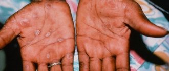
- Palmoplantar syphilide is a rash on the palms due to syphilis that also affects the plantar region. Syphilide has the appearance of dense papules, color from pale yellow to red-violet. It does not protrude above the skin, but has a surface of dense scales.
Papular syphilide on the soles and palms.
Pustular syphilide
Very rare. Patients with a malignant course of syphilis, with a weakened body and in the presence of chronic diseases are susceptible to it. In most cases, it is accompanied by a deterioration of the condition with a hyperthermic reaction and symptoms of intoxication.
Pustular syphilide has several manifestations:
- Acne - a profuse rash all over the skin. In appearance, these are papules the size of a pin's eye, with a clear outline and purulent contents. Very often it is a companion of roseola. Acne syphilide lasts up to 2 months and goes away without the formation of scar tissue.
- Smallpox syphilide - a rash on the hands due to syphilis, in the area of flexion of the limbs, also affects the torso and face. The smallpox form of pustular syphilide is pustules with a clearly defined depression in the center and a purulent infiltrate. It appears within 7 weeks and goes away without a trace.
- Impetiginous syphilide is a rash on the chest due to syphilis, also on the scalp, face and limbs. It manifests itself as papules with a diameter of up to 10 mm and a form protruding above the skin.
- Syphilitic ecthyma is a malignant manifestation of the disease in the form of a deep ulcer localized on the lower extremities, face, and torso. The erosion has great depth, purulent contents and necrotic masses. After healing it leaves a deep scar. Accompanied by severe deterioration of the condition with fever, muscle and joint pain.
Pustular syphilide is a purulent skin lesion caused by syphilis.
Vesicular syphilide
One of the most severe forms of skin manifestations. Occurs in patients with chronic and concomitant diseases. While most syphilitic rashes respond well to treatment or go away on their own without consequences, vesicular syphilide is difficult to treat and is prone to relapses.
It looks like red plaques of large diameter (up to 20 mm) on the surface of which bubbles with liquid contents form. During the ripening process, the blisters burst and turn into open ulcers with constant release of fluid.
Syphilitic leucoderma
Pigmented spots up to 1 cm in diameter. Localized in the neck, chest, back and abdomen. It appears gradually, starting with a few spots with their constant addition. Leucoderma does not cause discomfort, does not peel off and is not accompanied by deterioration in well-being.
Official medicine distinguishes three types of pigmentation:
- spotted;
- mesh;
- marble.
Leukoderma is localized on the back of the neck.
Syphilitic alopecia
Alopecia is a syphilitic lesion of the hair, with predominant baldness.
Official medicine distinguishes three types of syphilitic alopecia:
- Fine focal alopecia – small spots up to 1.5 cm in diameter with hair loss. They have an irregular shape and tend to clump together.
- Diffuse alopecia is a sharp decrease in the amount of hair throughout the scalp.
- Mixed alopecia combines small focal and diffuse forms.
Important. The secondary stage begins three months after the appearance of the first symptom - primary syphiloma and lasts from two to seven years.
Tuberous syphilide
Along with gummas, in tertiary syphilis, tubercular syphilide occurs, which is characterized by the appearance of more than 10 spherical-shaped dense tubercles on the skin and mucous membranes. The tubercles exist from several weeks to several months, then their reverse development begins. During outbreaks of the disease, new elements appear, as a result of which the patient simultaneously reveals tubercles that are at different stages of development, as well as pigmented spots and scars formed during healing. Syphilide is most often localized on the skin of the face in the forehead and nose, back, extensor surfaces of the limbs and mucous membranes. Does not cause any subjective sensations.
Histology
Infiltrate in tubercular syphilide is formed in the subpapillary and papillary layers of the dermis and is an accumulation of plasma and epithelioid cells, lymphocytes, eosinophils, fibroblasts and histiocytes. Polynuclear cells—mature neutrophilic leukocytes—appear. As a result of intimal swelling, the walls of the vessels thicken, and the lumen of the vessels narrows significantly. The processes of keratinization according to the type of parakeratosis are disrupted. The interpapillary processes lengthen - outgrowths of the epidermis and epithelium of the mucous membranes appear.
Appearance
Tuberous syphilide is located asymmetrically, usually hemispherical in shape, less often flat, has a copper-red color with a bluish tint, the size of a cherry pit, dense consistency and clear boundaries. The elements of the rash are located in a group, but never merge.
Decay
The tubercles undergo either dry necrosis or necrosis with the formation of ulcers. In the case of the development of dry necrosis, atrophic scars are formed, and when the tubercles disintegrate, sinking scars are formed. Each scar is surrounded by a pigment border. Ulcers formed as a result of decay have a rounded shape, smooth edges, a smooth, clean bottom, with a dense infiltrate around and at the base.
Differential diagnosis
Tuberous syphilide should be differentiated from tuberculous lupus, papulonecrotic tuberculosis, squamous cell skin cancer, erythematosus, small nodular sarcoid, discoid lupus erythematosus, cutaneous leishmaniasis, leprosy, pyoderma and varicose leg ulcers.
Rice. 9. Tertiary period of syphilis. Tuberous syphilide.
Skin manifestations of tertiary syphilis
During the third stage, skin lesions are more pathological and irreversible.
As such, there is no rash and skin lesions appear:
- Tuberous syphilide is a formation deep in the skin in the form of a node and with a diameter of up to 7 mm. At the moment of appearance, redness of the tissue is noted, then the node increases in size and begins to protrude above the skin. During the process of maturation, it turns into an open ulcer and an ulcer that does not heal for several months. It leaves behind deep scar tissue.
- Syphilitic gumma is a formation in the subcutaneous tissue. At the beginning of the manifestation it is mobile, but gradually fusion with the surrounding tissue occurs and mobility disappears. In the process of maturation it turns into an ulcer with purulent contents. The danger of syphilitic gumma is damage not only to the skin, but also to bone, cartilage tissue and internal organs.
The photo shows tubercular syphilide and scar tissue after its healing.
Important. Tertiary syphilis occurs 7-10 years after infection. Its distinctive feature is the alternation of exacerbations and a latent period, which sometimes drags on for years.
Signs and types
The disease is characterized by long periods of latency. Tertiary syphilides (gummas, tubercles, roseola) develop over many years. The patient does not experience discomfort. Clinical signs are as follows:
- Rashes appear on the skin - a specific type of tubercles (tubercular syphilide);
- A gummous ulcer develops (gummy syphilide);
- The cardiovascular system is affected (myocardial infarction, aortitis);
- The structure of bone tissue changes (osteoporosis, osteomyelitis);
- There are problems with the liver (chronic hepatitis);
- Gastritis and stomach ulcers form;
- There are difficulties in the functioning of the kidneys, intestines, lungs, and nervous system (neurosyphilis).
Ulcers (gummas) and tubercles disfigure the patient’s appearance. Such manifestations of tertiary syphilis are most unpleasant for women.
Most often they appear on the face, hands, and armpits. If the disease is not treated, it becomes irreversible and leads to the death of the patient. Both secondary and tertiary syphilis are successfully treated, the clinical picture of the disease is difficult, but modern drugs cope well with the disease at any stage.
Differential diagnosis
Syphilis is a disease very similar to a large number of other diseases, especially when skin rashes appear as a secondary disease. Therefore, sometimes even experienced doctors are confused in the diagnosis.
Table No. 1. Differential diagnosis of syphilitic rashes and similar diseases:
| Differential diagnosis | Differences between another disease and skin manifestations of secondary syphilis |
| Syphilitic roseola and measles | The skin manifestations are very similar, but measles is accompanied by fever, symptoms of intoxication, rhinitis, attacks of nausea and vomiting. |
| Papular syphilide and psoriasis | With psoriasis, the rashes have a chronic course with frequent exacerbations. Papules tend to grow peripherally and unite. Damage to the nail plates is observed. |
| Vesicular syphilide and vesicular lichen | The rash is accompanied by itching, burning and pain. The rash appears after swelling and inflammation of the skin. |
| Syphilitic leukoderma and vitiligo | Rashes of various shapes and ivory colors tend to grow peripherally and unite. |
| Diffuse syphilitic alopecia and favus of the scalp | Spots appear followed by transformation into a dry round element of a yellow hue. Prone to hair spreading and atrophy. |
Forecast
It all depends on the stage of development of the disease and the treatment method. If therapy was started in the early stages of the disease (primary, secondary and early latent syphilis) and is carried out using treponemocidal antibiotics, then in almost all cases without exception, a complete clinical cure occurs, and relapses of early syphilis and the occurrence of late forms of syphilis are prevented.
Treatment of syphilis in pregnant women in the first half of pregnancy in most cases guarantees the birth of a healthy baby. In the case of congenital syphilis, the prognosis is favorable if treatment of the disease was started in a timely manner. Treatment of later forms of the disease is less successful, since it only slows down the progression of the disease, but in all cases it can restore the impaired function of the affected organs and lead to negative serological reactions.
Treatment of syphilitic rash
Treatment of syphilitic rashes during all periods of the disease is not carried out separately, since it is only a separate symptom of the disease. Along with it, the entire body is affected and complex therapy for the disease is required.
To treat the disease, antibacterial agents from the penicillin series are used; if they are intolerant, other antibiotics from the group of macrolides, tetracyclines and cephalosporins are prescribed. The schematic prescription of drugs will depend on the stage of the disease and the general condition of the patient.
Treponema pallidum is most sensitive to penicillin-containing drugs that are administered by injection.
Table No. 2. Treatment at all stages of syphilis:
| Stage of syphilis | Features of treatment |
| First | Therapy at the first stage is carried out using water-soluble penicillins. Drugs in this group are most active against the causative agent of syphilis. Treatment is carried out in a hospital setting, since the drugs require frequent injections to maintain the required therapeutic concentration. The penicillin group is the drugs of choice, not only because of its high efficiency, but also because of its availability. The price of medicines is much lower than similar ones. Medicines:
|
| Second | Treatment of the second stage does not differ significantly from the treatment of primary syphilis; penicillin drugs are also prescribed. If they are intolerant, an alternative replacement is selected from the group of macrolides, tetracyclines and cephalosporins. Replacement drugs:
|
| Third | Therapy of the third stage is complicated by pathological lesions of the body, so treatment is prescribed based on the degree of damage to the body and the patient’s condition. As with the first two stages, the main thing is to take antibacterial drugs, but according to a specific regimen prescribed by the doctor. Further, complex treatment includes taking bismuth-based drugs and medications for the symptomatic treatment of damaged organs. |
Important. The instructions for bismuth preparations prohibit the use of the drug by patients with renal and liver failure.
The video in this article is about skin lesions caused by secondary syphilis.
Prevention
Standard preventive measures include avoiding casual sex, using condoms, and to prevent occupational syphilis, wearing disposable latex gloves before examination, manipulation and surgery.
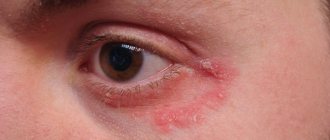
Condoms are not 100% protection - the chancre can be located extragenitally (pubis, perineum), and with secondary syphilis, a “necklace of Venus” is formed on the skin. In these cases, the infection from syphilis is transmitted by contact to the partner’s skin.
With syphilis, lifelong immunity is not formed. Having successfully recovered from this disease, you can become infected and get sick again. In this case, the disease will be just as severe. Therefore, there are no vaccinations against syphilis, and there cannot be.
Frequently asked questions to the doctor
Rash on the penis
Good afternoon, I discovered a red rash on the head of my penis. Tell me, could it be syphilis?
Hello, rashes on the head with syphilis are one of the first symptoms of the disease, but this does not mean that the diagnosis has been confirmed. Also, the cause may be other pathogens or an allergic reaction to hygiene products or underwear. It is better for you to consult a doctor and take all the necessary tests.