How does your typical morning start?
You wake up, have a quick breakfast or just drink a cup of coffee, get behind the wheel, and now - you are already in the office and the usual hectic working day begins. After a busy day at work, many women try to go to a beauty salon or fitness club in time, because everyone wants to be charming or beautiful in the eyes of others and the man they love. The modern rhythm of life is a harsh phenomenon that sometimes does not allow you to take a time out, that desired respite that you can devote to yourself. A common and normal phenomenon observed among modern women is the desire to prolong their youth through plastic surgery.
What is contopexy
Every woman wants her eyes to be alluring and captivate any man. In youth, it’s easy to maintain the beauty of your eyes: properly moisturize this delicate area, which is prone to the earliest appearance of signs of aging. But the skin around the eyes itself is thin and contains a small amount of collagen. Tonic, cream, mask - with the help of this complex the area around the eyes will be in normal balance until a certain point. With age, irreversible changes occur in the skin and soft tissues of the upper and lower eyelids, as a result of which blepharoplasty surgery becomes the only radical way to rejuvenate the area around the eyes. Today, there are many types of eyelid surgery, and now we will focus on one of them. We will talk about canthopexy.
Rehabilitation after surgery
Do not think that canthopexy is just a cosmetic procedure, after which no rehabilitation is needed. A rehabilitation period is required . You will have to spend several days at home - you cannot bend over, and under no circumstances lift anything heavy. In other words, it is necessary to adhere to all the requirements and rules that are indicated during the rehabilitation period after blepharoplasty.
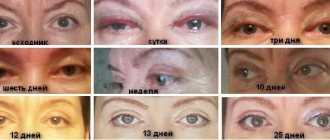
And, of course, after canthopexy, postoperative bruises cannot be avoided, but they will all go away within 7-10 days. Contopexy can be performed at any time of the year. In this regard, the most suitable time for surgery is spring, summer and early autumn. You can safely walk around in dark glasses without feeling awkward.
A week with dark glasses, and now everyone can see that your look has changed and your eyes have begun to smile.
Canthopexy to correct the shape of the eyelid
Canthopexy is a common operation that is used if there is a need to slightly change the shape of the eye shape and pull the outer corner of the eye up. Canthopexy is also performed to strengthen and tighten the “weak” lower eyelid, when it is everted or sagging, when a “round eye” is formed. This wonderful procedure will help make your look renewed, remove the pained, age-changed expression of the eyes, and the consequences of a previously unsuccessful lower eyelid surgery. This operation can be performed as an independent operation or as an addition to classic lower blepharoplasty.
There are two types of canthopexy, which differ in the type of surgical intervention: the most commonly used is minimally invasive eyelid correction (cantomyopexy) and a more simplified supportive canthopexy. The choice of technique depends on the degree of age-related changes in the lower eyelid (weakness - decreased elasticity, sagging and the presence of a “negative vector”).
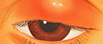
Weakness of the lower eyelid is a common condition in older people. In addition to the aesthetic defect, the lower eyelid moves away from the eyeball and turns out, which ultimately leads to disruption of the protective function of the eyelids and the development of corneal pathology. This is manifested by dryness of the cornea and conjunctiva of the eyes, redness and a feeling of sand in the eyes, or, conversely, increased lacrimation due to displacement of the lacrimal punctum, infection, the development of chronic blepharitis and conjunctivitis, the formation of erosions or ulcers of the cornea, etc.
Reviews about canthopexy
“I want to say a huge thank you to plastic surgeon A.I. Lightner. This is not the first time I have had surgery. The first operation, blepharoplasty, was 4 years ago. I was very pleased. The second operation took place on November 3, 2022 - face and neck lift and fractional rejuvenation. Everything went great. The attitude of the staff is wonderful. I'm very pleased with everything. Thank you very much!" Valentina, 50 years old.
Other reviews about eyelid lift at the Abrielle aesthetic surgery clinic.
Preparation
If canthopexy is required, preparations should be made before surgery. They first visit an ophthalmologist, he diagnoses the pathology of the organ of vision, determines contraindications and indications for surgical intervention. Be sure to visit a therapist, he will determine the general condition and identify diseases of the internal organs. Before the operation, laboratory and instrumental studies are performed.
General blood test - determine the indicators of formed elements, ESR. It is used to judge the presence or absence of inflammation, anemia, and disorders in the circulatory system.
A general urine test allows you to assess the condition and functioning of the kidneys; since surgery is ahead, they should be normal. Protein, red blood cells, white blood cells, mucus, epithelium, and the presence of ketone bodies are determined.
Blood biochemistry – evaluates the condition of internal organs: liver, pancreas, gall bladder, kidneys. Indicators are determined: liver enzymes, creatinine, bilirubin, c-reactive protein, albumin.
A coagulogram is done to determine bleeding time and blood clotting, this is important during surgery.
Perform an analysis for infections: HIV, syphilis, hepatitis.
From instrumental studies, an ECG is performed to assess the functioning of the heart, since surgery and anesthesia are to be performed.
During the week before surgery, it is not recommended to drink alcoholic beverages or smoke.
The surgery is performed in the plastic surgery department. Beforehand, on a computer, using a special program, the patient is shown what he will look like after the operation.
Indications for canthopexy
- Reduced elasticity and “stretching” of the lower eyelid, i.e. its horizontal elongation due to stretching of the canthal ligaments (mainly lateral);
- decreased tone of the orbicularis oculi muscle and its displacement below the edge of the cartilaginous (tarsal) plate;
- weakening or separation of the orbicularis oculi muscle, which is responsible for contraction of the lower eyelid with age or due to injury;
- Age-related depression of the eyes due to atrophy of the retrobulbar (behind-the-eye) fatty tissue (age-related enophthalmos).
Figure 1. The diagram shows that the lateral canthal ligament consists of an upper and lower part. Just below there is a thin ligament called the inferior retinaculum (retinaculum inferior).
In addition to external signs of lower eyelid weakness, you can perform a pinch test to determine its laxity. It is necessary to take the fold of skin of the lower eyelid in its central part, pull it down and release it. Normally, the eyelid immediately returns to its original position. If the lower eyelid can be pulled back by more than 6 mm and it slowly, “reluctantly” returns to its original position, this indicates weakness of the lower eyelid.
In most people, the most prominent point of the cornea and the zygomatic eminence are at the same level - this is called the “neutral vector”. If the zygomatic eminence protrudes forward in relation to the eye - “positive wind”.
In Figure 2 “Neutral vector” - red dotted line - the eyeball and the zygomatic eminence are on the same line.
In Figure 3, the black dotted line depicts a “positive vector” - the cheekbone protrudes forward in relation to the most protruding point of the cornea of the eyeball. The “neutral vector” is marked with a red dotted line.
“Neutral” and “positive vectors” indicate good development of the skeleton and soft tissues of the face, providing good support and support to the lower eyelid.
When measuring with a ruler the distance from the outer orbital edge to the most protruding point of the cornea, the result will be 15-17 mm. Ophthalmologists call this position of the eyeball normophthalmos. If the value is less than 15 mm, the eye is located inside relative to the inferior orbital margin, i.e. in the enophthalmos position.
In Figure 4, the black dotted line shows a “negative vector” - in profile, the eyeball protrudes forward in relation to the cheekbone, the eyes seem bulging. The “neutral vector” is marked with a red dotted line.
The “vector” is “negative” if the orbital edge is flat, smoothed and the eye protrudes forward relative to it, in which case the lower eyelid has no support and, in turn, does not provide sufficient support to the eyeball.
The “negative vector”, as a rule, is caused by underdevelopment of the facial skeleton and a deficiency of soft tissues in the midface. In such people, under the influence of gravity, “weakness” of the lower eyelids, ptosis (drooping, sagging) of the lower eyelids and cheeks appears quite quickly, compared to patients who have more developed bones of the facial skull.
In the presence of a “negative vector”, the eyeball is moved forward in relation to the cheekbone by more than 17 mm, this is called exophthalmos. The latter can be congenital (with the so-called “shallow orbit”, characteristic mainly of the Negroid race), or also appear as a result of age-related changes, endocrine disorders or volumetric processes in the brain skull (in such cases, it is most often one-sided).
Performing classic lower blepharoplasty when weakness of the lower eyelid is detected, especially in combination with the presence of a “negative vector,” is dangerous, since even minor removal of eyelid skin can lead to eversion - ectropion. In such cases, eyelid surgery is complemented by canthopexy or endoscopic midface lift.
Canthopexy technique
Rice. 2. The changes in the lower eyelid that occur with age are schematically depicted. In addition to excess skin, a fine network of wrinkles and “hernial sacs” of varying degrees of severity, a decrease in the tone and elasticity of the lower eyelid is noteworthy. The “weakness” of the lower eyelid manifests itself as follows: it does not fit tightly to the eye, moves forward a little, sags, and seems to be “pulled” down. The outer corner of the eye drops below the inner one (normally, in young people, the outer corner of the eye is at the same level or 2 - 3 mm higher than the inner one). As a result, the eyes appear bulging, rounded, and a white stripe of sclera is visible between the cornea and the edge of the lower eyelid. When viewed directly, the slightly expanded costal edges of the lower eyelids are visible. Progression of lower eyelid laxity can lead to ectropion.
Rice. 3. The dotted line shows preoperative markings.
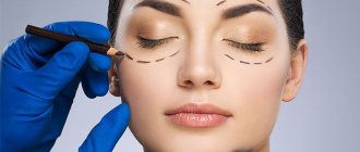
The markings for canthopexy are performed in the operating room and are similar to those for lower blepharoplasty. The line is drawn 1-2 mm away from the ciliary edge of the lower eyelid. The main difference is a small 0.7-10 mm incision, which will be located in the sub-eyebrow area.
Rice. 4. Making an incision with a scalpel along the lines of preoperative markings.
Rice. 5. Fixation with an absorbable thread of the orbicularis muscle of the lower eyelid to the periosteum of the upper orbital edge (subeyebrow region).
Rice. 6. Application of intradermal sutures.
Even if eyelid correction is performed under general anesthesia, the tissues of the lower eyelid are first infiltrated (flooded) with saline solution with the addition of an anesthetic and vasoconstrictor drugs. Then the skin is dissected with a scalpel along the lines of preoperative markings. In most cases, the plastic surgeon begins with a standard lower blepharoplasty: peeling away the skin, highlighting and removing the hernia bags.
After this, through an additional incision in the sub-eyebrow area, the periosteum is sutured with an absorbable thread, which is passed under the canthal ligament, the orbicularis oculi muscle is captured in the area of the outer corner, and then the thread is drawn in the opposite direction under the canthal ligament and finally fixed to the periosteum with a U-shaped suture. An internal suture placed in a similar manner ( canthopexy)
) will firmly hold the lower eyelid in the desired position.
The eyelid correction ends with excision of excess skin and application of an internal suture with a non-absorbable thread so that only two nodules are exposed to the skin: at the beginning and at the end of the incision. The stitches are removed on day 5. Postoperative scars will quickly fade and become invisible.
Rice. 7. The figure schematically shows a supporting canthopexy.
Supportive canthopexy differs in that it involves only the canthal ligaments, which are sutured together and fixed to the periosteum of the orbit above the lateral ligament of the eyelids.
The eyes are the area of our face that receives the most attention. When meeting for the first time, the first visual contact occurs using the eyes. The eyes most quickly display age or a hint of it. You can convey many feelings and emotions through your eyes: disappointment, joy, hope, regret.
If they say: “The eyes glow”, then they mean a cheerful, radiant look. In order for this look to remain as long as possible, you need to approach this problem competently. The canthopexy operation is absolutely safe and is performed in a short time, and the effect will last from 5 to 10 years.
Features of eye anatomy
The area of the organ of vision has structural features: the skin in this area is thin. It contains a small amount of collagen fibers. Therefore, here, before wrinkles form in other areas, skin tone is lost.
Under the influence of ultraviolet radiation, the skin quickly loses fluid.
Under the skin is the periorbiatle muscle. Due to it, the eyelids open and close. When stressed, skin creases appear in the corners of the eyes; at a young age they quickly return to normal; with aging, they remain and wrinkles and “crow’s feet” form.
Under the muscle there are plates of cartilage, the muscle fiber is attached to them by tendons and ligaments. The ends of the tendons of both eyelids are attached to the edge of the cartilage. They connect with each other, forming the corners of the eyes - external and internal.
The cartilage is located on the periosteum. The tendons of the outer corner are longer and stretched than the inner one. During life, they stretch even more, the outer canthus begins to descend, sometimes this is a congenital feature.
Contraindications
Contraindications to canthoplasty are:
- diabetes mellitus of any type;
- chronic pathologies of the eyelids and conjunctivitis in the acute stage;
- blood clotting disorder;
- pronounced myopia;
- sudden jumps in eye pressure;
- thyrotoxicosis;
- dry eyes;
- infectious diseases in the acute stage, as well as the presence of chronic infection in the body;
- exacerbation of any chronic pathologies.
Possible complications
The following complications are possible after canthoplasty:
- seal in the seam area;
- swelling and hematomas;
- dry eyes, irritation, discomfort.
Serious complications are:
- non-closure of the eyelids - too much skin excision;
- eversion of the eyelid - too much tension in the tendons or muscles;
- suppuration of sutures;
- bleeding - the stitches are not placed tightly enough;
- significant dryness of the eye - damage or blockage of the tear duct.
Prices for services
| Price | On credit* | |
| Lateral canthoplasty | 25,000 rub. | from RUB 2,498/month |
| Consultation with a plastic surgeon for surgery | 0 rub. | — |
Average cost of services
You can find out how much our plastic surgery services cost on average by calling in St. Petersburg: +7
SM-Clinic surgeons will develop an individual operation plan for you, since surgical body correction is often planned and carried out in a complex manner. The exact cost is calculated individually after consultation with a doctor.
Plastic surgery on credit and in installments
Postoperative period
Canthopexy and canthoplasty are minimally invasive procedures and can be performed on an outpatient basis. In the postoperative period, the doctor prescribes antibiotics to the patient to prevent the development of infection, analgesics if pain occurs, and eye drops to prevent irritation of the conjunctiva.
During the first few days after surgery, patients are concerned about swelling and bruising in the intervention area. On the first day, it is recommended to apply an ice pack every hour for 5-10 minutes.
In addition, special regenerating and antiseptic creams and ointments are prescribed to speed up the healing of the wound.
Recommendations in the postoperative period
- Try not to be in open daylight for more than 5 minutes in summer and spring; it is recommended to wear glasses with sun lenses in the first months
- Limit, or better yet completely eliminate, rest in front of the TV or laptop screen for a while, and, again, temporarily minimize reading books
- Avoid decorative makeup and wearing corrective lenses for a period of time.
- Avoid going to the sauna or working out in the gym, so as not to provoke excessive sweating and inflammation of the postoperative wound.
The stitches usually heal quickly. The effect of the operation is noticeable already in the first or second week. Final recovery occurs after 2-3 months, when the scar is completely regenerated and becomes invisible. The result lasts for 10-15 years. A very respectable period for the low price offered. The muscular-ligamentous frame of the eyes is ultimately securely attached to the periosteum.
