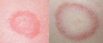
The structure of the formations, their size and type of contents vary, depending on the location and duration of the onset of the pathological process. They are true (inside lined with epithelial cells) and false (without lining). Based on the type of appearance, neoplasms are divided into:
- retention (usually formed due to a disruption in the process of outflow of secretions in tissues and organs, occurring in the mammary glands, uterus, cervix, ovary, etc.);
- ramolitic (usually found in the ovaries, brain and spinal cord, can form in areas of tissue necrosis, for example after a stroke);
- parasitic (appears in different organs, is the shell of the parasite);
- traumatic (due to bruises);
- dysontogenetic (usually congenital, include different tissues of the embryo);
- tumor (formed due to metabolic disorders, have cavities filled with physiological fluids).
Cysts in women and men: symptoms and treatment
In the initial stages of development, cystic elements do not have symptoms. When they grow and suppurate, they manifest themselves differently, depending on the etiology, location, composition of the contents and size. For example, when the ovary is damaged, women complain of a feeling of heaviness and compression of the lower abdomen, pain during menstruation, cycle disruption, etc. With kidney pathology, the lower back hurts and aches, it is difficult to empty the bladder, and jumps in blood pressure are noted.
Diagnostics
Diagnostic methods depend on the location of cystic formations. Most often this is an ultrasound, but MRI, CT, and radiography can also be prescribed. Laboratory tests are also carried out - blood and urine.
Surgery
The intervention tactics are determined by the doctor based on the location, size of the cystic formation and the patient’s complaints. The classic technique is scalpel excision of the tumor along with adjacent tissues. Today, such an operation is performed mainly laparoscopically using an endoscope. Laparoscopy of a cyst is less traumatic, the patient’s recovery lasts one day. You can also choose the method of aspiration (suction) of the contents with further cauterization of the capsule with a laser, liquid nitrogen, radio wave or a special drug for sclerosis of the walls. The removed lesion must be sent for histological examination.
Is surgery necessary?
When making such a diagnosis, many patients wonder whether the cyst needs to be removed. Some tumors, if they are small and do not bother the person, can remain under observation. Others, such as cervical cysts, can have consequences: bacteria accumulate in them, which sooner or later will lead to inflammation or suppuration. The need for removal should be discussed with your doctor.
What does a cyst look like on an MRI of the brain?
Cystic structure of brain metastases on MRI
The brain is studied using high-field equipment with a power of 1.5 Tesla, which allows creating detailed and clear images. With magnetic resonance imaging, monochrome sections of the area under study are obtained in increments of 1-2 mm. On MRI images, brain cysts appear as round formations of varying diameters with clear contours. The deviation can be determined by the change in the signal. The intensity of the latter will be different compared to healthy areas. The difference is clearly visible in the photographs. Special scanning modes allow you to obtain more detailed information about the cyst and determine its type:
- Epidermoid. It is usually located in the area of the cerebellopontine angle, prepontine cistern, quadrigeminal tract, ventricles, and less commonly in the hemispheres. Growth is slow, with the risk of compression of the brain stem, entrapment of nerves and blood vessels. There are signs of inflammation around the epidermoid cyst. The intensity of the signal from the formation is usually heterogeneous, but corresponds to the cerebrospinal fluid (if there is no fat inside). On DWI and FLAIR images, the cyst appears lighter than the brain fluid. Epidermoids with fat inside T1-weighted images give a hyperintense signal, on T2 - hypointense (compared to the cerebrospinal fluid).
- Dermoid. Usually the formation is located in the midline of the skull. Due to the high lipid content, T1 imaging gives a hyperintense signal. Unlike the epidermoid, it always has a heterogeneous structure.
- Lipoma. Fatty cysts are most often located in the area of the corpus callosum, interhemispheric fissure, pituitary infundibulum and hypothalamus. MRI scans reveal clear contours of the tumor. Mass effect and swelling of surrounding tissues are not observed. On T1 WI the cyst is sharply hyperintense. On T2 VI it is the opposite (compared to cerebrospinal fluid). Calcifications and vessels look like hypointense areas in the structure of the formation.
- Ependymal. A rare type of cyst that forms under the membrane lining the brain cavity. On MR scans it looks like a formation with clear contours, the signal intensity corresponds to the cerebrospinal fluid.
- Arachnoid. It is formed as a result of the accumulation of cerebrospinal fluid between the sheets of the arachnoid membrane of the brain, most often in the middle cranial fossa, interhemispheric fissure, cerebellopontine angle, at the level of the quadrigeminal. On T1, the VI looks somewhat lighter than the cerebrospinal fluid. In FLAIR mode it produces a hypointense signal.
Arachnoid cyst (indicated by arrow) on an MRI scan
- Neuroglial. Found in the cerebral parenchyma, in the area of the choroid plexus of the ventricles. In the photographs it looks like a round formation with liquor contents, often combined with anomalies of brain development.
- Colloidal. Usually located in the anterior superior part of the third ventricle, between the foramina of Monroe. It has a round shape, a fibrous capsule, and clear contours. In the presence of protein in the contents, it gives a hyperintense signal on T1 VI and a hypointense signal on T2 VI. Contrast does not accumulate.
- Rathke's pouch cyst. Rarely seen. Located in the area of the sella turcica. The signal intensity depends on the nature of the content. When serous, it gives a typical fluid response. Mucoid contents are characterized by a hyperintense signal on T1-weighted images. When contrasting, accumulation of the substance is observed in the capsule area.
Arachnoid cyst (circled in red) on MRI
An experienced radiologist knows exactly what a cyst looks like in an MRI photo. An ordinary person will not be able to independently identify and differentiate education. In doubtful cases, it makes sense to show the research results to several specialists.
The main types of cystic formations
Ovarian
This is a fluid-filled bladder that causes the ovary to enlarge, leading to pain and sometimes infertility. The main reason for its appearance is hormonal imbalance. The disease often occurs without symptoms; when the formation is large, the cycle is disrupted, pain is felt in the lower abdomen, a false urge to urinate occurs, and bleeding occurs outside of menstruation. Methods of surgical treatment: oophorectomy (complete removal of the ovary), laparoscopic cystectomy (excision of the cyst), adenxectomy (surgery to remove the uterine appendages). In most cases, the choice is not classical surgery, but gentle endoscopic removal of the cyst while preserving the patient’s reproductive function.
Read more about ovarian cyst removal
Coccygeal
Most often occurs in men aged 15-30 years. It is an opening in the area of the gluteal fold, approximately 10 cm from the anus. Externally it looks like a fistula. It can be congenital or acquired - due to too much hair in this area. It manifests itself as pain when walking, sitting, redness of the tailbone, a feeling of discomfort and the presence of a foreign body in the area where it is located. At a later stage, pus is released from the hole.
Read more about the treatment of coccygeal cyst
Bartholin gland
Appears due to blockage of the gland duct due to infection or chronic inflammation. The formation has a capsule and is filled with secretion, which gradually gels. If it is large, it interferes with walking and sitting, makes intimate intimacy inaccessible, and can become infected and lead to an abscess. Usually reaches 2 cm, but there are formations up to 9 cm. The main causes are chronic bartholinitis, candidiasis, bacterial vaginosis, decreased local immunity.
Where and how to remove a Bartholin gland cyst
Eye
A hollow formation filled with non-inflammatory fluid - products of the activity of the cornea or conjunctiva. May originate from the cornea, iris, conjunctiva and other eye membranes. Causes: inflammation and trauma, congenital anomalies. The disease is manifested by pain, a feeling of fullness, blurred vision, and the presence of translucent dots in the field of vision.
Maxillary sinus
A cavity containing fluid and having a membrane attached to the wall (usually the lower) of the maxillary sinus. A cystic formation can be true (its walls consist of mucous membrane) or pseudocyst (the mucous membrane is split and fluid accumulates in it). It manifests itself as headache, difficulty breathing through the nose, a feeling of fullness and heaviness in the eye and cheek area, mucus discharge from the nose and its flow down the wall of the pharynx, discharge of yellow transparent fluid from the nose, frequent sinusitis with suppuration. It is formed due to the peculiarities of the anatomical structure of the nose, blockage of the excretory duct of the glands of the maxillary sinus, inflammation of the teeth, spreading to the roots.
Read more about the treatment of abscesses and cysts of the ENT organs
Mammary gland
A cavity bounded by a capsule of connective tissue and filled with fluid is formed in the ducts and can be single or multiple. It is formed due to an increase in the duct of the mammary gland, the accumulation of secretions in it. The lesion may be round, oval, or irregular in shape. The disease is asymptomatic for a long time; over time, pain and burning appear in the mammary gland, and may be accompanied by suppuration and inflammation. One of the varieties is a fatty cystic formation that occurs when the sebaceous gland of the skin is blocked and filled with secretions. The main provoking factors are mastitis, thyroid disease, inflammation of the genital organs, and ovarian dysfunction.
Learn more about symptoms and treatments
Uterus and cervix
These are dilated and clogged glands, inside of which secretions (mucus) accumulate. The disease occurs against the background of endocervitis, cervitis. Provoking factors: abortion, childbirth, infections, menopause, hormonal imbalance, use of an intrauterine device, infections. Nabothian cystic formations are localized in the vaginal area of the uterus and are not removed until they reach a certain size. Retention occurs due to an excessive amount of secretion in the gland duct. The disease is often asymptomatic; its indirect signs are frequent inflammation.
Read more about surgical treatment of uterine and cervical cysts
Brain
A cystic formation is an accumulation of fluid in the substance or membranes of the brain. When large in size, it entails intracranial hypertension and puts pressure on the surrounding brain structures. Can form at any age. Depending on the location, cerebral (intracerebral) and arachnoid formations are distinguished. The first are found in the internal structures of the brain, in areas of necrosis. The second ones are in the meninges. Provoking factors: inflammatory diseases, trauma, including birth, cerebrovascular accident, parasites, complications after surgery. They manifest themselves as nausea, a feeling of pressure on the eyes, decreased performance, sleep disturbances, pulsation or noise in the head, and visual impairment.
Thyroid gland
Small formations (up to 5 mm) may not have pronounced symptoms. If the thyroid cyst has dense inclusions and a complicated structure, then special studies (for example, ultrasound), tests and biopsy are needed, because such a condition may be a sign of malignant degeneration.
Read more about surgical treatment of thyroid cysts
Larynx
Cysts of the larynx can be located in any part of the larynx. They do not grow into the mucous membrane, but grow towards the lumen of the larynx and thereby narrow it. The cause of retention cysts is blockage of the excretory ducts of the laryngeal glands. There are cysts of the vocal cords that arise due to constant irritation.
More information about surgical treatment of laryngeal cyst (laryngocele)
You can find out the prices for cyst removal endoscopically or by another method, as well as the cost of preoperative diagnostics on our website or by calling honey. +7 (812) 435 55 55.
MRI of the brain showed a cyst, what does it mean?
Colloid cyst in the third ventricle of the brain (circled with a dotted line)
As a result of magnetic resonance imaging, multiple layer-by-layer images of the anatomical area are obtained. Sections are made in three planes, which allows for a detailed study of each area of the skull. The fact that the MRI showed a cyst in the brain is reported when the results are issued. In conclusion, the doctor specifies the location and type of formation, size, and relationship of the cavity with surrounding structures. If tissue metaplasia is suspected (the appearance of cells in the study area that are not typical for this area), it is recommended to perform an MRI with contrast enhancement. The distribution of the drug can suggest the nature of the tumor.
Detecting a brain cyst on an MRI means that there is a round-shaped cavity filled with fluid in the skull. Based on specific signs, the radiologist determines the type of the latter and describes in detail the changes in the surrounding tissues. Detecting a pathology is not a reason to panic. Most cysts are congenital, grow slowly, and do not become malignant. With the results of the study, you need to contact your doctor (who initiated the tomography), neurologist or neurosurgeon. The specialist will determine how dangerous the deviations are, make a prognosis for the development of the pathology, and prescribe additional examination.
SYMPTOMS, COURSE
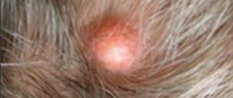
It can occur on any part of the body, but the predominant localization is the scalp, face, back, neck, and genital area.
On the head, single atheromas occur in 30% of cases, multiple in 70%, and 10% of patients may have more than 10 atheromas.
Patients complain of a tumor-like formation, superficially located, densely elastic, usually well mobile (partially displaced when pressed with a finger), painless. The skin over the formation is most often unchanged, but with inflammation it can be red, and sometimes, with rapid growth, the skin over the atheroma can ulcerate. In some cases, an enlarged obstructed excretory duct of the sebaceous gland is visible on the skin near the center of the formation. The tumor may remain small for many years or grow larger. Sometimes the atheroma communicates with the surface of the skin through a small hole, through which cheesy white or yellowish masses, usually with an unpleasant odor, can be separated.
Differential diagnosis is carried out with soft tissue tumors (fibromas, lipomas, dermoid cysts, osteomas).