What is lichen?
Ringworm is a group of dermatological polyetiological diseases, which is characterized by the appearance of a rash and itchy dry/weeping elements. There are several varieties of lichen, which differ from each other in:
- the type of rash present;
- pathogen;
- contagiousness;
- localization.
Among the most common types of lichen in humans:
- multi-colored (pityriasis);
- encircling;
- red flat;
- ringworm (trichophytosis);
- pink (pityriasis).
The course of the disease is long. There is always a danger of secondary infection.
Contraindications and side effects
The use of salicylic ointment is not advisable for pregnant women, lactating women and small children - it is better to choose an approved medicine for the fungus at the pharmacy. It is not advisable to use acid for people with kidney failure and patients prone to allergies. Salicylic acid thins the epithelial layer, so people with rosacea and thin skin should avoid using acid.
When the solution is used externally in humans, the following side effects may occur:
- allergic reactions;
- burning, itching, redness in the treated area;
- in very rare cases, individual intolerance to the component is possible.
Causes of lichen in humans
Ringworm is usually caused by a fungal or viral infection. The mechanism of infection is not fully understood, since not all people, even those at risk, suffer from lichen. Presumably, fungal infection of the skin is promoted by the simultaneous influence of the following factors:
- stress;
- reduced immunity;
- hereditary predisposition;
- emotional/physical fatigue;
- infectious diseases.
The incidence of different types of lichen may vary depending on gender and age.
Causes of pink lichen Zhibera
Pityriasis rosea is of an infectious-allergic nature.
Causes of herpes zoster
Shingles develops through secondary contact with the Herpes zoster virus. It can also be caused by activation of a latent herpetic infection. Its causative agent is a virus of the Herpesviridae family.
Causes of tinea versicolor
Pityriasis versicolor or pityriasis versicolor affects the stratum corneum of the epidermis. Its appearance is promoted by sweating, seborrheic skin condition, and hot climate. Infection occurs through household contact, that is, through the use of common household items, through direct contact with a sick person. The causative agents of lichen versicolor are fungi:
- Pityrpsporum orbiculare;
- Malassezia furfur;
- Pityrosporum ovale.
Causes of ringworm
Infection with ringworm occurs as a result of contact of a child or adult with a person or animal infected with trichophytosis, as well as household items on the surface of which the fungal spores have fallen.
Causes of lichen planus
Lichen planus belongs to the category of a multi-cause disease that develops under the influence of internal/external factors that cause disruption of immune and metabolic processes. There is a hereditary predisposition to the disease. It can also occur as a result of contact with certain medications and chemicals.
Properties and effect of salicylic alcohol
Salicylic alcohol is a good antiseptic solution with the following benefits:
- successfully fights bacteria and fungal infections on the epithelium, disrupting protein synthesis in fungal spores;
- used in the treatment of acne, blackheads and other inflammatory skin diseases, due to the anti-inflammatory properties of alcohol;
- in a small concentration, it has an exfoliating effect on the upper layer of the epidermis and helps its normal formation;
- suppresses the secretion of the sebaceous glands, cleanses the dirt accumulated in them;
- suppresses the secretion of sweat glands and is used for hyperhidrosis as a separate remedy or in combination with boric acid.
The undeniable advantage of salicylic alcohol is that it does not cause allergies, unlike other multicomponent drugs.
What does lichen look like in humans - symptoms
Symptoms of pityriasis rosea
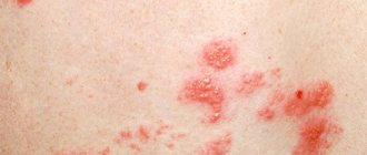
Pityriasis rosea begins with the appearance of a bright pink plaque on the skin, the diameter of which is up to 5 cm. If you look at a photo of the disease, it will become clear that this is a spot that peels off in the center. Within 1-2 weeks, the lichen begins to peel off more strongly along the periphery, its center becomes like tissue paper.
A week after the first spot appears, round pink spots appear on the back, stomach, and shoulders, located along the lines of skin tension. They can merge with each other. After recovery, dark areas may remain on the body for a long time.
Symptoms of herpes zoster
Symptoms of shingles include:
- soreness of the affected area of the skin and a slight burning sensation that occurs several hours before the first rash appears;
- pain in muscles, joints;
- numbness of the affected tissues;
- the appearance of bubbles of different sizes, the formation of edema in places where they are localized;
- enlarged lymph nodes.
In the photo, shingles is usually shown as a unilateral rash that is located on the scalp, along the intercostal nerves, on the abdomen or chest in the direction from the spine to the sternum. Shingles seems to encircle the torso, which is why it is called that.
A few days after the bubbles appear, the liquid contained in them becomes cloudy. They dry out and become crusty. After the latter falls off, pigmentation remains in place of the lichen. A second wave of rashes may then follow. The incubation period for shingles is about 2 weeks.
Symptoms of pityriasis versicolor (lichen versicolor)
Lichen versicolor most often affects the skin of the back, shoulders, neck, chest, and rarely - the head, thighs, groin, forearms, hands, shins, and behind-the-ear folds. First, the patient notices small pink spots, which gradually enlarge, merge, turn brown and turn white after the fungus dies. This creates:
- itching;
- burning;
- peeling of the skin at the site of the lesion.
Tinea versicolor can last for several years.
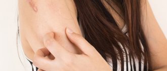
Symptoms of ringworm
Symptoms of ringworm, which is usually pictured as a bald patch on the scalp, include:
- itching in areas of rash;
- oval and round pink spots without clear boundaries, rising along the periphery like a ridge (may be covered with crusts and blisters).
Ringworm rashes often coalesce to form large lesions. The incubation period is 5-7 days.
Symptoms of lichen planus
Lichen planus in children and adults manifests itself:
- itching;
- skin lesions in the area of the anterior surface of the leg, genitals; torso, flexor surfaces of the forearms;
- the appearance of spots on the nails, oral mucosa, palms, face, soles.
In the photo, lichen planus looks like dark brown bumps. If the fungus infects the nail plates, they darken and become covered with white stripes, along which the nail soon splits.
If you experience similar symptoms, consult your doctor
. It is easier to prevent a disease than to deal with the consequences.
Treatment of versicolor
To treat the disease, ointments, sprays, shampoos, tablets and capsules with an antifungal effect are used. In particular, products based on fluconazole, imidazole or selenium sulfide are used.
Antifungal medications prevent further growth and reproduction of the fungus. Local remedies are quite effective, but they cannot penetrate deep into the hair follicles, sebaceous and sweat glands, where the infection can persist and cause relapses of the disease. Therefore, local treatment is sometimes combined with the administration of systemic antifungal drugs in the form of tablets or capsules.
The destruction of the fungus must be combined with the treatment of concomitant pathologies. It is possible to use drugs that normalize metabolic processes and strengthen the immune system. To successfully treat lichen versicolor, you should spend more time in the fresh air and sunbathe. The house must be wet cleaned daily and the rooms must be ventilated regularly. This will help eliminate fungal spores that remain on clothing and household items.
Diagnosis of lichen
Diagnosis of all types of lichen includes:
- listening to patient complaints;
- anamnesis analysis;
- inspection of rashes to determine their color, size, shape, location, etc.
Further, the dermatologist’s actions depend on the preliminary diagnosis he made.
Diagnosis of pityriasis rosea
To diagnose pityriasis rosea in a child or adult, in addition to a medical examination, during which the final diagnosis is usually determined, the following is additionally carried out:
- microscopy of scales to exclude syphilis;
- serodiagnosis of syphilis.
A biochemical and general blood test is also taken, and a general urine test is given.
Diagnosis of herpes zoster
Usually, a doctor can accurately determine whether a patient has pityriasis rosea during an examination. But in some situations additional research methods are required:
- detection of viral DNA by polymerase chain reaction in the contents of vesicles and in the blood;
- determining the presence of antibodies to the pathogen;
- identification of the pathogen virus genome in cells/tissue sections using in situ hybridization.
Diagnosis of pityriasis versicolor (lichen)
Diagnosis of versicolor versicolor pumps in:
- Balzer test (iodine test). The affected areas of the skin are treated with iodine and immediately wiped with alcohol. Pityriasis versicolor always turns brown;
- examination under a Wood's lamp (lichen looks like a brown or yellow glow);
- microscopic examination of scales (allows one to identify hyphae (curved short filaments of mycelium), oval and round spores).
Diagnosis of ringworm
To determine ringworm, the following are carried out:
- examination of lesions using a Wood's lamp (lichen becomes green);
- microscopic examination;
- sowing the discharge on various nutrient media (to determine the sensitivity of the pathogen to antibiotics);
- biochemical and general blood tests, urine analysis.
Diagnosis of lichen planus
To diagnose lichen planus in an adult/child, a small affected area of skin is taken for histological examination. The patient is also prescribed a general urine and blood test and biochemistry.
What to replace it with?
If for some reason the acid cannot be used against lichen, then its main analogue is salicylic ointment, which is used to get rid of dermatological infections and is available in concentrations of 2%, 5%, 10% and 60%. The following ointments are full-fledged analogues, used instead of acid: salicylic-sulfur and sulfur-tar ointment - each of them contains salicylic acid, but in a lower concentration. Sometimes boric acid is prescribed for treatment, but with caution, because its antiseptic effect is strong and sometimes it greatly dries out damaged areas on the body.
Treatment of lichen
Treatment of pityriasis rosea
Usually, pityriasis rosea does not require drug treatment, as it goes away on its own. During illness you need:
- follow a hypoallergenic diet;
- limit water treatments;
- do not use cosmetics;
- wear underwear made from natural fabrics.
If the patient complains of itchy skin, he is prescribed:
- antihistamines;
- hormonal creams and ointments containing boric acid/zinc;
- hyposensitizing drugs.
Effective methods of non-drug therapy include exposure of the skin to UV radiation with a wavelength of 280-320 nm.
Treatment of herpes zoster
Treatment of herpes zoster in children and adults involves the use of Virolex, Acyclovir, Zovirax. These drugs are effective at the very beginning of the disease. Additionally, Curantil and Furosemide are prescribed. Intravenous administration of immunoglobulin, which activates immunogenesis, helps to achieve good results.
Depending on the severity of the disease, the patient may be recommended symptomatic therapy, which consists of taking:
- general health-improving medications (B vitamins);
- analgesics;
- antipyretic tablets.
Sedatives help with sleep disorders. In some cases, taking antidepressants is indicated. In case of pronounced intoxication of the body, detoxification measures are carried out with forced diuresis.
Locally, lesions of herpes zoster can be treated with a solution of brilliant green, five percent dermatol ointment. For a sluggish form of the disease, metacil ointment and Solcoseryl are used. Areas of skin with rashes can be affected locally using quartz, UV irradiation, and laser.
Treatment of pityriasis versicolor
Treatment of pityriasis versicolor is carried out on an outpatient basis. Antifungal agents are effective against the pathogen:
- salicylic alcohol (3-5%);
- salicylic ointment (5%);
- sulfur ointment (5-10%);
- resorcinol alcohol (3-5%);
- Bifazol;
- Miconazole;
- Cycloperox;
- Naftifin;
- Lamisil et al.
If the lesions are spread throughout the body, oral administration of Orungal, Itraconazole, and Ketonazole is preferable.
Treatment of ringworm
If possible, the treatment of ringworm should be limited to the use of topical medications. If only smooth skin is involved in the pathological process (hair follicles are not affected), you can alternately treat the lesions with an iodine-containing solution and antifungal ointment (Microspor, Lamisil, Exoderil).
For severe skin inflammation, oral antifungal medications are recommended. The optimal triazole derivatives are Itrazyl, Irunin. It is best to shave or trim the hair in the ringworm area.
If suppuration forms on the surface of the lesion, it must be removed by first softening the scabs with salicylic ointment or a solution of potassium permanganate. At the infiltrate stage, ichthyol ointment and Liniment according to Vishnevsky can be used.
Additional methods of treating ringworm include vitamin therapy, as well as taking medications aimed at strengthening the immune system.
Treatment of lichen planus
Treatment of lichen planus is aimed at eliminating itching and burning. The patient may be prescribed:
- Tavegil;
- Suprastin;
- Zyrtec.
Selective phototherapy or PUVA therapy using photosensitizers helps achieve good results. It is also possible to take combined antimalarials (Plaquenil, Delagil) and corticosteroids (Diprospan, Prednisolone).
Local therapy for lichen planus is usually not carried out. If mucous membranes are involved in the pathological process, they should be treated with Rotokan, Solcoseryl, corticosteroid ointments and vegetable oils. If a secondary infection occurs, antibiotics are prescribed.
Which is more effective?
Acid is more effective in treating lichen than alcohol.
In terms of indications and action, both solutions are identical. Salicylic alcohol for ringworm is not as effective as acid: it is much more effective and acts faster than alcohol. The acid kills fungi and spores, preventing them from reaching the incubation period. Therefore, to quickly get rid of lichen, it is advisable to use salicylic acid or other preparations with it: solutions or ointments based on it. If you can’t get acid, then the use of alcohol is allowed. There is only one drawback - you will have to wait longer for the effect.
Diet for lichen
Nutrition during treatment of lichen depends on the type of disease, but there are foods that are equally useful for skin lesions of any type of fungus:
- green vegetables, herbs;
- dairy products;
- breakfast cereals;
- mineral water;
- natural honey;
- foods high in iron.
Pityriasis rosea requires adherence to a dairy-vegetable diet.
For shingles, ringworm, lichen planus and multicolored lichen, it is necessary to enrich the diet:
- foods rich in vitamin E (peanuts, sea buckthorn, rose hips, eel, walnuts, viburnum, sorrel, squid, hazelnuts, prunes, salmon, oatmeal/barley, seeds, vegetable oils);
- products that are sources of antioxidants and bioflavonoids (apples, grapes, apricots, blueberries, cherries, prunes, blueberries, carrots, kiwi, cherries, sweet red peppers, barley, raisins, etc.).
The list of prohibited products for the treatment of any type of lichen includes:
- spices (mustard, pepper, horseradish);
- spicy dishes, pickles;
- alcohol;
- tangerines.
It is necessary to limit consumption:
- offal;
- smoked meats, mushroom and meat broths;
- fatty fish;
- canned food;
- salty and spicy cheeses;
- cream cakes;
- legumes;
- products with preservatives.
Drink coffee, strong black tea and cocoa in small quantities.
Prevention of pityriasis versicolor
To prevent the disease, you need to monitor your health, avoid stressful situations and do not forget about the rules of personal hygiene. You should not use other people's washcloths, clothes or towels. To strengthen the immune system, you need to eat more fruits and vegetables, take vitamins in winter, exercise and do active recreation.
If the disease still makes itself felt, contact the SANMEDEXPERT clinic for the treatment of pityriasis versicolor, and experienced specialists will select an effective therapy that is suitable for you.
Why is lichen dangerous?
The harm that lichen can cause to health depends on its type. Pityriasis rosea always occurs without complications. The prognosis of the disease is favorable for ability to work and health.
Shingles is more dangerous. It can lead to:
- decreased motor activity in the limbs;
- partial loss of sensation in the limbs;
- inflammation of the membranes of the spinal cord/brain (encephalitis, meningitis);
- viral damage to the lungs and liver;
- the addition of a secondary infection;
- inflammation of the cornea, eyeball, loss of vision.
The most common consequence of shingles is postherpetic neuralgia. This disease develops after an illness and is manifested by severe pain. Only after a few months the pain goes away.
Pityriasis versicolor and ringworm can lead to a secondary infection. This makes the treatment process more complicated and requires the use of broad-spectrum antibiotics. Trichophytosis can also lead to the formation of bald patches that will last a lifetime.
Complications of lichen planus include:
- addition of bacterial infections;
- inflammation of the oral mucosa.
Overdose symptoms
An overdose is fraught with bleeding of various types.
An overdose of such a drug seems impossible, but some sources record cases of poisoning and overdose, characterized by the following symptoms:
- wounds on the mucous membranes, the appearance of ulcers on them;
- severe, burning pain;
- hearing loss and ear damage;
- Bleeding from the nose, gums, and sometimes even internal organs—the stomach—may occur.
Introduction
Pityriasis ruber pilaris (PRP) is a rare chronic papulosquamous disease with keratinization disorder with an incidence of 1 in 5000 to 1 in 50,000, with no gender predisposition (1).
The pathogenesis is still unclear and has been hypothesized to be caused by an abnormal immune response to various antigenic stimuli such as infections, trauma, and malignancies (2, 3).
Most cases are sporadic, but familial forms of the disease have been described, particularly those associated with mutations in the CARD gene. In PRP, the epidermis is in a hyperkinetic state with increased proliferation of follicular keratinocytes.
A pathogenetic role has been suggested for vitamin A deficiency or dysfunction (7) or decreased serum levels of retinol binding protein, which carries vitamin A (8), along with some clinical similarities to phrynoderma (a cutaneous manifestation of vitamin A deficiency) (9).
Clinical picture
Typically, PRP is characterized by small follicular papules ~1 mm in diameter with a central keratotic plug, confluent scaly yellow-pink macules, and palmoplantar keratoderma.
The lesions are symmetrical and diffuse and appear first on the extensor surfaces of the extremities, shoulders and buttocks, usually spreading caudally with the possible development of erythroderma (1, 7).
Differential diagnosis with psoriatic erythroderma is carried out by identifying areas of unaffected skin. Peeling is common and is small-plate in nature, scaly on the face and scalp and large-plate in the lower part of the body.
The skin on the palmoplantar areas tends to thicken and appear yellow-orange in color with well-defined borders. Sometimes patients complain of fever, chills, itching, and malaise (1, 7).
Nail lesions are also very often accompanied by subungual hyperkeratosis and yellow-brown spots (10).
The oral mucosa may also be covered with white spots and lines: erythematous, painful rashes with white streaks may appear on the mucosa of the cheeks, gums, and tongue, mimicking the features of lichen planus (11).
In adults, skin lesions appear first on the face and scalp, spreading caudally, while in children, PRP usually appears on the lower body. However, the clinical presentation and evolution of PRP are highly variable.
Classification
In 1980, Griffiths (13–15) classified PRP into the following five types based on clinical features, age of onset, and prognosis: classic adult type I, atypical adult type II, classic juvenile type III, limited juvenile type IV, and atypical juvenile type V.
Type VI, associated with human immunodeficiency virus (HIV), was later added ( 16 ). Classic adult type I is the most common with acute onset and accounts for ~50% of patients. It usually begins with the appearance of a single erythematous patch on the upper half of the torso. Consistently, the skin lesions spread caudally over weeks to months and may develop into erythroderma with typical islands of palmoplantar yellow-orange hyperkeratosis. The duration of this form is ~3-4 years (13-15). Atypical adult type II, which develops in 5% of patients, has a chronic course for up to 20 years. It is characterized by ichthyoid lesions, especially on the skin of the lower extremities, in association with alopecia and areas of eczematization (13–15). Classic juvenile type III is similar to adult type I but affects children (10% of patients) and has a more favorable clinical course than in adults, usually remitting after 1 year (12). The described juvenile type IV affects children as well as young adults (25% of patients); follicular hyperkeratosis and erythema develop, usually only on the knees and elbows with clearly defined boundaries. Palmoplantar lesions are also characteristic of this form, as well as dorsal lesions of the hands and feet. In most cases it remains localized, but can be characterized by remissions and exacerbations (12, 17, 18). Atypical juvenile type V occurs in the first few years of life in 5% of patients; it is chronic and characterized by follicular hyperkeratosis and scleroderma skin lesions of the upper and lower extremities, while erythema is not pronounced (13-15).
HIV-associated form VI is similar to type I, with a symmetrical pruritic rash consisting of erythematous and scaly follicular papules, but it is more severe and generally refractory to treatment.
Marked plugging of follicles is another common finding in this form; however, other follicular manifestations may be associated with HIV-associated PRP, such as acne conglobata, hidradenitis suppurativa, and lichen spinosa (16).
This classification is arbitrary: in fact, it is not always possible to classify each form of PRP into one type, since intermediate forms and transition from one type to another can occur (15).
Diagnosis
Diagnosis is mainly clinical, based on the above criteria. However, sometimes, especially when PRP is in the form of erythroderma, it is quite difficult to establish a clear clinical diagnosis and can be misdiagnosed as psoriasis, follicular eczema, follicular ichthyosis, generalized hypersensitivity reaction, T-cell lymphoma and lichen planopilaris (1-7).
In the last few years, dermoscopy has also been used to evaluate inflammatory skin diseases and has been proposed as a possible adjunctive tool in clinical diagnosis to better differentiate PRP from psoriasis: psoriatic lesions typically present as evenly distributed punctate vessels on a light red background, while as PRP lesions present with linear and punctate vessels and round/oval yellowish areas (19–21).
Recently, two cases of PRP in an adult and an adolescent were also studied using confocal microscopy, which may represent an additional tool to improve diagnostic confidence (21).
In fact, clinicopathological correlation remains the “gold standard” for the diagnosis of PRP, to the exclusion of other dermatoses, especially psoriasis and cutaneous lymphoma.
Typical histopathological features of PRP, although not pathognomonic, are alternating orthokeratosis and parakeratosis in both vertical and horizontal directions, forming a checkerboard pattern; other common findings are hypergranulosis, follicular occlusion, wide reticulate ridges, narrow dermal papillae, and a sparse superficial perivascular lymphocytic infiltrate (22).
Therapy
Treatment of PRP includes local and systemic therapy depending on the extent and severity of the disease (Table 1).
Local treatment
Local treatment is indicated for localized forms, such as PRP type III, and is always recommended also in combination with systemic therapy for severe forms to reduce skin symptoms such as itching and burning (1, 23, 24).
For localized forms of PRP, the main therapeutic choice (23, 24) is topical therapy, including moderate-to-high potency corticosteroids, keratolytics, emollients, and vitamin D derivatives such as calcipotriol (25), tretinoin, and tazarotene (18, 26).
Topical treatment in combination with systemic therapy is useful for the treatment of hyperkeratosis and palmoplantar keratoderma, as well as for improving skin texture, reducing peeling, erythema and infiltration, in particular the use of keratolytics (5-10% urea or 5% salicylic acid) and emollients are recommended (27).
Systemic treatments
Systemic treatment methods are indicated for moderate to severe diseases. Traditional systemic therapy is based mainly on the use of retinoids, which are actually considered as first-line therapy in both adults and children (1, 27).
Unfortunately, there are refractory cases that cannot be treated or recur after stopping the drug. In these cases, treatment is complex and a standard therapeutic protocol does not yet exist.
Treatment is mainly based on case reports or case series because controlled randomized trials have never been conducted due to the rarity of the disease (27).
However, in recent years, numerous case reports have demonstrated the effectiveness of new therapeutic agents such as biologics, particularly in refractory forms of PRP.
Retinoids
Systemic retinoids are considered as first-line systemic therapy for adults and children with PRP, as most case reports and case series demonstrate their effectiveness (27–39).
Clinical response to retinoids usually occurs after 3-6 months of therapy, but some patients require longer treatment. Isotretinoin and acitretin are the most commonly used, but there is some evidence that alitretinoin is also effective for PRP (27–30, 35–38).
In a retrospective study, Eastham et al. (27) All 12 patients receiving acitretin, eight patients receiving acitretin alone at a dose of 25–50 mg/day, two patients receiving acitretin in combination with methotrexate, and two patients receiving acitretin in combination with cyclosporine reported partial or significant improvement during treatment.
In a prospective study, isotretinoin (2 mg/kg/day) was used in 45 patients with PRP, and after 4 weeks of therapy, 28 (62%) patients showed significant improvement (29).
In another retrospective study of 15 patients receiving isotretinoin (primarily 40 mg twice daily), 10 (67%) patients had complete clearance within a median of 25 weeks (16–44 weeks), but only three patients Refractoriness to treatment was observed (30).
In clinical practice, the recommended dosage of isotretinoin for adults with PRP is 1 mg/kg/day, while acitretin is used at a dosage of 0.5–0.75 mg/kg/day. However, high doses of retinoids are poorly tolerated due to frequent mucocutaneous side effects.
The most common side effects of retinoids are dry skin and mucous membranes, hyperlipidemia, increased transaminase levels, and blurred vision or ossification.
In children, retinoids can cause hyperostosis and premature closure of the pineal gland, especially in prepubertal children receiving high doses. In addition, retinoids should be avoided in women of childbearing potential due to their teratogenic effects: pregnancy should be avoided during and for 3 years after treatment with acitretin and during and for 6 weeks after therapy with isotretinoin.
Due to the rarity of the disease and the lack of standardized studies, there is no consistent data on the duration of treatment, the risk of relapse if treatment is stopped, or the need for maintenance treatment.
Phototherapy
Ultraviolet B (UVB) phototherapy and psoralen ultraviolet A (PUVA) therapy have been shown to be successful treatments in some patients suffering from PRP (39, 40), although the response to light is quite variable. Conversely, exacerbations of PRP have been described in some patients (8, 41, 42).
Given reports of photoaggregated PRP, phototesting should always be performed prior to treatment. A combination of retinoids and phototherapy may be an acceptable alternative to treatment for refractory forms.
Acitretin has been reported to be used as a combination therapy with narrowband UV (43), ultraviolet A1 (UVA1) (44), and PUVA (28).
A recent case report of pediatric PRP demonstrated that narrowband UV phototherapy was successful after 2 months of treatment in combination with topical emollients, hydrocortisone cream, and calcipotriene, achieving >90% clearance (45).
Methotrexate
Methotrexate is an alternative treatment option for PRP as a second-line treatment (27, 34). Retrospective studies have demonstrated the effectiveness of weekly doses of 5-25 mg. Another retrospective study demonstrated a favorable response in 8/14 patients with PRP type I: median treatment duration was 6 months (range 1 to 12 months) (30).
Another retrospective study demonstrated the effectiveness of methotrexate in combination with a retinoid, despite an increased risk of hepatotoxicity (33). Another study including five patients found retinoid-refractory methotrexate to be effective (34).
Gastrointestinal upset is a common side effect. Additional serious side effects that should always be monitored are pancytopenia, hepatotoxicity, and pneumonitis. Additionally, it should be avoided by women of childbearing age due to its teratogenic effects.
Other immunosuppressants
Other systemic drugs such as azathioprine (46, 47) and cyclosporine (48–50) have been reported to be effective in the treatment of PRP in isolated cases, especially in refractory forms.
In particular, cyclosporine can be considered an acceptable therapy for patients who do not respond to retinoids or methotrexate, and can be used in both adult and juvenile forms. Additionally, fumaric acid esters may be an alternative option (51).
Biological drugs
Given the clinical and histological similarities between psoriasis and PRP, all biologics approved for the treatment of psoriasis have also been used in the treatment of refractory forms of PRP or in patients resistant to or contraindicated to standard systemic treatments (27, 34).
Since the pathogenesis of PRP is still unclear, it has been proposed that TNF-alpha is a key cytokine (52).
More recently, inhibitors of the IL-23/IL-17 pathway have also been successfully used in PRP, and elevated levels of IL-17 have been found in patients with PRP, providing a rationale for the use of these new therapies (53).
However, clinical experience with biologics in the treatment of PRP is limited to case reports, and case series and multicenter randomized clinical trials have not been conducted due to the low incidence of PRP.
In contrast, cases of PRP refractory to biologics have also been observed, although these are rarely reported, such as recently described for type IV PRP resistant to anti-TNF-α and IL-23 inhibitors ( 54 ).
Anti-TNF-alpha (infliximab, etanercept and adalimumab)
Over the past 10 years, retrospective studies and case reports have shown that anti-TNF-alpha agents are effective in the treatment of PRP in both adults and children (53–64).
A systematic review of type I PRP including 15 patients treated with infliximab, etanercept, or adalimumab found a complete response in 12/15 patients and a partial response in 2/15 patients. In only one case was there no improvement. Six patients received anti-TNF-α as monotherapy, while nine patients were treated in combination with acitretin or methotrexate (63).
In most cases, TNF-alpha inhibitors have been used as second-line agents for PRP refractory to conventional systemic treatment. In a case series of seven patients with refractory onset in adults, PRP was effective against infliximab (three patients) and etanercept (five patients), with 5/7 patients receiving combination therapy with low-dose acitretin (0.2 mg/kg/day).
All patients had significant clearance at week 12, and infliximab was associated with a faster response than etanercept. During follow-up, only the PRP type II patient relapsed 2 months after stopping treatment (51).
In a recent retrospective study of 40 patients with PRP conducted at a tertiary center, nine patients were treated with anti-TNF-alpha agents with favorable responses within 5–7 weeks.
Despite this, most of them received initial therapy with another systemic drug (methotrexate, acitretin, or prednisone) (27).
In other case reports of refractory PRP treated with adalimumab, rapid and durable remission was observed after only 4 weeks: no relapses were reported after treatment cessation (60–62).
A patient with PRP type I treated with adalimumab who achieved clinical remission after 4 months had elevated levels of TNF-alpha mRNA in the affected and surrounding normal skin.
This finding is consistent with the observed clinical remission and supports the use of anti-TNF-α for the treatment of PRP (64).
IL-23 and IL-17 inhibitors (ustekinumab and secukinumab)
Several case reports have described the use of ustekinumab in type I PRP refractory to conventional systemic therapy or anti-TNF-alpha agents (65–69).
Ustekinumab has demonstrated significant and rapid efficacy in reducing signs and symptoms of the disease, as well as long-term disease control (69).
In a recent case series, a biopsy of the affected skin was taken from three patients suffering from refractory PRP and the mRNA expression of cells producing innate pro-inflammatory and T-cell cytokines was studied.
Gene expression analysis revealed an increase in T helper (Th) 1 cytokines and in particular Th17 cytokines such as IL-17A, IL-22 and IL-23. In one patient, IL-17A levels in lesional skin samples were measured before and after ustekinumab treatment and decreased after skin improvement.
This case report suggests a role for the IL-23/Th17 axis in PRP, providing a rationale for targeted therapy targeting this pathway as a treatment option for refractory PRP (53). Secukinumab was recently shown to be effective for the treatment of PRP in two case reports (70, 71).
In the first case, it was used in a patient with type I PRP refractory to acitretin (70).
The second case was proposed as an acceptable treatment option for refractory type II PRP, with improvement in erythema and palmoplantar keratoderma observed after 2 weeks of treatment and no recurrence after 6 months of follow-up (71).
Other treatments
A recent case has proven the effectiveness of apremilast in the treatment of refractory PRP: after 4 weeks of therapy, the patient reported significant improvement, and after 6 months of follow-up, he/she experienced complete resolution of the skin lesions (72).
Extracorporeal photochemotherapy has also been used to treat two patients with erythrodermic PRP type I in combination with systemic retinoids and cyclosporine, with good results (73). Another report reported that a case of adult type II PRP was successfully treated with intravenous immunoglobulin (74).
Conclusion
PRP is a rare inflammatory skin disease that can have a serious impact on the quality of life of patients, especially in chronic and refractory forms. Many therapeutic options have been tested, but we do not have a standard protocol for PRP treatment.
Treatment of refractory forms remains challenging. Biologic agents appear to be an effective new treatment for PRP, although multicenter randomized clinical trials should be conducted before considering them as a first-line treatment option.
Further understanding of the pathogenesis of the disease may allow us to identify some clinical and phenotypic features of various forms of PRP as prognostic factors allowing individual therapeutic choices to be made.
Pityriasis rosea in children
Pityriasis is diagnosed in children over 5 years of age. The dermatologist prescribes antihistamines and multivitamins to patients, which restore the immune system. For children under 2 years of age, the doctor prescribes antiseptics and zinc ointment.
Children with weak immunity are susceptible to pathology in the fall and spring. The disease is diagnosed more often in girls, less often in boys. Rashes are rare in infants. The reason is overheating or hypothermia of the baby.
Causes of skin disease:
- allergic reactions to foods or fabrics;
- intestinal disorder;
- depression, emotional turmoil;
- insect bites.
The specialist will recommend medications to speed up recovery. The disease period is up to 2 months. To destroy microorganisms, it is necessary to take antibiotics, and for allergies - antihistamines. At high temperatures, the doctor prescribes antipyretic tablets.