Vulvar neoplasms are tumor diseases of the external genitalia. The formation of foreign bodies may be asymptomatic. If the tumor develops rapidly, it increases in size, the woman feels discomfort and pain. Accompanied by bloody or purulent discharge from the perineum. Causes itching and irritation of the vulva. Diagnosed during examination by a gynecologist. Various studies are being carried out. Vulvar neoplasms are recognized by smears and biopsies.
The type of tumor is determined and treatment is prescribed. Benign ones are excised or enucleated. Special technologies and individual therapy are used to treat malignant tumors.
The collection of external female genitalia is called the vulva. This includes the pubis, clitoris, labia, and vestibule of the vagina. The organs serve as a protective barrier for the genitourinary system. The hymen is the boundary between the reproductive system and the vulva. The vulva has a sensitive function. Her external organs are responsible for arousal during sexual intercourse. The signal is transmitted to the secretion of the glands, thereby moisturizing the vaginal slit. Congenital defects of the vulva include a violation of the development of any organ, for example, hermaphroditism. A visual examination of the vulva can be performed by a gynecologist. You can do this yourself with the help of a mirror or a sexual partner.
Damage to the organs of the vulva is possible during childbirth and injury. The process is accompanied by pain, swelling, and wounds. Bleeding is observed, the area of the clitoris and vestibule suffers. The vulva is subject to a number of inflammatory diseases, such as gonorrhea, vulvovaginitis, vulvitis, trichomoniasis, and candidiasis. There are growths: papilloma, condyloma, kraurosis, leukoplakia. These diseases are frequent precursors to cancer. Treatment includes surgical radiation or medication.
Vulvar neoplasms are found in the following places:
- Pubic area.
- Labia majora and labia minora.
- Cliteral part.
- Perineum and posterior commissure.
- The opening of the urethra.
Benign neoplasms
Frequently encountered benign formations are fibroma, papilloma, and fibroids. Benign tumors are not dangerous. They differ in the following characteristics:
§ slow growth;
§ do not damage other organs with metastases;
§ germination into neighboring organs is not observed;
§ the large size of the growth can compress nearby organs;
§ have clear boundaries.
The danger of neoplasms is that they continue to develop even after all provoking factors have been eliminated. There are several types of benign neoplasms in gynecology:
Myoma. This neoplasm, consisting of muscle fiber, is divided into 2 subtypes: rhabdomyoma and leiomyoma. One is caused by transverse, and the other by smooth muscle fibers. The main characteristics are mobility, dense consistency, independent existence without connection with neighboring tissues. Fibroids are most often diagnosed on the surface of the labia majora.
Fibroma. Consists of connective tissue processes. It consists of bundles of collagen fibrils. Has no adhesions with nearby tissues. It has a leg and a main body. Differs in slow growth. The density of the consistency depends on the degree of compaction of the cytoplasm and collagen fibrils. Location deep in the muscle tissue of the labia. Sometimes found in front of the vaginal opening.
Vulvar fibroids. This neoplasm is found in the large ligamentous muscles. Has combined characteristics of previous types.
Papilloma (condyloma). The formation consists of an epithelial cover. Associated with human papillomavirus. It has a multi-layer structure and rich pigmentation. Mostly brown, sometimes white. External growth. Spreads singly and multiple times. The leg can be wide or thin. Externally it resembles a papillary shape. Localized on the labia majora and in the vestibule of the vagina. A dangerous type of neoplasm. After contact with the vaginal mucosa, there is a high risk of degeneration into a malignant tumor.
Lipoma. Fatty structure interspersed with connective tissue. This is a capsule that has a round shape. The consistency is soft. The neoplasm has little mobility. Location: lips or pubis.
Lymphagioma. Consists of lymphatic fibers. There are many cavity grooves on the surface. Visually it resembles a bluish nodule. The consistency is soft. Contains compactions and protein accumulations. Localized in the labia majora.
Myxoma. The tumor is located on the skin of the labia or pubis. It is diagnosed more often in older women. On microscopic examination it has a round shape. The capsule is filled with a light yellow color. The consistency is jelly-like.
Hemangioma. Located on the labia. These are small nodules of a reddish or bluish tint that hang over the skin or mucous membrane. Hemangioma has a capillary structure. It grows quickly. Intensively affects the vaginal tissues, and also enters the uterus and cervix.
Benign tumors, if they do not cause harm to health, can be under medical supervision. Non-progressive and asymptomatic neoplasms are not dangerous. Surgical intervention is used in case of malignancy, exacerbation of symptoms, causing discomfort, acceleration of growth.
If the neoplasm has a stalk, then excision of the base is performed. The tumor is removed from the tissue, and the bed is sutured. If the location is near the urethra, the operation is performed extremely carefully, with the involvement of a urologist. When exposed to a tumor, the urethra can be damaged.
When removing any type of formation, various treatment methods and surgical operations are used. Typically, small tumors do not cause symptoms. Muscle, fibroid and fatty tumors protrude to the surface due to rapid growth. This causes the sensation of a vaginal foreign body. In the perineal area, a woman feels discomfort, restriction of movement and sexual intercourse. If the integrity of certain types of neoplasms is violated, bleeding occurs. If the tumor affects the bladder, urination is impaired. When pressure is placed on capillaries and vessels, necrosis and hemorrhage appear. Swelling and pain appear in the vulva area. Swelling changes the color of tissues.
Can vulvar cancer be prevented?
The main measures to prevent vulvar cancer are to prevent infection with the human papillomavirus. It is necessary to limit the number of sexual partners (ideally there should be one), use condoms. The vaccine effectively protects against dangerous strains of HPV.
Since vulvar cancer is more common among women who smoke, quitting the bad habit is an effective preventive measure.
Even if a woman leads an absolutely “correct” lifestyle, this does not guarantee that she will not get sick. Therefore, early diagnosis is important and you need to regularly visit a gynecologist.
Diagnostic procedures
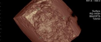
The vulva is examined during a pelvic examination. A gynecologist performs vaginal examinations. A transvaginal ultrasound is prescribed. If necessary, vulvoscopy and colposcopy. A bacterial smear is examined to exclude infectious diseases. Clarification of the nature of the neoplasm using histological examination. For biological analysis, scrapings and punctures of tumors are taken.
Removing the tumor often causes a new growth to appear. Removing papillomas requires long-term treatment. Surgeries and the postoperative period may occur with complications. Hematomas may form, the urinary system may be disrupted, and severe bleeding may occur.
Prices for treatment of vulvar cancer at Euroonco
The cost of treatment for vulvar cancer depends on the stage of the tumor, the extent of surgery, and whether it needs to be supplemented with chemotherapy, radiation therapy, and other types of treatment. At Euroonko you can get care at the level of leading Western oncology centers, but at a lower price.
Appointment with an oncologist-gynecologist - 6,900 rubles. Photodynamic therapy - from 170 rubles (calculated depending on the patient’s weight).
Book a consultation 24 hours a day
+7+7+78
Types of surgery
Based on the characteristics of the tumor, its treatment is prescribed. Surgical interventions to eliminate this disease:
- Cryodisruption. Nitrogen exposure is carried out. Freezing tissue stops cell development. During the operation, low-temperature liquid nitrogen is applied to the condyloma. The operation is performed without anesthetic. Not painful. The operation was well tolerated. Removing tumors with liquid nitrogen is a special technology; not every clinic performs these manipulations. The operation has minimal complications. There may be a small scar or burn.
- Radio wave excision. A directed flow of low frequencies dissects the tumor. Radio wave is the most gentle method in relation to neighboring tissues. There is no pain. There is no risk of scarring, bleeding, suppuration, or necrosis. Healing occurs in the shortest possible time.
- Laser. A directed laser evaporates the tufted growths. Laser ablation is used to excise resistant condylomas. The skin is not injured, and under the influence of the laser, epithelial cells grow. The depth of laser exposure is controlled. Does not leave scars. No contact with tissue occurs.
- Electrical coagulation. Electric current is used to cauterize the damaged area. The removed material is sent for histological analysis. The process has a number of contraindications. For example, herpes, oncology, hemostasis, inflammation of neoplasms. There are cases of relapse. There may be a small scar on the surface of the vulva.
- Chemical exposure. The destruction method involves applying a special drug to the affected area. A special composition with organic and inorganic acids, nitrates causes burning and death of condyloma cells. It is not accompanied by pain and is used without anesthesia.
- Plasma coagulation. An arc discharge containing gas penetrates the tissue and vaporizes areas with growths. Cauterizes blood vessels. The method has a high level of efficiency. Thermal damage to tissue occurs at a minimum depth, which promotes rapid healing. No scars are formed and pain is minimal.
Prevention of inflammation of the labia
To prevent vulvitis, it is necessary, first of all, to maintain intimate hygiene. It is recommended to use products designed specifically for caring for intimate areas. Ginocomfort washing gels for intimate hygiene, which were developed by specialists of the pharmaceutical company VERTEX, taking into account the characteristics of the microflora of the genital organs, are well suited for this purpose. They do not dry out the mucous membrane and do not disturb the acid-base balance. These products have a package of necessary documents and quality certificates.
Other measures to prevent inflammatory processes in the labia area include the following:
- Proper drying of the genitals after hygiene procedures. It should be carried out with a clean towel or napkin in the direction from front to back or with gentle blotting movements.
- Careful selection of underwear. It is optimal if it is underwear made of natural and soft material that will not squeeze or rub the perineum.
- General maintenance of immunity, which will allow the body to independently fight infection and suppress the proliferation of pathogens.
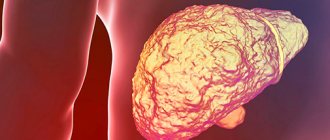
Malignant neoplasms
The external part of the female genital organs is susceptible to cancer. Young women are diagnosed with papilloma, which most often develops into a malignant neoplasm. Penetrating to the cellular level, the pathogen causes gene changes. This leads to abnormal protein release and accelerated cell proliferation.
Various factors for the development of vulvar cancer are known:
1. Age-related changes. More than half of women over 70 years of age have this diagnosis.
2. Bad habits. Smoking has a detrimental effect on blood vessels. Increases the risk of developing cancer.
3. HIV. The infection weakens the woman's immunodeficiency. The risk of contracting papillomavirus increases.
4. Vulvar neoplasia. Precancerous condition. An abnormal number of epithelial cells are found in the subcutaneous layers, then transform into cancer.
5. Cervical cancer. Oncology spreads to the vulva.
The appearance of malignant tumors is caused by the occurrence of adenoma-carcinomas. They are located in the thickness of the labia. Tumors are mistakenly confused with cysts. Adenocarcinoma can arise in the Bartholin glands and sweat gland cells of the vulva. Basal cell carcinoma sometimes occurs in this area. Melanomas arise from cells that produce melanin. It most often occurs in areas of the skin exposed to sunlight.
Vulvar cancer is divided into 4 stages:
1. The first stage of the tumor is caused by tumor growth within the vulva. It does not affect the lymph nodes and neighboring organs.
2. The second stage tumor grows into nearby organs. This can be the vagina, anus and urethra.
3. Neoplasms at the third stage affect the lymph nodes.
4. The last stage is characterized by damage to the lymph, vagina, bladder, and pelvic bones. Metastases penetrate to distant areas.
The stage of the tumor determines the further prognosis of the patient’s life activity and treatment. The growth of the tumor deeper causes manifestations; before this, no prerequisites may appear. The color of the skin differs significantly from healthy tissues; a pink or red tint predominates. A skin lump appears; it could be a wart, papilloma, or ulcer. The occurrence of pain, burning and itching.
Many women experience bloody discharge outside the menstrual cycle. These symptoms indicate the presence of a malignant neoplasm of the vulva. Adenocarcinoma is located deep in the labia majora and can be palpated, creating the sensation of a foreign body.
Radiation therapy for vulvar cancer
Radiation therapy is used for vulvar cancer in the following cases:
- Before surgery in combination with chemotherapy. This helps to reduce the tumor and convert inoperable cancer into operable one.
- After surgery to prevent relapse.
- Radiation therapy can be used as the only treatment for inguinal and pelvic lymph nodes.
- It is used alone or in combination with chemotherapy to treat women who are contraindicated for surgery.
Diagnostics
Cancer is diagnosed using a biopsy. High accuracy of the study helps to distinguish a malignant tumor from a benign one. A gynecologist examines a woman using a colposcope. The skin is treated with a special solution, which makes the lesions appear more pronounced. If the tumor spreads beyond the vulva, perform the following steps:
- Research of the genitourinary system and rectum.
- Detection of tumor growth and penetration into lymph nodes and organs using CT or MRI.
- Search for metastases using radiography.
Surgical treatment of vulvar cancer
Surgery is the main treatment for vulvar cancer. During the operation, the doctor tries to completely remove the tumor tissue, while maintaining the aesthetic appearance of the genital organs, normal evacuation of urine and stool (this is especially important when the tumor is close to the urethra and anus). Depending on the stage, one of the following surgical options is used:
- Local excision of the tumor. Possibly in the early stages. The surgeon excises the tumor and approximately 1.3 cm of surrounding tissue and subcutaneous fat. The removed tissue is sent to a laboratory for microscopy. No tumor cells should be found at the edges of the incision (negative resection margin) - this will indicate that the tumor has been completely removed, and most likely there are no more cancer cells left in the body.
- Vulvectomy . The surgeon removes all or most of the vulva. There are different options for vulvectomy. In some cases, after surgery, it is possible to maintain the normal appearance of the genital organs; in some women, it is subsequently necessary to resort to reconstructive plastic surgery.
- Evisceration (exenteration) of the pelvis is the most complex and traumatic operation; it is performed if the cancer has spread to the pelvic organs. The bladder, lower part of the colon and rectum, and the uterus along with the cervix and vagina are removed. The volume of intervention depends on where the tumor has managed to grow.
- Removal of lymph nodes . Previously, it was always carried out - just in case, since the surgeon could not know for sure whether the tumor cells had spread to the nearest lymph nodes. Currently, it is possible to conduct a sentinel biopsy - a study of the lymph node, which is the first on the path of lymph outflow from the tumor. If there are no cancer cells in it, then it makes no sense to remove the remaining lymph nodes.
Sometimes, if there is a high risk of relapse after surgery, surgical treatment is combined with a course of radiation therapy and chemotherapy.
Write to an oncologist
Inflammation of the labia in women, video
Gynecologist Irina Vladimirovna Garyaeva about vulvitis in women.
Source - KVD - dermatovenerological dispensary Sources:
- ROLE OF INFECTIONS IN THE GENESIS OF VULVA DISEASES. Reutskaya M.A., Kulinich S.I. // Siberian Medical Journal (Irkutsk). – 2010. – No. 6. – pp. 239-242.
- CLINICAL AND MORPHOLOGICAL PRINCIPLES OF TREATMENT OF CHRONIC VULVITIS. Kulinich S.I., Reutskaya M.A., Pokinchereda T.V., Ezhova I.V. // Acta Biomedica Scientifica. – 2013. – No. 5 (93). – P.42-48.
- Diseases of the cervix, vagina and vulva: Clinical lectures. Ed. V.N. Prilepskaya. // M.: MEDpress. - 1999. - P. 432.
- Recurrent vulvovaginal candidiasis: etiology, pathogenesis, treatment. Levonchuk E.A. // Med. news. - 2001. - No. 4. — P. 40-43.
- Dystrophic diseases of the vulva. Diseases of the cervix, vagina and vulva. Ed. V.N. Prilepskaya. // M.: MEDpress. - 1999. - pp. 326-336.
- https://simptom-lechenie.ru/en/vulvit-u-zhenshhin-i-devochek-simptomy-i-lechenie.html
- https://www.thenakedscientists.com/science-articles
- https://woman-centre.com/vlagalische-i-vulva/vulvit/vulvit-u-zhenshhin.html
- https://simptomer.ru/bolezni/zhenskie-zabolevaniya/864-vulvit-simptomy