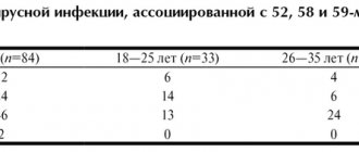
Papilloma viruses are the only group of viruses today for which scientists have proven the induction of cancer tumors. Moreover, HPV is the most common sexually transmitted infection; more than half of adults are infected with it. Find out how the papilloma virus manifests itself and how to treat it.
Appointment with a gynecologist - 1000 rubles. Appointment with a urologist - 1000 rubles. Removal of tumors - from 500 rubles. Consultation with a doctor based on the results of ultrasound and tests - only 500 rubles. (at the request of the patient).
MAKE AN APPOINTMENT, TEST OR ULTRASOUND
What is papillomavirus, papillomas, condylomas
The content of the article
Human papillomavirus infection (HPV) belongs to the genus A of the Papovaviridae family and is transmitted primarily through sexual contact, infecting epithelial (cover) cells - the skin, mucous membrane and urogenital area. Infection of the skin and genital organs occurs through microtraumas. The papilloma virus is contained in the urethra, Bartholin's glands and seminal fluid. The development of malignant tumors of the penis, vagina, and cervix is associated with human papillomavirus.
HPV strains, of which science knows more than 100, behave differently in the cell. Some exist separately from chromosomes, others are able to integrate into the cell genome (they most often cause complications). When the viral DNA is inserted into the nucleus of the host cell, it controls the cell's genetic material, leading the cell to malignant transformation. During the replication cycle, the human papillomavirus genome expresses 8–10 protein products. Oncogenicity is determined by proteins E6 and E7. It is difficult to kill the infection - the virus remains viable at a temperature of 50 degrees. From 30 minutes.
The virus infects stem cells located in the basal layer, and they transmit the infection to the surface epithelial cells. Superficial dividing immature cells are especially susceptible to HPV, which explains the high risk of oncogenes affecting the vulva, lower part of the vagina and cervix.
The most common visual manifestation of papillomavirus is papillomas. The formation got its name from the Latin papilla - nipple + Greek oma - tumor. Papillomas can develop into cancer. The development of oncology can be prevented by monitoring the development of papillomas and removing them in a timely manner.
A type of papillomavirus in women and men is genital and flat condylomas that develop on the genitals. They must also be removed.
A complication of the virus is a precancerous condition - cervical dysplasia and its logical conclusion - cervical cancer. You can avoid cancer by treating dysplasia. Treatment also involves removing the affected layer of cells.
Papillomas on the cervix: causes and treatment
Speaking about the reason for the appearance of papillomas, we are entering one of the most mysterious areas of medical knowledge - the nature of viruses.
These microscopic formations have a non-cellular structure, unlike most organisms living on Earth. They exist exclusively by parasitizing living cells. The papillomavirus we are interested in (HPV or HPV) penetrates skin cells, integrates into chromosomes and changes the tissue growth program. Cells begin to rapidly divide, growing to form papillomas. More than a hundred strains of HPV have already been identified, but not all of them are well studied.
How papillomavirus is transmitted, risks
Papillomavirus enters the human body in different ways, the probability of infection is 60%. You can get an infection in the following ways:
- Sexually, regardless of the type of sexual relations - oral, genital and anal. The presence of genital warts in a partner guarantees infection by 98-100%.
- Contact household - in the case of using a shared towel, washcloths, etc. The virus can also be transmitted through saliva during a kiss.
- Through wounds - a violation of the integrity of the skin and mucous membranes - an open gateway to the body.
- Infection of a child during the passage of the birth canal - children suffer from rare forms, papillomas grow in the nasopharynx and sinuses. Recent studies have shown that children born by cesarean section also become infected - this may indicate that the virus is able to penetrate the placenta.
Infection does not always guarantee the development of the disease. HPV causes disease in the body in 50% of cases, the rest are limited to carriage of the infection: a strong immune system can keep the virus in numbers that are not dangerous to the body. The incubation period ranges from a year to 20 years, with an average of 3-5 years.
The development of the disease is provoked by hormonal imbalances, immunodeficiency states, and sexually transmitted infections (STDs, STIs). Any condition that reduces immunity increases the risk - pregnancy, bad habits, chronic diseases, stress, etc.
It should be understood that infection of epithelial cells is a necessary but not sufficient factor for the development of oncology. According to Professor V.A. Molochkov, a well-known and respected scientist in the world of medicine, a number of other factors are necessary for the development of irreversible neoplasia:
- active expression of genes E6, E7 of highly oncogenic types hpv16 and hpv18;
- induction of estradiol metabolism to 16-OH;
- multiple damage to chromosomal DNA in an infected cell.
The first stage of CIN I neoplasia is expressed by active copying of the virus and its asymptomatic course. Tumor development is stimulated by the interaction of papillomavirus with cytomegaloviruses, trachomatis, mycoplasmas, ureaplasmas, and herpes simplex virus type 2.
Important numbers: the statistics are scary
- In the last 10 years, the number of people infected with hpv
has increased 12-fold. - HPV ranks second after genital herpes among all female infections and is found in 70% of adult women.
- Papillomavirus is the cause of all cases of cervical cancer.
- HPV is associated with 50% of anogenital cancers.
- The greatest risk of infection is between the ages of 18 and 25. The peak age for development of cervical dysplasia is 30 years, and cervical cancer is 45 years.
Global burden of cervical cancer
Cervical cancer is the fourth most common cancer among women globally, with an estimated 570,000 new cases in 2018 and accounting for 7.5% of all cancer deaths in women. It is estimated that more than 85% of the more than 311,000 annual deaths from cervical cancer occur in low- and middle-income countries. Women with HIV infection are six times more likely to develop cervical cancer than women without HIV infection, and an estimated 5% of all cervical cancer cases are HIV-related (2).
High-income countries have programs that provide girls with opportunities for HPV vaccination and women with regular screening. Screening can detect precancerous lesions at stages when they can be easily treated.
In low- and middle-income countries, access to such preventive measures is limited, and cervical cancer is often detected only at advanced stages, when symptoms have developed. In addition, access to treatment for disease at such advanced stages (eg, surgery, radiotherapy and chemotherapy) can be very limited, leading to high mortality rates from cervical cancer in these countries.
The high global mortality rate from cervical cancer (age-standardized rate: 6.9/100,000 in 2022) can be reduced through effective interventions.
Types of HPV
Scientists know more than 100 types of papillomaviruses. A third of them infect the human urogenital tract, affecting the skin and mucous membranes of the genital organs. A fifth of the viruses in this group have not yet been studied and may well present new unpleasant surprises.
The papilloma virus is classified according to its oncogenicity and area of damage:
- Non-genital - nasopharynx, mouth, sinuses, vocal cords, lungs;
- Affects the organs of the urinary system - ureters and bladder, urethra, renal pelvis;
- Genital in women - affects the mucous membranes of the external genitalia, the vestibule of the vagina and the vagina itself, the perianal area, the cervix, the perineum;
- Genital in men - affects the glans penis, foreskin, frenulum, coronary sulcus, shaft of the penis, scrotum, groin skin, pubis, perineum, perianal area. The external opening of the urethra in men is affected in 20-24% of cases.
Based on oncogenicity, HPV can be divided into:
- HPV low risk - type 6,11,42,43,44;
- HPV average risk 31,33,35,51,52,58;
- HPV high risk 16,18, 45, 56.
The most dangerous types of HPV papillomavirus (hpv) - those belonging to the high cancer risk group - are 16, 18, 31, 33, 35, 39, 45, 51, 52. They cause genital cancer.
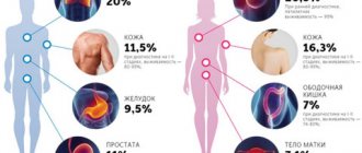
How dangerous the papillomavirus is for men and women can be understood by looking at the table of diseases associated with HPV activity.
Human papillomavirus is the cause of warts, condylomas and papillomas
Human papillomavirus is a family of viruses that cause warts, papillomas, condylomas, dysplasia or cancer of the cervix and genital organs in humans. General family: Papillomaviridae . Latin name: Human Papillomavirus . Abbreviation: HPV or HPV (as written in tests). 1. Over 50 years, more than 100 types of human papillomavirus have been discovered. Pathogenic for humans - 80 types. 2. According to WHO, 70% of the world's population is infected with HPV. 3. HPV types 16 and 18 are more likely than other types to lead to cervical cancer. 4. HPV is the overwhelming cause of genital cancer in women and men. 5. The most effective prevention against cervical and genital cancer throughout the world is considered to be a vaccine against types 6, 11, 16 and 18 of papillomaviruses. Infection.
The source of the virus is the skin cell or mucous membrane of a sick person. If a patient has a papilloma, even a small one, it is the direct source of the virus! However, upon examination, the patient may not yet have a wart or condyloma. The changes may still be microscopic and not visible to the eye (subclinical stage of the disease). But such a person can already transmit the virus to another person. Infection usually occurs in childhood. Through microdamage to the child's skin (scratches, abrasions), the papillomavirus penetrates the skin and causes the appearance of warts. In adults, certain types of the virus (discussed below) cause the development of anogenital warts, or genital warts, on the genitals. The transmission mechanism of these types is predominantly sexual. But contact-household transmission is also theoretically possible - through general hygiene items, the toilet rim, taking a bath, visiting a bathhouse, swimming pool, etc. Through microtraumas of the genital organs, the virus is transmitted from one sexual partner to another. In this case, the patient may also not have any changes visible to the eye. But there may be microscopic changes in the mucous membrane of the genital organs. And these altered cells are the sources of the virus. Next, the virus penetrates the skin or mucous membrane and is met by various cells of the human immune system. In most cases, immune cells destroy the virus. But if the immune system is weakened, the virus manages to penetrate the cells of the basal layer of the epithelium of the skin or mucous membranes, integrates into the chromosomes of the cells and changes the functioning of these cells. The cells begin to divide excessively and grow in a limited area, externally turning into warts and papillomas. Remember: - HPV types that cause warts enter the body during childhood, - HPV types that cause genital warts enter the body primarily through sexual contact. In rare cases, the development of human papillomavirus infection in the human body can lead to malignancy (that is, degeneration into cancer). Therefore, all types of papillomaviruses are classified according to the degree of oncogenicity (that is, according to the degree of possible cancer development). Classification of HPV types by oncogenicity (according to studies by McConcl DJ, 1991; Lorincz A. T., 1992; Bosch E X. et al., 2002; Kozlova V. I., Puchner A. F., 2003; Syrjanen S., 2003; Shakhova N.M. et al., 2006;). 1) Types of papillomaviruses that never cause cancer: 1, 2, 3, 4, 5, 10, 28, 49 2) Types of low oncogenic risk (very rarely cause cancer): 6, 11, 13, 32, 34, 40, 41, 42, 43, 44, 51, 72. 3) Types of average oncogenic risk (average percentage of cancer degeneration): 26, 30, 35, 52, 53, 56, 58, 65. 4) Types of high oncogenic risk (of all types of the virus, these are the types that most often give rise to degeneration): 16, 18, 31, 33, 39, 45, 50, 59, 61, 62, 64, 68, 70, 73. This is especially important in women. By the way, sometimes the classification changes. For example, HPV type 58 in women is no longer highly oncogenic. It began to be classified as a type with average oncogenicity. Occurrence in diseases: • In 73-90% of cases with cervical cancer they find: 16, 18 and 45 types of HPV • In 77-93% of cases with cervical cancer they find: 16, 18, 45, 31 and 59 types of HPV • B In 80-94% of cases of cervical cancer, the following are found: HPV types 16, 18, 45, 31, 33 and 59 • Precancerous conditions in urology and gynecology are often combined with HPV types 61, 62, 68, 70, 73. The most common types found in tests are: • human papillomavirus 16 (written HPV 16) - 50% • human papillomavirus 18 (HPV 18) - 10% Symptoms and clinic Symptoms and manifestations of HPV infection are warts, papillomas and cervical dysplasia. A) Warts. They are caused by the following types of HPV - 1, 2, 3, 4, 5, 10, 28, 49. • juvenile (or flat) warts - caused by types 3 and 5 of the virus. These are small flat elevations on the skin that occur mainly in children. • spinules (or plantar warts) - caused by types 1 and 2 of the virus (you can read more about them. • vulgar warts on the fingers - caused by type 2 viruses.
These are flat warts on the face
These are vulgar warts on the hand
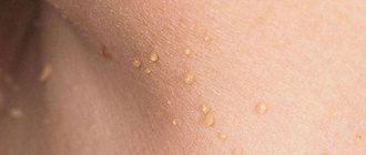
B) Genital warts. Localization : on the genitals, in the anus, in the oral cavity and on the lips (types - 6, 11, 13, 16, 18, 31, 35).
These are genital warts
The main mechanism of transmission of this disease in adults is sexual. Very rarely, a contact route of transmission can occur - through shared toilet items, through a dirty toilet rim, using a shared bathroom, in a bathhouse, etc. If a mother suffering from genital condylomatosis gives birth to a child, he will also become infected and subsequently may also develop genital warts or papillomatosis of the larynx and respiratory tract (discussed above). However, the frequency of such symptoms in infants is extremely low. Children have a fairly high level of immunity, which protects them from such manifestations of infection. B) Laryngeal papillomatosis. Multiple papillomas appear on the vocal cords. Caused by virus type 11. Sometimes it appears in children born to women with genital warts.
This is laryngeal papillomatosis
Remember: - cervical erosion and HPV are FAR from the same thing. A detailed article about what cervical erosion is and how it differs from dysplasia and HPV is here. Modern medicine declares with 100% certainty that cervical cancer is caused exclusively by papillomavirus types 16, 18, 31, 33, 35, 39, 40, 42, 43, 55, 57, 59, 61, 62, 66, 67. In the diagram - development of HPV infection over the years
E) Skin cancer of the penis (Bowen's disease). Caused by virus types 16 and 18. G) Today, some foreign scientists believe that the human papillomavirus is the cause of cancer of any location. Since cancer is a malignant tumor of the epithelium of the skin or mucous membrane, therefore, the HPV virus, which causes dysplastic phenomena in the epithelium, causes the appearance of cancer. And with cervical cancer this has been proven 100%. There is evidence for breast cancer and laryngeal cancer, although it has not yet been formalized into global recommendations. And, according to some cancer researchers, the day is not far off when cancer of other locations (for example, intestines) is also recognized as the result of the activity of the human papillomavirus in the human body. Remember: - any viral infection that is constantly present in the human body (and HPV is one of these) is activated only when immunity decreases. Diagnostics 1) PCR analysis. The main method for diagnosing papillomavirus is the PCR reaction. The most common types of HPV tests are virus types 16, 18, as well as a number of other highly oncogenic types. Material for analysis is taken from the mucous membrane of the woman’s vagina and cervix. In men - with the mucous membrane of the penis. The PCR reaction can also give a false result, both a false positive and a false negative result, especially if the conditions for its implementation are violated (even a push of the table on which the study is being carried out can lead to such a false result). Thus, according to modern researchers in the West, up to 20% of all PCR results for papillomavirus were false. And this fact did not depend on the complexity of the equipment and the quality of the reagents. 2) Digene test. New research gaining popularity in the medical community. This test is used to determine the presence of clinically significant concentrations of the virus. Thanks to this test, it is possible to identify whether the viruses in the patient’s body have a high or low degree of oncogenicity. The Digene test is used in conjunction with a cytological examination of the cervix, and they are also evaluated comprehensively. 3) Examination by a gynecologist and/or urologist. 4) Cytological examination. A smear taken during a gynecological examination is examined. This study is often called “liquid-based cytology”, or simply “cytology”. In this case, a laboratory doctor, under a microscope, determines the presence or absence of pathologically altered cells, which should not normally be present, but they appear only with the development of the disease. The presence of such altered cells may indicate the presence of CIN (or cervical dysplasia) in a woman. 5) Histological examination. A microscopic piece of tissue is examined, also taken during a gynecological or urological examination. Another name for this test is “biopsy.” Under a microscope, the doctor evaluates the degree of change in the tissue taken for examination.
How to interpret an HPV test?
The unit of measurement is the number of genome equivalents (in simple terms, the number of viruses) per 100,000 human epithelial cells (that is, by 10 to the 5th power). Abbreviated form: Lg Gradations: 1. < 3 Lg, that is, the number of viruses is less than 3 per 10 to the 5th power. This is a good indicator, the viral load is small, that is, the concentration of the virus is insignificant, the risk of developing the disease is low. 2. 3 – 5 Lg. This is a clinically significant indicator. The risk of developing the disease is average. It is necessary to undergo examination by a doctor. 3. > 5 Lg. High viral load. You should definitely undergo a full examination to exclude cervical dysplasia. What is a reference value? This means the average statistical indicators for a given study in a given age group. That is, in simple terms, reference values are the norm. For HPV, the reference values are negative. That is, normally there should not be HPV in the tests. What is KVM? KVM is the control of material taking. Normally, the doctor should take a scraping so that the sample of material contains at least 10,000 (or 10 to the power of 4, or 4Lg) epithelial cells. If the CME value is less than 4Lg, this means there are few cells for analysis. The analysis is not recommended, as it will be uninformative, and the doctor is recommended to repeat the collection of material. Treatment When treating human papillomavirus, you need to know: the virus may not be completely removed from the body. The main goal of treatment is to increase immunity, stabilize the virus, remove manifestations of the virus and reduce its concentration in the body so that the human immune system itself suppresses the virus. 3 areas of treatment are required (carried out by a specialized infectious disease specialist, immunologist, dermatologist or gynecologist): • taking antiviral drugs • strengthening the immune system • removing manifestations - warts, condylomas, dysplasia (erosion) or cervical cancer. All 3 areas are effectively carried out by modern medicine. Self-medication has low effectiveness and can lead to progression. Self-medication for diseases of the genital area is especially dangerous. 1) Antiviral drugs • Isoprinosine (or groprinosin), Allokin-alpha, • 5% Aldara cream. The active ingredient is imiquimod. 2) Drugs that enhance immunity Polyoxidonium, Reaferon, Roncoleukin, Immunal and others. The main drug for advanced forms at the moment is roncoleukin, which is used according to a certain scheme (prescribed by an immunologist or infectious disease specialist) 3) Removal of papillomas, warts, condylomas can be - Scalpel - classical surgery, electrocoagulation or an electric loop knife, radio wave removal, liquid nitrogen. These are outdated techniques that are traumatic, not always effective and can lead to relapses and post-burn scars at the sites of removal. -Laser - today this is the best method in terms of efficiency, safety, aesthetics. It is not recommended to use locally necrotizing drugs (acids, alkalis): Superchistotel, Solcoderm, Duofilm, Collomak, Verrukatsid, Ferezol, Condilin - and a number of others, since their application to the skin can contribute to the spread of the virus to healthy, previously uninvited areas of the skin, and also leads to skin burns and subsequent scarring
Required: a healthy lifestyle that improves immunity. Remember: First the doctor must make the correct diagnosis, and this is already half the treatment!!! Including treatment of human papillomavirus. Therefore, in the presence of multiple warts or relapses, it is recommended to first conduct a course of antiviral and immunomodulatory therapy under the supervision of an infectious disease specialist or immunologist!
Prevention of HPV Prevention is the best cure. Remember this phrase, especially when it comes to the sexual sphere. Nature has come up with a wonderful healing and prevention mechanism for humans, which then helps him not to get sick again. This is the immune system. If a person has already had warts or papillomas once, then he subsequently develops immunity to this type of virus. Therefore, juvenile warts, spinules and warts vulgaris very rarely appear in adults. This is why it is SO IMPORTANT to maintain your immunity at a high level. Let us list the main directions for the prevention of papillomavirus infection in humans: • Personal hygiene measures in public places • A healthy lifestyle that maintains immunity at a high level • Correct work and rest regime • Moderate physical training • Taking vitamins, fruits, juices • Only one sexual partner (in ideally) • Using a condom during sexual intercourse
We bring to your attention 3 videos on the removal of papillomas and warts at the Aurora clinic!!!
Find out more about the service and cost of tumor removal
Diseases caused by HPV (table)
| Disease, clinical manifestation | Type hpv |
| Skin diseases | |
| Plantar warts | 1,2,4 |
| Common (simple) warts | 2, 4, 26, 27, 29, 57 |
| Butcher's warts | 7 |
| Flat warts | 3, 10, 28, 49 |
| Verruciform epidermodysplasia (hereditary disease - verrucous dysplasia) | 2, 3, 5, 8, 9, 10, 12, 14, 15, 17, 19, 20, 36, 37, 46, 47, 50 |
| Diseases of the genital mucosa | |
| Flat condylomas, cervical dysplasia | 6, 11, 16, 18, 30, 31, 33, 39, 40, 42, 43, 51, 52, 55, 57, 61, 62, 64, 67 |
| Condylomas acuminata | 6, 11, 42, 54 |
| Cervical cancer, genital cancer, vaginal cancer, anal cancer | 16, 18, 31, 33, 35, 39, 45, 51, 52, 54, 56, 66, 68 |
| Diseases of the mucous membranes | |
| Epithelial hyperplasia of the oral mucosa | 13, 32 |
| Neck, lung and head cancer | 2, 6, 11, 16, 18, 30 |
| Respiratory tract papillomatosis | 6, 11, 30 |
One patient can be infected with several types of papillomavirus at the same time, which is usually the case.
Types and symptoms
Neoplasms caused by HPV infection can form on the skin and mucous membranes of various parts of the body, including the face, neck and décolleté. They can also form on the arms, legs, back, genitals, including the perineum, labia minora and majora, vulva, vagina, cervix, penis, especially along the coronary sulcus and frenulum. Damage to the mucous membrane of the oral cavity, tongue, nasopharynx, esophagus, bladder, conjunctiva of the eye, trachea and other internal organs is possible.
The human papillomavirus can lead to the appearance of neoplasms of various types. In general, they can be divided into 3 groups, although in all cases the cause of their appearance is the same - infection with the human papillomavirus.
- Papillomas are benign neoplasms of pink, white, pearl or light brown color, most often forming on the eyelids, lips, chest, armpits, and neck. They are located singly and usually do not tend to merge even with multiple lesions. Papillomas are usually round or lumpy, resemble the head of a cauliflower, and often have a stalk.
- Condylomas are benign formations of a dirty brown or paler color in the form of a cock's comb or many villi united by a common base. They are most often found in the genital area, anus and near the mouth. They tend to merge with each other and, as a result, cover large areas of the body. Their appearance is caused by infection with HPV types 6 and 11. There are pointed, flat and intraepithelial condylomas.
- Warts are uneven, light, benign tumor-like formations in the form of a plaque or small nodule on the surface of the skin of the hands, near the nails, feet, face, and front of the body. Warts can be similar to papillomas, but differ from them in that they have a wide base. They usually occur when infected with HPV types 1-5, 7-10, 12, 14, 15, 17, 19-24.
Such tumor-like formations can vary in size from a few millimeters to large growths covering large areas of the skin or mucous membranes.
Also, neoplasms may differ in appearance, which directly depends on the type of HPV that entered the body. The most common ones are:
- Vulgar or ordinary - protrusions of dense consistency with a diameter of more than 1 mm. They tend to merge and be located in groups.
- Plantar warts are protruding above the surface of the skin, often painful bumps with a shiny surface and rim. A characteristic feature is the absence of a skin pattern. They provoke the formation of HPV types 1, 2, 4.
- Flat papillomas are soft, smooth, flat, usually round growths that have a normal skin color or slightly yellowish, pinkish. They can provoke itching, so they are often injured, painful and inflamed. The reason for their formation is HPV strains 3 and 10.
- Filiform (acrochordas) are one of the most common papillomas, especially among elderly patients. Most often they are found on the face, around the eyes, in the groin, armpits, and neck. They are yellowish in color and tend to gradually enlarge, turning into bumps with a dense but elastic consistency.
- Genital warts in the perineal area, genitals.
Papillomas can be visible to the naked eye or located deep in the skin or mucous membranes. In the latter case, they are called endophytic and one of their manifestations is cervical dysplasia. Damage to the female internal genital organs by papillomatosis may be indicated by:
- itching, burning, weeping in the perineal area;
- profuse leucorrhoea;
- spotting, particularly after sexual intercourse;
- discomfort during intimacy.
Sometimes papillomatosis can provoke pain in the back and pelvis, weakness, swelling of the legs and causeless weight loss. Such signs are among the most alarming, as they may indicate the development of complications of HPV infection.
Symptoms of papillomavirus
HPV infection can be asymptomatic or give the following symptoms:
Genital warts (genital warts)
Fibroepithelial (skin) neoplasms with a thin stalk or a broad base. They can be single or merge, forming a growth that looks like a cauliflower head. Condylomas can become inflamed and bleed when injured, as they contain blood vessels that feed them.
Condylomas can be found on the clitoris, labia minora, urethra, vagina, cervix, around the anus and in the anus. Exophytic forms of OC are a consequence of the activity of benign types of the HPV virus - 6, 11. Endophytic condylomas (flat and inverted) grow on the cervix and initially do not give symptoms. Detected during extended colposcopy. Genital warts affecting the lips, tongue and palate are visible during routine examination.
People with HIV and during pregnancy develop very large genital warts. Giant Buschke-Levenshtein condyloma is not uncommon.
- Anal warts.
Anal warts can be found up to the dentate line of the rectum. At first they do not cause discomfort, but over time they itch, hurt, and smell unpleasant. - Urethral warts.
In women, the external urethral opening is affected in no more than 8%. Such warts are easily identified by a gynecologist. Deep damage to the urethra (urethra) cannot be determined visually, but the disease gives symptoms of sluggish urethritis. Urethral warts in men cause a split urine stream associated with a narrowing of the urethral opening.
Flat condylomas
Flat condylomas do not protrude above the surface of the mucous membranes, which is why they got their name. These formations have a high oncogenic potential. Most often, flat condylomas are located on the cervix and vaginal mucosa. Flat condylomas can only be detected by colposcopy.
Dysplasia, cervical cancer
Dysplasia is a tissue pathology associated with the modification and degeneration of cells. This is a precancerous condition. There are 3 degrees of the disease, all of which are detected by colposcopy. Stages 2 and 3 require surgical treatment. Cervical dysplasia is preceded by cervical erosion.
Cervical cancer is a consequence of dysplasia. It is the most common tumor of the female reproductive organs. It may be asymptomatic or cause pain, bleeding and other symptoms characteristic of problems with the female reproductive system.
Prevention measures
Although there are no treatments targeting HPV, there are effective prevention methods. It can be primary and secondary.
Primary prevention aims to prevent HPV infection. The most effective method is vaccination. In Russia, two vaccines against HPV types 16 and 18 are currently registered: Gardasil and Cervarix. They contain virus-like particles that do not cause infection, but trigger an immune response so that the body becomes protected when it encounters the real virus. In accordance with current international recommendations, all boys and girls aged 11–12 years should be vaccinated against HPV (you can start at 9 years old). To create reliable immunity, two doses of the vaccine must be administered. If a teenager has not been vaccinated before age 15, he will need three doses. Unfortunately, the vaccine will not be effective if a person is already infected. Therefore, people over 26 years of age who are sexually active can, of course, be vaccinated, but the benefits of vaccination for them are questionable.
In 2022, scientists proved that HPV vaccines prevent not only precancerous changes in the cervix, but also invasive cancer.
Other measures for primary prevention of malignant tumors caused by HPV:
- Sex education, taking into account the age of children and the cultural characteristics of society.
- Using condoms. Doctors should tell the public that this method of contraception helps protect against dangerous infections, including reducing the risk of HPV infection.
- To give up smoking. In this regard, it is also important for doctors and scientists to conduct educational work.
- Male circumcision. Penile cancer often develops in the foreskin area, so this is an effective preventative measure. So-called female circumcision is unacceptable even under the pretext of protecting against infections - in any form it is clearly a mutilating operation that deprives a woman of the opportunity to have a full sex life.
Secondary prevention aims to prevent the development of cancer in people who are already infected with high-risk HPV. All women who are sexually active (including those who have been vaccinated) should be regularly screened for cervical cancer:
- Up to 65 years - PAP test (Pap smear) every 3 years or PAP test in combination with HPV tests every 5 years. At least once a year it is necessary to undergo preventive examinations with a gynecologist.
- Over 65 years: If a woman has been screened regularly for the past 10 years and has not had any cancer or precancerous lesions (CIN 2 intraepithelial neoplasia or more severe) in the past 25 years, screening can be stopped.
- If a woman has had her uterus and cervix removed (not due to cancer or severe precancerous lesions), screening may not be necessary. If the uterus was removed, but its cervix was left, screening must be done like everyone else.
Research also shows that people at higher risk (who have anal sex, those with HIV) may also benefit from anal Pap smears. A malignant tumor or precancerous changes in the oral cavity or oropharynx can be detected by a dentist - this is another reason to undergo regular preventive examinations.
Screening helps to detect cancer at an early stage and begin treatment in a timely manner, thereby significantly improving the prognosis.
Of course, it is important to pay attention to any suspicious symptoms and not delay visiting a doctor. For example, cervical cancer can manifest itself in the form of abnormal vaginal bleeding (between periods, during sex, postmenopause), pain in the pelvis, and pain during sexual intercourse. These symptoms do not necessarily indicate that a woman has a malignant tumor; most often they indicate other diseases. But only a doctor after an examination can establish an accurate diagnosis.
In the clinics of the federal network "Euroonco" you can get a consultation with an oncologist-gynecologist, learn about your risks, and about screening tests that are recommended in your case. Our doctors have extensive experience in the treatment of benign neoplasms and malignant tumors of the female reproductive organs. We also have a comprehensive program “Women’s Health” - it includes all the necessary laboratory and instrumental tests, helps to identify many diseases in the early stages.
Book a consultation 24 hours a day
+7+7+78
Papilloma virus - diagnosis
Human papillomavirus can be detected in its early stages only by laboratory methods. The infection can be detected visually only when condylomas or papillomas appear. If HPV is suspected, the following are prescribed:
- Gynecological examination
or examination by a urologist with taking smears for an HPV test. If condylomas are detected, urethroscopy is performed. In case of cervical erosion, the gynecologist must take a smear for oncocytology. - If HPV is detected, a colposcopy is required - examination of the vagina and cervix with a gynecological microscope - colposcope. The doctor uses special tests to exclude hidden pathologies.
- Colposcopy with biopsy.
Indicated for all women with neoplasia. At the same time, coloring and exposure to suspicious areas is carried out. A sign of APC may be whitish areas formed after treatment with vinegar, uneven accumulation of iodine when exposed to Lugol's solution, a mosaic pattern, and protrusions of the epithelium. - Histological and cytological examination
- assessment of the cellular composition, and the cells themselves, for atypicality (cancer). - PCR – search for traces of papillomavirus DNA. This is the most accurate and diagnostically informative analysis that detects the papilloma virus in men and women and specifies its type.
To assess the viral load, a quantitative HPV test is recommended, which determines the critical concentration of the virus associated with the risk of malignancy of tumors. The analysis is also carried out to assess the effectiveness of treatment.
Treatment of papillomavirus
Unfortunately, medicine is not yet able to completely rid the body of the virus. Therefore, the task of the doctor and the patient is to deal with the consequences in a timely manner. It is recommended to remove all warts and treat precancer and cancer stages. According to recent studies in the USA, the human immune system is able to cope with HPV on its own within 2 years after infection in 90% of cases. If this does not happen, treatment is strictly necessary. Papillomas are removed using the following methods:
- Surgical removal
is an outdated but effective method. Recommended in exceptional cases. - Electrocoagulation
– cauterization of affected areas with electric current. Not everyone likes the method, as it can also affect healthy tissue. - Laser coagulation
- laser cauterization - is the most modern and effective method that gives a minimum of complications. - Cryodestruction with liquid nitrogen
differs from other methods in that the pathological growth is affected by cold. Requires a lot of experience from the doctor. - Chemical cauterization
- the doctor acts on the diseased area with concentrated acids or alkalis. The method can also affect healthy skin - the chemical will leave a scar. - The radio wave method
is the most expensive, but the best. Does not cause pain, complications, bleeding. Does not leave scars.
You can read more about methods for removing tumors in the “Low-traumatic operations” section.
After removal, antiviral treatment and means to restore and strengthen the immune system are prescribed.
Treatment and removal of papillomas
Treatment of human papillomavirus infection is always complex. Of course, you can simply remove the disturbing papilloma, but in this case there is a high risk that a new one or even several will soon form in its place. Since the main reason for the formation of papillomas is a decrease in the body’s own defenses, which allows HPV dormant in the body to become more active, patients with papillomatosis are first prescribed drug therapy. It includes taking immunomodulators and antiviral drugs. For extensive lesions of the skin and mucous membranes by papillomas, cytotoxic drugs can also be prescribed. But their use is carried out under the supervision of a doctor, since they can provoke serious side effects.
If a patient is diagnosed with concomitant infections or diseases, he must be prescribed treatment appropriate to the situation, and sometimes it is additionally necessary to obtain advice from a specialized specialist and undergo therapy under his supervision.
Condylomas of all types are subject to mandatory removal, as well as papillomas, which are often injured and inflamed. In other cases, removal is carried out at the request of the patient. But it is possible to begin eliminating visible manifestations of HPV infection only after completing treatment for concomitant diseases, if any have been identified, and while antiviral therapy is continued.
In general, all modern methods of destruction or removal of papillomas can be divided into 2 large groups:
- chemical - involve the use of various chemical compounds to remove papillomas, including trichloroacetic acid, Solcoderm, Ferezol, Collomak, etc.;
- physical - involve the removal of papillomas surgically, using electrocoagulation, cryodestruction, laser, radio wave or plasma coagulation.
After removing the papilloma by one method or another, it is important to use topical products prescribed by your doctor to speed up healing and eliminate the risk of infection.
The success of treatment and especially the removal of papillomas depends on the strength of the immune system. In its normal state, in 90% of cases, within 2 years from the moment of infection, HPV is suppressed or even completely destroyed. But this does not guarantee that there is no risk of re-infection or the formation of new papillomas. If immunity is reduced due to certain factors, papillomatosis becomes chronic, periodically relapses and can lead to serious complications.
In order to prevent infection with dangerous strains of HPV and the development of severe complications, it is recommended, especially for girls, to be vaccinated with Gardasil or Cervarix vaccines between the ages of 9 and 25.
Surgical removal of papillomas
The essence of the method is to remove the tumor using a scalpel, which is associated with the formation of scars and an increase in healing time. Therefore, it is used only when it is necessary to remove a large papilloma or in cases where it is necessary to conduct a histological examination, since the formation of malignant cells is expected in it.
The procedure is performed under local anesthesia, which ensures its painlessness. The doctor uses a scalpel to remove the tumor itself, including surrounding healthy tissue. This is necessary to eliminate the risk of re-formation of papilloma in the same place. If it has a pedicle, it is cut with surgical scissors, and the blood vessel that fed the neoplasm is “sealed” using an electrocoagulator. The remaining wound is sutured, treated with an antiseptic and covered with a sterile bandage.
Today, surgical removal of papillomas is mainly carried out if there is suspicion of the formation of cancer cells.
Cryodestruction
The method involves the use of low temperatures to destroy papilloma cells. This is achieved through the use of liquid nitrogen, the temperature of which is -196° C. The essence of the procedure is to use a special attachment or touch the papilloma with a cotton swab dipped in liquid nitrogen. Under its action, the water contained in the cells instantly turns into ice crystals, which destroy them from the inside. The procedure is performed under local anesthesia or without it, since the exposure time to liquid nitrogen does not exceed 5-20 seconds.
The complexity of the method lies in choosing the correct duration of exposure in order to remove the papilloma to its full depth and not harm healthy tissues, which will lead to scar formation.
Immediately after treatment with liquid nitrogen, a white spot forms at the site of the papilloma. Subsequently, a small blister with clear or pinkish liquid contents forms on it, and the surrounding skin turns red and may swell slightly. This may be accompanied by minor discomfort such as a burning or tingling sensation.
After 3-4 days, the blister bursts, and in its place a crust forms, which itself falls off after a few days, revealing healthy young skin. Under no circumstances should you pierce the bubble yourself or injure it or the crust in any other way.
Cauterization of papillomas with liquid nitrogen is possible only in cases where the possibility of the formation of malignant cells in it is completely excluded. This method is most often used to remove papillomas and condylomas on:
- centuries;
- face;
- small joints;
- genitals.
Laser removal of papillomas
The use of a laser to remove papillomas is also possible only in cases where their malignancy is completely excluded. The procedure is carried out under local anesthesia and consists of evaporating moisture in papilloma cells using thermal laser energy. Thus, a focused laser beam is directed at the tumor. The duration of exposure does not exceed 1 minute.
As a result, a crust forms at the site of the papilloma, and the surrounding skin turns red and swells. The crust will come off on its own within a week. It should not be wetted and steamed for 3 days, nor should it be torn off or injured, as this can lead to the formation of scars. In addition, for 2 weeks after removal of the papilloma, you should protect the treatment area from sunlight. Otherwise, there is a high probability of hyperpigmentation of this area.
When removing papillomas on the face with a laser, decorative cosmetics should not be applied to the affected area until complete healing.
After the crust falls off, healthy pink skin is exposed, which gradually acquires a normal shade. This is the main advantage of laser removal of papillomas, since it is not associated with the formation of scars. They can only form when large tumors are removed and the rules of care in the postoperative period are violated. Also, laser removal completely eliminates the risk of wound infection and is an absolutely bloodless method, since under the influence of thermal energy, instant coagulation of small blood vessels occurs.
Using a laser, papillomas are mainly removed on:
- hands;
- legs and feet;
- face;
- centuries;
- neck;
- genitals.
Laser is the only reliable way to remove plantar warts, as their roots can go 1 cm or more deep into the tissue.
Electrocoagulation of papillomas
The essence of the method is to use electric current to remove papillomas. Using a special device called an electrocoagulator, the doctor captures the papilloma and cuts it off within healthy tissue. In this case, bleeding is completely absent, since the thermal energy of the current is enough to coagulate small blood vessels. But the procedure can be painful, especially if it is necessary to remove a tumor in areas of the body with delicate skin.
After electrocoagulation, a crust also forms. And the entire recovery period takes 7-10 days. After the crust falls off, the skin underneath should be protected from damage and exposure to ultraviolet radiation.
The method is used to remove papillomas in different parts of the body. Moreover, electrocoagulation can also be used in cases where it is necessary to conduct a histological examination of the neoplasm and accurately determine its nature. But the result of the procedure completely depends on the qualifications and experience of the doctor, however, as in other cases, since if the removal is not deep enough, the papilloma can form again in the same area.
Radio wave surgery
This method is one of the most advanced in removing papillomas of any kind. It involves the use of a special Surgitron device. But at the same time, it has much in common with laser removal of papillomas. The procedure is carried out under local anesthesia, and the actual removal occurs due to the effect of radio waves on the tumor. Thanks to them, it is possible to completely cut off papilloma of any size and location, and also avoid the formation of scars.
Prevention of papilloma virus
You can prevent the appearance of warts by maintaining personal hygiene. You should not touch other people's papillomas, condylomas, etc. You can protect yourself from genital warts by barrier contraception (using a condom), but if the formations are on external tissues, this will not help. The most effective method of protecting against HPV is strengthening the immune system.
Girls and boys are recommended to get vaccinated against HPV. The vaccine prevents the occurrence of cervical cancer, vulvar cancer, vaginal cancer, genital warts and other diseases associated with papillomavirus. There are three types of vaccines against papillomavirus. In Russia, the drug Gardasil is recommended, directed against 4 types of hpv (16, 18, 6, 11).
The vaccine is given three times, starting at age 9 and before the start of sexual activity. Women can get vaccinated up to 26 years of age, but the drug will not work against viruses that are already in the body.
Fighting cervical cancer: an integrated approach
The Global Strategy to Accelerate the Elimination of Cervical Cancer as a Public Health Problem, adopted by the WHA in 2022, recommends a comprehensive approach to cervical cancer prevention and control. The recommended set of measures includes activities covering the entire life cycle.
| Primary prevention | Secondary prevention | Tertiary prevention |
Girls 9-14 years
| Women 30 years or older | All women as needed |
Girls and boys as appropriate
|
| Treatment of invasive cancer at any age
|
The fight must be multidisciplinary and include components of community education, social mobilization, vaccination, screening, treatment and palliative care.
Primary prevention begins with vaccinating girls against HPV between the ages of 9 and 14 years, before they become sexually active.
Sexually active women should be screened for abnormal cervical cells and precancerous lesions starting at age 30. Screening of sexually active women with HIV infection should begin at an earlier age, immediately after testing positive for HIV.
To treat precancerous lesions, removal of the abnormal cells or lesions is recommended, which is done on an outpatient basis (the abnormal cervical tissue can be destroyed using cryotherapy or thermal ablation).
If signs of cervical cancer are present, treatment options for invasive cancer include surgery, radiation therapy and chemotherapy, and patients need to be referred to the correct level of care.
Where is papillomavirus treated in St. Petersburg, prices
You can contact the Diana private clinic in St. Petersburg for diagnosis and treatment of papillomavirus. We use modern methods approved in the best European clinics, and the prices for specialist services are quite affordable.
Here you can get tested for the human papillomavirus and remove any tumors. Removal of formations associated with HPV is carried out using the latest Fotek radioknife with a coagulator that prevents bleeding. Price for removal of condylomas, papillomas, etc. — from 500 rub. We also offer the HPV vaccine with Gardasil.
Can an HPV infection be cured?
There are currently no treatments that specifically target HPV. Papillomas and condylomas, as well as precancerous changes caused by HPV, can be removed surgically, using a laser, or cryosurgery. When the cervix is affected, loop electrosurgical excision (removal of a pathological formation using a wire loop heated by an electric current) and conization (removal of a cone-shaped section of cervical tissue) are used. However, it is important to understand that surgery cannot remove the virus from the body.