03/07/2018 There is a risk of a scar appearing on the skin after surgery. The smaller the area and depth of the damage, the less likely it is to occur. Is it possible to guarantee that a scar will not appear or to cure it completely?
This is an article by Mikhail Gusarov, member of the European Academy of Dermatology and Venereology (EADV), International Dermatoscopy Society (IDS), author of more than 10 scientific publications.
What determines the risk of scarring?
The appearance of a scar and its nature depends on a number of factors:
- Depth and area of damage: if they are small, a scar may not appear at all.
- Tissue healing abilities.
- The presence of chronic diseases (for example, diabetes).
- Time for complete healing of the wound (it may increase if the wound has become infected).
The process of complete wound healing, which lasts for 21 days, ends with pathological scarring in only 33% of cases. If the process lasts more than 21 days, the probability increases to 78%.
Rules of care after wart removal
To avoid complications and speed up the healing of an injured area of skin after removing a plantar wart or located in any other place, it is necessary to follow certain rules.
Upon completion of the wart removal procedure, medical doctors must instruct the patient in detail about the procedure and standards for wound care and select the optimal medications. Our clients can receive a full range of high-quality services in Moscow, at the clinic on Kashirskaya metro station, and in Ramenskoye, on the street. Chugunova, 21-A.
How does a wound heal?
The following phases of wound healing are distinguished:
- Inflammation. It begins immediately after the injury and lasts about 2-3 days, during this phase it is necessary to clean the wound and carry out anti-infective measures.
- Proliferation. From 3 days to 2 weeks. It is necessary to maintain the absence of infection and adequate hydration.
- The appearance of a scar. From 2 weeks to several years. During this phase, measures are taken to correct the scar depending on its type. [12]
What happens if you tear off a wart (cut it off, rip it off), consequences, what to do
Warts are not only a cosmetic defect, but can also cause a lot of inconvenience to a person. They can often become injured and inflamed. At first glance, the most convenient way to deal with them is to tear them off. Such a procedure, performed at home without following hygiene rules, can be dangerous.
Is it possible to pick a wart?
A wart is a benign skin growth. It has a root, which is responsible for its growth and spreads the pathological process to neighboring, undamaged tissues. A person cannot remove the root of a wart on his own. After its removal, active growth of a new growth will be observed, because the virus remains in the skin and continues to be active.
Self-removal of warts is prohibited. It does not bring the necessary result, since after tearing off benign skin formations, they continue to grow with possible generalization.
In case of injury to a skin tumor, it is recommended to pay attention to its appearance.
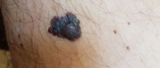
If the wart has not previously been examined by a dermatologist and oncologist, an emergency indication for consultation with the listed specialists is the dark brown or black color of the wart, the glossy appearance of its surface, as well as drop-shaped discharge of blood.
A wart can fall out on its own due to an accidental blow or clothing rubbing against it. If it was in natural skin folds, it can cause swelling, itching, burning, and hyperemia.
The greatest danger facing a person is the degeneration of a skin growth into a malignant neoplasm. This can happen when an infectious process begins to develop in the wound. The degeneration of cells occurs because they have already lost some of their original functions, and after injury it is difficult to predict their behavior.
With human papillomavirus, it is prohibited to use any folk remedies to combat them. At home it is impossible to check for the degree of malignancy.
The use of tinctures and decoctions can speed up the process of wart malignancy and cause skin cancer. The use of acids and alkalis at home is strictly prohibited: these substances can cause severe chemical burns.
It must be remembered that self-medication of skin growths can be dangerous. To remove them, you must consult a doctor who will choose the most effective and safe method of treatment.
What happens if you tear off a wart?
If its leg comes off, inflammation of the epidermis begins. An open wound is a gateway for infection to enter the body. Inflamed skin changes color and swells. These are accompanied by itching, burning and an increase in local body temperature.
Cutting off warts yourself can cause consequences such as:
- Excessive bleeding as a reaction to mechanical trauma to the skin. The appearance of hemorrhage is caused by the intensive proliferation of blood vessels at the site of wart formation.
- The spread of virus to healthy areas of the skin, which contributes to the formation of new growths.
- The appearance of scars and cicatrices on the skin. They are difficult to remove, and they can be noticeable on the face, which spoils a person’s appearance.
- Infection of the wound surface. An open wound in a person infected with the papilloma virus is prone to infection due to decreased immune defense. Infection of the wound is manifested by swelling, hyperemia, and suppuration. Often, wound infection can cause general sepsis.
If after cutting off the wart there is no suppuration, redness of the skin, or pain, then this may indicate that there is no papillomavirus in the body. But even in this case, there is no absolute guarantee that a malignant process will not develop on the affected area of the skin.
Sometimes a wart can masquerade as a malignant tumor, such as melanoma or basal cell carcinoma. If you tear it off, it will be an impetus for the intensive growth of a cancerous tumor.
Treating it with home remedies can only increase the progression of cancer. In advanced cases, the tumor spreads metastases to nearby lymph nodes or to distant organs of the body.
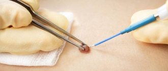
What to do if the wart has been cut off
In case of accidental injury, the damaged growth must be treated using available antiseptic drugs.
Under no circumstances should the torn part of the wart be thrown away: it must be placed in saline solution and sent for histological examination. It will help prevent malignant cell degeneration. Histological examination of wart tissue helps to choose the most appropriate treatment method.
Is a scar a reason to go to the doctor?
Reasons for visiting a doctor:
- aesthetic dissatisfaction;
- itching and pain in the scar area;
- the formation of deformities and contractures, which can lead to disability.
If scar tissue begins to develop pathologically and causes a keloid scar, it is better to consult a doctor.
There are factors that increase the risk of developing a scar:
- genetic predisposition;
- race (black patients are more likely to develop keloids);
- localization of injury (when removing tumors in the chest, neck and shoulders, the likelihood of keloid scars is higher);
- age (the formation of keloid scars is typical for young people). [3]
Before and after. How we remove scars
Types of benign neoplasms
The most common benign skin tumors are:
- moles and nevi - congenital or acquired hyperpigmented spots
- papilloma (wart) – represented by a papillary growth or growth on a stalk of viral etiology (it is provoked by the human papillomavirus)
- lipoma (wen) – formed from the fatty layer
- atheroma - a cyst from the epithelial layer, prone to inflammation and suppuration
- dermatofibroma - a nodule of connective tissue
- hemangioma - pathological proliferation of vascular tissues
Borderline neoplasms include freckles and xeroderma pigmentosum.
How to prevent the appearance of a keloid scar?
There are several ways to prevent the appearance of a keloid scar.
- If surgical excision of the tumor is performed, techniques that minimize tension on the wound edges should be used during suturing.
- Mechanical stabilization - the use of special silicone plates or the use of silicone-based gels.
- The use of various medications containing flavonoids - substances obtained naturally from various plants (for example, onion extract). However, data on the effectiveness of this group of drugs is contradictory.
- Pressure therapy (compression) – within 15–40 mm Hg. Art. for 23 hours a day for 6 months. Data on the effectiveness of this method are also conflicting.
- Glucocorticosteroids (GCS) - can be used as creams or ointments in the initial stages of scar formation and to prevent its formation, as well as in the form of injections into scar tissue. [4]
Complications of benign neoplasms
Even if a benign neoplasm is not accompanied by unpleasant symptoms such as pain, growth or suppuration, it is necessary to be regularly examined by a dermatologist. The following types of formations are prone to malignancy:
- moles and nevi
- papillomas (in the form of a growth on the leg)
- condylomas (warts in the genital area)
- dermatofibroma (in rare cases)
Frequent damage and infection in the wound leads to inflammatory processes, which may involve surrounding tissues. Inflammation is accompanied by pain, if an area of exposed skin is affected, this is accompanied by severe psychological discomfort.
Timely removal of skin lesions prevents the risks of complications and reduces the volume of surgery.
What if a scar appears?
Once scar tissue has formed, unfortunately, it will not be possible to completely get rid of it. All medical actions in this case will be aimed at minimizing the manifestations of the scar. The following methods are used to treat formed keloids:
- GCS injections are a popular and effective method for treating keloid and hypertrophic scars. A GCS solution is injected into the scar tissue 1–2 times a month until the expected results appear. Unfortunately, with excessive use of the drug, skin atrophy may develop at the injection site, telangiectasia or an atrophic scar may appear. [4]
- Surgical excision of the scar - you should not resort to this method as the only remedy, since it always ends in a relapse that exceeds the existing scar in area. This method can be resorted to after previous long-term therapy carried out in various ways. [4]
- Cryotherapy is the treatment of scars with liquid nitrogen. There are various methods of its use: spray, contact method and intralesional cryoprobe. The latter method is more effective. In addition, the combination of cryotherapy with GCS injections gives the best result compared to others. [4]
- Laser treatment – various lasers are used. Nd:YAG laser with a wavelength of 1,064 nm is used to target the vessels of scar tissue. In addition, CO2 and Er:YAG lasers are actively used in fractional mode. But it is not recommended to use ablative lasers (CO2 and Er:YAG) as the only treatment method, since in this case there is a high probability of relapse. [5]
A dye laser (PDL) with a wavelength of 585 nm is also considered effective. [6]
There is evidence of the effective use of CO2 laser in fractional mode during surgery to improve the cosmetic outcome of healing. [7]
- Ointment with 5-fluorouracil for the treatment of scars, but it is not sold in Russia. [4]
- Botulinum toxin type A is used to weaken the tension of the wound edges by reducing muscle activity. Injections are given immediately after surgery. [8]
- Imiquimod cream is recommended for use after surgical excision of a scar to minimize the likelihood of recurrence. [9]
- Other methods - bleomycin, verapamil, TGF-β, interferon, tacrolimus, sirolimus, tamoxifen, epidermal growth factor, retinoic acid, tamoxifen and other drugs are also considered effective for the treatment of scars. [10]
When should warts be removed?
Removing warts on the face is not in doubt, since they are in plain sight and often cause psychological discomfort. For medical reasons, it is recommended to remove long-standing warts, as well as those located in places with regular mechanical or chemical irritation. It is better to remove the formation if it is located:
- in places where belts, straps, shoulder straps, collars fit;
- wearing rings and chains;
- friction of the straps of bags, backpacks, etc.;
- large folds of skin in obese people;
- under the breasts, if they are large;
- on the scalp;
- in places where hair is regularly removed by shaving or depilation.
The latter include the areas of the armpits, bikini, mustache and beard in men. Warts are removed not only for medical reasons, but also for cosmetic reasons, if their appearance causes discomfort in a person.
Which method is the most effective?
Unfortunately, there is no most effective treatment for scars. The most effective is considered to be a combination of various methods, which is always selected individually. Injections of corticosteroids into hypertrophic or keloid scars are considered an accessible, effective and inexpensive method.
Atrophic scars are distinguished separately: for their correction, planar ablative lasers, fractional ablative lasers (CO2 and Er:YAG), various combinations of lasers (fractional +PDL, Qsw+fractional lasers), collagen, fillers with hyaluronic acid, PRP therapy, microneedling are used. , peeling and a number of other methods [11, 12]
Previously, the dermabrasion method was used to correct atrophic scars, but now it is practically not used, since it is associated with the risk of side effects: bacterial or viral complications, telangiectasia, hyperpigmentation, a long rehabilitation period, as well as hypertrophic or keloid scars.
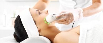
How is the laser mole removal procedure performed?
At the PRAGA clinic, the laser removal procedure for nevi is carried out under the guidance of a highly qualified dermatologist Elena Ivanovna Karpenko. At the initial consultation, she performs an examination and identifies contraindications. The doctor will calculate the cost of mole removal based on their size.
If there is no danger of complications, pre-treatment of the skin is carried out. A special nozzle cools the area where the mole is located. Then the neodymium laser precisely targets this area. The duration of laser irradiation is only about three seconds. The mole begins to darken, fade, and after a week disappears without a trace.
It is important to know that the laser beam does not cut the skin and does not cause infection. This is a non-contact sterile instrument.
How not to treat scars
Incorrect treatment, in addition to wasting money and time, can lead to enlargement of the scar. What could cause this:
- non-compliance with doctor’s recommendations after removal or self-medication;
- traumatization and irritation of the wound can lead to relapse;
- non-compliance with prescribed antiseptic methods;
- removal of the crust that has formed at the site of removal: under it the restoration process is more efficient.
There is not enough information in scientific sources to evaluate the effectiveness of folk remedies in the treatment of scars. From my experience I can say that I have not seen a single patient for whom folk remedies produced any significant positive changes. And in fairness, it’s worth saying that I rarely encounter the use of folk remedies in the treatment of scars.
Surgery to remove a tumor: preparation
Before deciding on the removal of the formation, the surgeon performs dermatoscopy, excluding the malignant nature of the pathology.
When using low-traumatic methods (laser, radio wave, electrocoagulation), the patient does not need to take tests or undergo a comprehensive examination. If we are talking about an operation under general anesthesia, it is necessary to undergo diagnostics that exclude contraindications to this intervention.
Comprehensive preoperative diagnostics includes laboratory tests and instrumental methods. Mandatory laboratory tests include:
- clinical blood and urine tests
- blood for infections, group and Rh factor, coagulation
Instrumental studies:
- ECG
- fluorography
Before the operation, the patient is examined by an anesthesiologist.
Bibliography:
- J Am Acad Dermatol. 2016 Apr; 74(4): 607–25; quiz 625–6. doi: 10.1016/j. jaad. 2015.08.070. Wound healing and treating wounds: Chronic wound care and management. Powers JG, Higham C, Broussard K, Phillips TJ.
- J Am Acad Dermatol. 2016 Apr; 74(4): 589–605; quiz 605–6. doi: 10.1016/j. jaad. 2015.08.068. Wound healing and treating wounds: Differential diagnosis and evaluation of chronic wounds. Morton LM, Phillips TJ.
- Ann Hum Genet. 2022 Feb 27. doi: 10.1111/ahg.12245. Gene-based evaluation of low-frequency variation and genetically-predicted gene expression impacting risk of keloid formation. Hellwege JN, Russell SB, Williams SM, Edwards TL, Velez Edwards DR.
- Int. J. Mol. Sci. 2022, 19(3), 711; doi: 10.3390/ijms19030711. Recent Understandings of Biology, Prophylaxis and Treatment Strategies for Hypertrophic Scars and Keloids. Ho Jun Lee, Yong Ju Jang.
- Br J Dermatol. 2011 Nov; 165(5): 934-42. doi: 10.1111/j.1365-2133.2011.10492.x. Laser and intense pulsed light therapy for the treatment of hypertrophic scars: a systematic review. Vrijman C, van Drooge AM, Limpens J, Bos JD, van der Veen JP, Spuls PI, Wolkerstorfer A.
- Laser Ther. 2013 Dec 30; 22(4): 255–260. doi: 10.5978/islsm.13-OR-20 Laser Scar Management Technique. Takafumi Ohshiro, Toshio Ohshiro, and Katsumi Sasaki
- Arch Dermatol. 2011 Sep;147(9):1108-10. doi: 10.1001/archdermatol.2011.248. A randomized split-scar study of intraoperative treatment of surgical wound edges to minimize scarring. Ozog DM, Moy RL.
- Plast Reconstr Surg. 2022 Mar;141(3):646-650. doi: 10.1097/PRS.0000000000004110. Effects of Botulinum Toxin on Improving Facial Surgical Scars: A Prospective, Split-Scar, Double-Blind, Randomized Controlled Trial. Hu L, Zou Y, Chang SJ, Qiu Y, Chen H, Gang M, Jin Y, Lin X.
- Curr Pharm Des. 2017;23(15):2268-2275. doi: 10.2174/1381612822666161025144434. The Effectiveness of Topical Anti-scarring Agents and a Novel Combined Process on Cutaneous Scar Management. Fang QQ, Chen CY, Zhang MX, Huang CL, Wang XW, Xu JH, Wu LH, Zhang LY, Tan WQ.
- Dermatol Surg. 2022 Jan; 43 Suppl 1:S3-S18. doi: 10.1097/DSS.0000000000000819. Keloids and Hypertrophic Scars: Pathophysiology, Classification, and Treatment. Berman B, Maderal A, Raphael B.
- Clinical and Experimental Dermatology, June 1, 2013. Combination of intense pulsed light and fractional CO2 laser treatments for patients with acne with inflammatory and scarring lesions. B. Wang, Y. Wu.
- Skinmed. 2022 Aug 1; 15(4):271-276. eCollection 2022. Techniques for Optimizing Surgical Scars, Part 1: Wound Healing and Depressed/Atrophic Scars. Konda S, Potter K, Ren VZ, Wang AL, Srinivasan A, Chilukuri S.
Is a wart on the head dangerous and how to remove it?
Warts are skin growths that can appear on different parts of your body. They are caused by the human papillomavirus (HPV). Although more than 100 types of HPV have been identified that can be trusted, only certain types of HPV cause warts. Warts are common and are estimated to affect about 10 percent of people.
Most warts are benign (noncancerous). However, some types of HPV can cause cancer in areas such as the cervix, penis, anus and throat. Warts can appear on any part of the body, including the scalp. Warts on the scalp are often just a nuisance, mostly leading to cosmetic problems.
What Causes Warts on the Scalp?
Scalp warts are caused by a virus called HPV. You may be familiar with HPV in the context of genital warts. However, the types of HPV that cause scalp warts are different from those that cause genital warts.
HPV can be transmitted to other people, mainly through direct skin-to-skin contact.
It can also be spread by touching objects or surfaces that have come into contact with the virus, such as towels, razors, or the floors of public showers or locker rooms. HPV usually spreads through an open cut or scrape.
From there, the virus can affect host cells, causing increased cell growth. This leads to the formation of growths on the skin called warts. There are several types of warts.
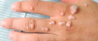
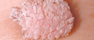
Common warts
Common warts can appear on any part of your body, including your scalp. However, they are more common on your hands and fingers. They can range in size from 1 millimeter to several centimeters. These warts are usually painless and often feel rough or scaly to the touch.
They may have a round appearance. Common warts can come in a variety of colors, including pink, beige, or brown. They may also be speckled with small black dots, which are tiny blood vessels that have become clotted (seminal warts).
Flat warts
Unlike regular warts, flat warts are smoother and smaller in size. They usually occur in multiples. Flat warts may be yellowish or brownish in color. They are usually found on the face and legs. In some cases, they may occur on the scalp.
Filiform warts
Filiform warts appear filamentous or bristle-like. They can grow quickly and often appear on the face around the mouth, eyes and nose. In more rare cases, they can be seen on the scalp.
Seborrheic keratosis
Seborrheic keratosis is a skin condition that can affect older people. It may start out as small bumps that eventually take on a warty appearance. These growths can appear anywhere on the body.
Common locations include the scalp, as well as the chest, back and neck. The color of seborrheic keratoses can vary and may include white, brown, or black. Although it may look like warts, seborrheic keratoses are not caused by HPV and do not spread to others.
Its exact cause is unknown.
Other options
There are other skin conditions that can also occur on the scalp and can potentially resemble warts. These include: Moles are collections of pigment-containing skin cells. They are round or oval in shape and can be flat or raised.
Although moles can appear anywhere, they often appear in areas exposed to the sun, such as the scalp, back, and arms. Actinic keratosis. Actinic keratoses occur on sun-damaged areas of the skin. It is often found on the scalp, face and hands.
Areas of actinic keratosis have a rough, scaly appearance and may be itchy. Nevus sebaceous. A sebaceous nevus is a rare birthmark that can appear on the scalp, face, or neck. It often becomes more noticeable during puberty and may take on a warty appearance.
Could it be cancer?
Skin cancer often occurs in areas that are frequently exposed to the sun, such as the scalp, face and back. Some types of skin cancer may have a warty appearance. There are three types of skin cancer: Basal cell carcinoma (BCC). SCD often appears as a skin- or pearl-colored bump.
It may also appear as a pink patch on the skin. Squamous cell carcinoma (SCC). RBS may appear as a scaly patch, hard red lump, or ulcer. It can also develop from an existing actinic keratosis. Melanoma. Melanoma can develop in a new or existing mole.
Melanomas usually:
How exactly does cryodestruction help get rid of warts? | Blog MC Sanas
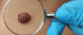
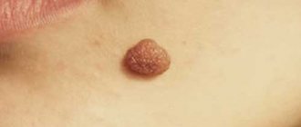
Warts are benign skin growths that appear due to HPV, the human papillomavirus, entering the body. They usually look like a nodule or papilla measuring 1-15 mm with a rough surface, but they can also be smooth. Color: flesh, gray or brown.
Infection occurs when the skin is damaged or through contact with objects contaminated with viruses.
Avoiding the formation of warts is quite simple. Prevention methods are basic and primarily related to personal hygiene:
- Wash your hands regularly
- Treat any skin damage with hydrogen peroxide, iodine or brilliant green
- Use individual slippers and towels when visiting the fitness club/swimming pool/sauna
- Support your immunity
- Avoid stressful situations
- Use contraception during sexual intercourse
Cryodestruction - what is it?
Cryodestruction is a method of removing pathological skin growths using low temperatures. Liquid nitrogen is used for cooling. It freezes the intracellular structure of benign formations. The virus dies, and the skin is painlessly cleansed. This method has been used in practice for a long time and has no side effects.
The cryodestruction procedure is very quick and painless. The doctor applies liquid nitrogen to the wart and presses it lightly for 5-30 seconds. Contact time depends on the size of the tumor. This is a comfortable procedure that does not require a rehabilitation period. Usually, patients do not even have time to understand that the treatment process has already ended, and they can return to their usual rhythm of life.
After the procedure, a small bubble forms at the site of the wart, which cannot be opened. It will burst on its own in 4-5 days. This area of skin must be treated with an antiseptic and sealed with a bactericidal plaster. Contact of water with skin is absolutely safe.
There are no restrictions from day one. A crust forms at the site of the bubble, which will disappear within 7-10 days. Complete restoration of the skin occurs within a period of up to six months.
As a result, no scars or scars remain on the body, and the skin tone is completely evened out.