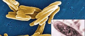
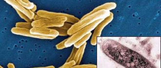
Tuberculosis is an infectious disease that affects the lungs (mainly) and other internal organs: mediastinum, stomach, muscles. The main reason for the development of tuberculosis is low immune status, in which mycobacterial infection (Koch bacillus) can cause an extensive infiltrative-inflammatory process.
Tuberculosis is transmitted by airborne droplets and is considered a potentially fatal disease. Until the 20th century, the disease was incurable, and it could be diagnosed (before the invention of X-rays) only at a late stage by external signs - coughing up blood, yellowing of the skin, visual deformation of bones and lymph nodes (formation of tubercles). Today, diagnosing tuberculosis in the early stages with identifying any changes in lung tissue and complications (including fibrocavernous process) is possible using CT.
Interesting fact: Italian Simonetta Vespucci, depicted in Sandro Botticelli's paintings "Portrait of a Young Woman", "Spring" and "Birth of Venus", died at the age of 23 from tuberculosis. In the painting “The Birth of Venus” you can see that the sitter’s left shoulder is drooping - this happens with tuberculosis in the shoulder girdle.
Today, tuberculosis can be successfully treated. In medicine, a separate specialty has emerged: phthisiology (translated from Greek as “treatment of consumption”). A phthisiatrician is a doctor who treats tuberculosis. In some countries there is a specialty “phthisiosurgery” (surgical treatment of tuberculosis). In Russia and the post-Soviet space, surgical treatment of tuberculosis is carried out by specialists of various specialties: thoracic surgeons (pulmonary tuberculosis), orthopedists (osseous and joint tuberculosis), gynecologists and urologists (tuberculosis of the genitourinary system). An important condition is the presence of a related specialization in the field of phthisiology. Tuberculosis is a serious disease that requires precise diagnosis and special treatment.
In this article we will talk about the causes, types and symptoms of tuberculosis, and consider the characteristic CT picture of the disease.
Primary tuberculosis in adults
This form of the disease accounts for up to 1% of all cases of registered respiratory tuberculosis in adults. The disease occurs at the first meeting of an infectious agent with the patient’s body, occurs with damage to the intrathoracic lymph nodes, which is why it has a protracted, chronic course. The infection can spread through the lymphatics and blood vessels into the lung tissue and pleura. The bronchus adjacent to the lymph node is often affected. When all the walls of the bronchus are affected, a fistula is formed.
It is necessary to clearly distinguish between primary and secondary forms of tuberculosis, since each of them has its own unique characteristics of course and treatment.
Contraindications to X-rays
Before talking about contraindications, it is worth noting the development of modern fluorography
, which allows patients to receive a minimal dose of radiation. Therefore, during the treatment of tuberculosis, especially miliary tuberculosis, an x-ray of the lungs is a mandatory and safe procedure: inaction is much more dangerous to life. X-rays of the lungs are absolutely contraindicated for children under 15 years of age and pregnant women. An obstacle to the study is the patient’s serious condition, in which he cannot stand for a long time or hold his breath.
Forms of pulmonary tuberculosis
Focal pulmonary tuberculosis
This form of the disease accounts for about 14% of all pulmonary tuberculosis. Complaints and bacilli excretion are extremely scanty. The disease occurs without pronounced symptoms of intoxication. There is no active release of mycobacteria into the external environment.
Due to the meager amount, it is sometimes not possible to determine the presence of MBT in sputum using laboratory methods, which means that the open form of tuberculosis is extremely rare. The radiograph reveals focal shadows up to 1 cm in diameter, most often in the upper parts of the lungs; decay cavities are determined extremely rarely.
Rice. 1. The photo shows the infiltrative form of the disease (plain radiograph). Focal shadows are visible in the upper lobe of the left lung in the subclavian region.
Infiltrative pulmonary tuberculosis
This form of tuberculosis accounts for 60 to 70% of all pulmonary forms of the disease. Clinical symptoms of the disease, the presence of decay cavities and the detection of MBT are always present and depend on the volume of the lesion. The smaller the volume of the lesion, the more often the closed form of tuberculosis is recorded and vice versa. On an x-ray, a focal shadow of 2–3 cm in diameter is determined in the lung tissue. In some cases, the volume of the lesion is much larger and can occupy an entire lobe of the lung.
Rice. 2. The photo shows the infiltrative form of the disease (plain radiograph). The pathological process involved the entire upper lobe of the right lung. A large decay cavity is visible.
Caseous pneumonia
Caseous pneumonia develops in individuals with significantly weakened immunity. The clinical symptoms of the disease are pronounced and correspond to the clinic of acute inflammation of the lung tissue. The x-ray shows intense shadows of caseous necrosis and multiple decay cavities. The process quickly spreads through the bronchi, affecting large areas of lung tissue. MBTs are released into the external environment in huge quantities. Caseous pneumonia is always an open form of tuberculosis. The patient's condition is serious. Caseous pneumonia often ends in death.
Rice. 3. The photo shows caseous pneumonia. The x-ray shows intense shadows of caseous necrosis and multiple decay cavities. The process spread through the bronchi and affected large areas of lung tissue.
Disseminated pulmonary tuberculosis
Disseminated forms of tuberculosis account for about 14% of all pulmonary forms. The infection spreads through the circulatory system, bronchi, and lymphatic tracts. The lungs are most often affected on both sides. There are numerous decay cavities. Clinical symptoms of the disease are significantly pronounced.
Acute tuberculous sepsis
The disease is the most severe form of disseminated tuberculosis. Develops in the complete absence of immunity in severe somatic patients (oncology, acute leukemia, AIDS, massive steroid therapy). MBT are freely located in body tissues. Due to the lack of immunity, immune cells do not accumulate around mycobacteria, which means that tuberculosis granulomas do not form. There are no changes on the chest x-ray. There is no release of mycobacteria into the environment. Patients die within 2–3 weeks. Often, due to diagnostic difficulties, the disease is registered only at autopsy.
Acute tuberculous sepsis is always a closed form of tuberculosis.
Acute miliary tuberculosis
In acute miliary tuberculosis, against the background of a sharp decrease in immunity, mycobacteria from the primary foci enter the bloodstream in large quantities. Internal organs are affected. In 100% the liver is affected, in 80% the lungs are affected. Slightly less common are the genitourinary system, the musculoskeletal system and the meninges. The disease is difficult to recognize. The clinical symptoms of the disease are pronounced. In half of the cases, the disease is registered as an open form of tuberculosis. The outcome of the disease has two options: either complete resorption of the lesions and recovery, or death.
Rice. 4. The photo shows an acute disseminated form (X-ray in direct projection). Multiple foci are visible throughout the lung fields
Subacute disseminated pulmonary tuberculosis
One of the most severe forms of the disease. Clinical symptoms of tuberculosis are pronounced. The disease occurs with damage to the larynx. Lung tissue is affected evenly throughout its entire volume. Often the foci and decay cavities are located symmetrically on both sides of the lungs. When cured, the lesions in the lungs become denser. The affected lung tissue is replaced by fibrosis. Often the disease takes on a protracted, chronic course. The closed form of tuberculosis is recorded extremely rarely. Almost all patients are extremely dangerous to others.
Rice. 5. The photo shows a chronic disseminated form of the disease, subacute course. X-ray in direct projection.
Rice. 6. The picture on the left shows extensive dissemination in the lungs. Multiple small foci (tubercles) are visible throughout the lung fields. On the right, the x-ray shows multiple foci against the background of fibrotic changes.
Fibrous-cavernous pulmonary tuberculosis
The most unfavorable form of tuberculosis. The disease always leads to profound disability of patients and is characterized by a large and constant release of MBT. The open form of tuberculosis is registered in almost all patients. The effectiveness of chemotherapy is extremely low. Up to 70% of all patients die from disease progression.
Rice. 7. The upper lobe of the right lung is destroyed. Huge cavities with irregularly shaped walls are visible. Severe fibrosis shifted the mediastinum to the affected side. The left lung is affected.
Different forms of tuberculosis are nothing more than different phases of one process.
Diet food
This disease is highly contagious, so it is important to protect others from infection. Is tuberculosis transmitted through skin? Yes, it is transmitted, but only if there are scratches, abrasions and cracks on the skin of a healthy person, so the patient needs to protect those around him and not come into contact with the skin of healthy people.
To recover faster, you need to change your diet from the first days, adding as much protein as possible to your diet. The menu should include: meat, fish, dairy products, milk, whole grain bread.
Be sure to follow the recommendations:
- food should be high in calories, but no overeating;
- be sure to consume fresh lard, butter and vegetable oil, but in reasonable quantities;
- eat as many fresh vegetables and fruits as possible;
- reduce the amount of consumption of baked goods, sugar, sweets;
- drink as many herbal teas, low-sugar compotes, and mineral water as possible.
You should completely avoid drinking alcoholic beverages; they, together with potent drugs, can lead to the death of the patient.
Bronchial tuberculosis
With pulmonary tuberculosis, the bronchi are always affected. As an independent disease, bronchial tuberculosis is rarely recorded. Damage to large bronchi is diagnosed by bronchoscopy. The disease always occurs with a cough due to the involvement of areas of cough centers in the process. Clinical symptoms are scant. The open form of tuberculosis is extremely rare.
When large bronchi are damaged, when the delivery of air to the lung tissue is blocked due to inflammation, atelectasis (collapse of lung tissue) may occur. Very quickly, nonspecific inflammation occurs in these areas or a large amount of MBT penetrates into the affected area, causing the development of caseous pneumonia. If bronchial patency is not restored within a week, then the airiness of the affected area of the lung tissue will not be restored. With a favorable development of the disease, the inflamed area is transformed into a fibrous cord, which, during its development, changes the location of the pulmonary structures and mediastinum, which is complicated by the development of respiratory failure.
Rice. 8. The photo shows ulcerative tuberculosis of the right main bronchus, which developed as a result of a breakthrough into the bronchus of caseous masses from the affected intrathoracic lymph nodes (the fistula opening is indicated by the arrow).
Rice. 9. The radiograph shows atelectasis of the upper lobe of the right lung. Due to damage to the bronchus, air delivery to the upper lobe is stopped. The lobe of the lung is significantly reduced in size and homogeneously darkened.
List of visualization research methods
to diagnose tuberculosis :
- fluorography;
- radiography;
- computed tomography (CT).
Tuberculosis is a disease that is often not accompanied by clinical symptoms at its onset. For this reason, pathological changes are often an incidental finding during chest radiography. Every doctor and x-ray technician should know what the lungs look like with tuberculosis on an x-ray so as not to miss this dangerous disease.
A separate article has been written about the diagnosis and differential diagnosis of tuberculosis.
Photo examples
For comparison, you can see x-rays of the chest before and after the disease:
Normal chest and lung x-ray. The lung fields are clean, the roots of the lungs are without any features.
Focal tuberculosis at the initial stage. A small focal change in the left lung, 1-1.5 cm in diameter, with an uneven and unclear contour, the structure is heterogeneous.
Residual calcifications after tuberculosis (52 years ago). Multiple rounded shadows in the right lung, strong intensity with a clear edge, homogeneous.
People with pulmonary tuberculosis may have a varied X-ray picture, which is associated with the variety of forms of the disease and stages of its course.
Tuberculous pleurisy
Pulmonary forms of tuberculosis are often complicated by pleurisy. Much less frequently, the disease exists as an independent nosological entity in the form of pleural tuberculosis.
The ways in which MBT is introduced into the pleura are very diverse. The clinical picture of pleurisy depends on the amount of accumulated fluid. The main complaints are pain and shortness of breath. If the patient does not immediately remove the pleural fluid, then on the 5th - 7th day pleural adhesions appear, which subsequently cause chest pain that accompanies the patient throughout his life. Due to limited mobility of the pleura, respiratory failure develops. Symptoms of intoxication are significantly expressed. When a nonspecific infection penetrates into the pleural cavity, pleural suppuration or tuberculous empyema develops. This is always a closed form of the disease, since there is no release of mycobacteria into the environment.
Rice. 10. The photo shows an accumulation of fluid in the left pleural cavity.
Fluorography
Fluorography is a method of x-ray examination that involves photographing an image from a fluorescent screen that appears on it after X-rays pass through the human body. There are several types of techniques:
- small frame;
- large-frame;
- electronic.
Small- and large-frame fluorography may not show tuberculosis if changes in the lungs are small in size or not clear enough. This is its main drawback, due to which it is impossible to make a diagnosis based on this study. Currently, only electron fluorography is relevant, in which the above disadvantages are eliminated.
It is used to detect tuberculosis during mass preventive studies. Modern fluorography reveals pulmonary tuberculosis thanks to the high quality of the resulting image, the ability to dynamically change the contrast and computer image editing. These features allow you to find the smallest changes.
Here are some examples of what tuberculosis looks like in a fluorography image:
Small-frame fluorogram.
Electronic fluorography. A large focal shadow in the region of the root of the left lung, heterogeneous in structure, has a clear contour.
Extrapulmonary forms of tuberculosis
In 90% of cases, extrapulmonary forms of the disease are combined with tuberculous damage to the respiratory system. The process most often affects the genitourinary system and peripheral lymph nodes.
Kidney tuberculosis
MBT penetrate through the bloodstream, much less often - through the lymphogenous route into the renal cortex (the zone of the vascular glomeruli), where the disease begins to develop. Often both kidneys are affected. First, the renal parenchyma (the actual kidney tissue) is affected. With tuberculous papillitis, the process involves the apexes of the papillae, which unite the renal tubules. Over time, the papillae die off. In their place, microdestructions (decay cavities) appear, filled with caseous masses. After the caseous masses are rejected, the decay cavities merge, forming either one cavity or a system of cavities separated from each other by thin connective tissue bridges. The open form of tuberculosis is registered when MBT is released into the environment with urine.
Rice. 11. The photo shows a macroscopic specimen of the kidneys. Arrows indicate areas of destruction.
Rice. 12. The photo shows a huge cavity in the upper pole of the left kidney.
Tuberculosis of peripheral lymph nodes
Tuberculosis most often affects the lymph nodes located in the neck. The axillary lymph nodes are affected somewhat less frequently. Generalized lymphadenitis is rare. As a result of specific inflammation, purulent melting of the lymph node tissue occurs. The lymph node increases in size, the skin over it turns red and thins. Caseous masses break out. A fistula forms and does not heal for a long time.
Rice. 13. The photo shows a lesion of the supraclavicular lymph node.
Spinal tuberculosis
The disease takes a long time, is difficult to treat, often ends in loss of ability to work and leads the patient to disability. Starting with damage to the body of one vertebra, the process gradually spreads to neighboring vertebrae. Their destruction leads to deformation of the spinal column and a number of serious complications. In 65% of patients who sought medical help for the first time, damage to 3 vertebrae was noted. Over the next few years, without treatment, the disease can affect up to 10 vertebrae.
Rice. 14. The photo shows an x-ray of the spine. Typical lumbar vertebral injury.
Rice. 15. The photo shows a pronounced curvature of the spinal column.
Lupus
Skin tuberculosis includes a whole group of skin diseases that vary in clinical features and morphology. The disease is diagnosed late in 80% of cases, always takes a long time and is difficult to treat. The skin is disfigured by scar tissue. Various forms of skin tuberculosis have their own symptoms and manifestations.
Rice. 16. The photo shows skin tuberculosis.
Treatment
There are several types of treatment for skin tuberculosis. Each of them is recommended for a specific form of the disease. Do not forget that self-medication can only worsen the patient’s condition. If you use traditional medicine methods, then only in combination with traditional ones and after agreement with the doctor.
The first and basic rule of treatment is to be under the close supervision of a specialist. General therapy can last from 9 months to one and a half years. Treatment can be divided into several stages:
- Up to 4 medications are prescribed and taken from two to four months.
- Without chemotherapy, treatment will not be effective.
- After a while, the number of drugs is reduced to two, and they are replaced with others. This system does not allow harmful bacteria to develop resistance to the active substances included in the drug.
- An important part of therapy is strengthening the immune system and improving the general condition of the body. To achieve this goal, it is recommended to take vitamin complexes, special diet foods high in protein and vitamin C. The doctor also recommends drinking water properly. To maintain normal water balance in the body.
Often the doctor prescribes electrophoresis to the patient using anti-tuberculosis drugs. This type of treatment gives maximum effect. In rare cases, the doctor may recommend surgery.
Main drugs for treatment:
- specific antibiotics “Rifampicin” + “Isoniazid”; in addition, “Pyrazinamidone” may be prescribed;
- at the second stage, the doctor prescribes medications of average effectiveness: “Ethambulon”, “PASK”, “Streptomycin”;
- ulcers are sprinkled with Isoniazid powder.
It is important to remember that oral medications must be taken daily, without missing a single dose. If you don’t take the medicine just once, it can result in mycobacterial resistance, in which case it will be more difficult to recover.
Patient's appearance
Patients with tuberculosis appear emaciated in appearance, as they lose a lot of weight. They also have pale skin and sharp facial features. Despite the pale face, a blush is always noticeable on the cheeks of such people.
Best materials of the month
- Coronaviruses: SARS-CoV-2 (COVID-19)
- Antibiotics for the prevention and treatment of COVID-19: how effective are they?
- The most common "office" diseases
- Does vodka kill coronavirus?
- How to stay alive on our roads?


