Etiology of the disease
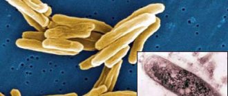
- The causative agent of skin tuberculosis is Mycobacterium tuberculosis, which was first discovered by R. Koch in 1882.
- The tuberculosis bacillus belongs to the family of radiant fungi, the genus of mycobacteria, which, in addition to it, includes the causative agents of leprosy, scleroma and more than 150 species of atypical mycobacteria.
- Mycobacteria reproduce by division and budding. This process lasts 24 hours.
- MBT exhibit significant stability in the external environment. They cannot be frozen out. They remain viable in boiling water for up to 15 minutes. They live in manure for up to 15 years, and up to 1 year in wastewater. When dried, the pathogen remains viable for 3 years.
- Mycobacteria are resistant to phagocytosis (macrophages cannot destroy the mycobacterium, although they begin to fight it - incomplete phagocytosis).
- There are human, bovine and intermediate types of MBT.
- The pathogen has the appearance of an elongated rod of a rather complex structure: a three-layer cell wall and intracellular membrane contain polysaccharides, lipoprotein complexes and proteins. Proteins are responsible for antigenic properties (tuberculin). Polysaccharides play a role in antibody detection. Lipid fractions help MBT resist acids and alkalis.
Rice. 2. Mycobacterium tuberculosis.
Ways of spreading skin tuberculosis
- Patients with tuberculosis spread the infection through sputum, urine, through fistulas and household items. The source of infection is also sick animals and food products contaminated with MBT from sick animals. During the formation of immunity, primary skin tuberculosis occurs (tuberculous chancre, lichenoid tuberculosis of the skin).
- There are a number of diseases that are associated with tuberculosis not directly, but indirectly. These are allergic vasculitis, resulting from allergic immune (“paraspecific”) inflammation in response to infection with mycobacteria (rosacea-like Lewandowski tuberculide).
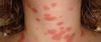
Rice. 3. The photo shows tuberculosis of the skin of the face and neck.
Introduction
Tuberculosis is an infection caused by mycobacteria, most commonly Mycobacterium tuberculosis.
This infection most often affects the lungs. When the skin is affected, it is called cutaneous tuberculosis. This is a rare disease. The history of cutaneous tuberculosis dates back to 1826, when it was first reported by Laennec. He reported lesions on his arm caused by penetration of the pathogen into the skin. However, the causative agent of the disease was not known until 1882, when M. tuberculosis was first discovered by Robert Koch. It was later isolated from a cutaneous lesion of an affected individual (1, 2, 3).
Classification
Although various classification systems exist, the most universally used classification of cutaneous tuberculosis variants is based on the following:
· Exogenous tuberculosis of the skin: Tuberculous chancre and warty tuberculosis of the skin
· Endogenous cutaneous tuberculosis: Contact or autoinoculation (scrofuloderma, official tuberculosis and some cases of lupus vulgaris); hematogenous dissemination (lupus vulgaris, tuberculous gumma and acute miliary tuberculosis)
· Tuberculides: Papulonecrotic tuberculosis; lichen scrofulous
· Cutaneous tuberculosis: Secondary vaccination with Bacillus Calmette–Guerin (BCG)
Etiology
Infection with M. tuberculosis causes cutaneous tuberculosis. This is a contagious disease that is transmitted from person to person. It can also be caused by Mycobacterium bovis and, less commonly, by the BCG vaccine (4, 5, 6).
Epidemiology
Unlike pulmonary tuberculosis, cutaneous tuberculosis is rare. Of all patients with extrapulmonary manifestations of tuberculosis, 1% to 2% suffer from cutaneous tuberculosis. It is more common in parts of the world where human immunodeficiency virus (HIV) infection is common or where people are immunocompromised for other reasons.
Pathophysiology
Skin invasion by M. tuberculosis leads to the development of the disease. This invasion can be exogenous or endogenous. Endogenous invasion is usually caused by the spread of pulmonary tuberculosis through hematogenous or lymphogenous dissemination. Exogenous invasion is caused by direct inoculation of bacteria. This leads to an immune cascade in skin cells that leads to the formation of granulomas characteristic of tuberculosis. They are present in various forms of clinical lesions.
Histopathology
Early lesions may present with superficial ulcerations, epidermal hyperplasia, and a dense dermal infiltrate consisting of neutrophils, lymphocytes, and plasma cells. Over time, cells involved in chronic inflammation and foci of granulomatous inflammation are replaced by neutrophils with variable caseous necrosis. Acid-fast bacilli are usually readily detected in early lesions but are rarely seen after granulomas have developed.
Clinical and physical examination
The patient's medical history may reveal various symptoms. The patient may have pulmonary tuberculosis along with cutaneous tuberculosis and may have a history of cough, sputum, night sweats, fever, weight loss, hemoptysis, chest pain and fatigue, among others, along with skin symptoms. In disseminated cutaneous tuberculosis, abdominal pain and diarrhea may indicate abdominal tuberculosis. Headache may indicate tuberculous meningitis. Direct inoculation of bacteria results in skin symptoms only.
A history may also reveal immunodeficiency due to underlying diseases such as AIDS, uncontrolled diabetes, malignancies, and end-stage renal disease (ESRD). Other causes of immunodeficiency include intravenous drug use and immunosuppressive therapy.
On examination, objective findings may include various types of skin lesions. There may be inflammatory papules, ulcers, nodules, pustules, verrucous plaques or any other type of lesion.
Survey
Assessing the symptoms of cutaneous tuberculosis requires a history and laboratory examination along with a complete examination. Testing may include tuberculosis skin test (TST) or Mantoux reaction, serum QuantiFERON-TB Gold (QFT-G), PCR, and skin biopsy (7, 8, 9).
The tuberculosis skin test (TST) involves inoculating 0.1 ml of a liquid containing a purified protein derivative (PPD) into the skin. The injection site is the skin of the outside of the wrist or the volar surface of the forearm. The test is interpreted after 48 to 72 hours. The diameter of the skin infiltrate is measured and recorded.
The QFT-G test is an alternative to the tuberculosis skin test (TST). This is a blood test that detects both active and latent tuberculosis using an interferon gamma release assay.
Skin biopsy is a reliable test for diagnosing cutaneous tuberculosis. The skin biopsy is assessed in two ways. First, sections are prepared and examined under a microscope to detect acid-fast bacilli. Next, the tissue is cultured at low temperature to detect the growth of M. tuberculosis.
Other tests that can help make a diagnosis include a chest x-ray and sputum examination. Sputum examination includes microscopy of acid-fast bacilli AfB and culture.
Treatment/Management
Treatment of cutaneous tuberculosis is similar to the treatment of systemic tuberculosis. It also includes treatment with multiple drugs. The most commonly used are isoniazid, rifampicin, pyrazinamide, ethambutol, or streptomycin. Treatment consists of 2 stages.
· Intensive phase, which includes a rapid decrease in the number of M. tuberculosis
Continuation phase, also called sterilization phase
The intensive therapy phase lasts about 8 weeks. After this phase, the patient is no longer contagious, but still requires additional treatment to achieve resolution of the infection. The continuation phase is designed to kill remaining bacteria and lasts 9 to 12 months. Treatment of tuberculosis requires the patient to strictly adhere to the treatment regimen.
Treatment results depend on the patient's immunity, general health, stage of the disease, type of skin lesions, patient compliance, duration of treatment and the presence of any side effects.
Differential diagnosis
Differential diagnosis of skin tuberculosis includes:
- Chronic ulcerative-vegetative pyoderma
- Blastomycosis (endemic deep mycosis - ed.)
- Chromomycosis (subcutaneous mycosis - ed.)
- Reactions to drugs
- Granulomatosis with polyangiitis (Wegener's granulomatosis)
- Granulomatous rosacea
- Syphilitic gumma
- Keratosis spinosa
- Papular eczema
- Coccidioidomycosis (endemic deep mycosis - ed.)
- Glossitis
- Atypical mycobacterial infection
- Chronic granulomatous disease
- Lupoid rosacea
- Nodular vasculitis (Weber-Christchen disease)
- Nodular chills
Many different diseases can be included in the list of differential diagnoses depending on the manifestations of cutaneous tuberculosis. It is difficult to diagnose this condition due to its similarity to many other skin lesions.
Forecast
The prognosis for cutaneous tuberculosis is favorable in patients who do not have a weakened immune system. However, even active treatment may not be effective in immunocompromised and multidrug-resistant patients.
Other aspects
Prevention
Prevention of cutaneous tuberculosis includes BCG vaccination, examination, isolation and treatment of the person who is the source of infection, as well as the use of sterilized instruments and other items. Since immunodeficiency is the main cause of skin tuberculosis, one should try to avoid it by managing diabetes and other diseases. In cases where drug therapy is the cause of immunodeficiency, preliminary testing and treatment of latent tuberculosis with antibiotics is indicated.
Compliance with recommendations
Following treatment recommendations is extremely important when it comes to treating tuberculosis. A patient who does not follow recommendations may develop drug resistance. Multiple treatment modalities may be required, and directly monitored treatment (DOT) is recommended in these patients.
Improving the performance of medical personnel
The diagnosis of cutaneous tuberculosis can be made by a specialist in the field. Since there are particularities in the diagnosis and treatment of skin diseases, the patient should be referred to a consultation with an infectious disease specialist, dermatologist or therapist. Treatment of cutaneous tuberculosis is similar to that of systemic tuberculosis. It also includes treatment with multiple drugs. The most commonly used are isoniazid, rifampicin, pyrazinamide, ethambutol, or streptomycin. Treatment consists of 2 stages.
· Intensive phase, which includes a rapid decrease in the number of M. tuberculosis
Continuation phase, also called sterilization phase
Treatment results depend on the patient's immunity, general health, stage of the disease, type of skin lesions, patient compliance, duration of treatment and the presence of any side effects.
Pathomorphology
With tuberculosis, a tubercle (tuberculum) appears around the embedded tuberculosis bacilli, the components of which are:
- inside the tubercle there are phenomena of caseous necrosis (damage) of tissues and Mycobacterium tuberculosis (a component inherent only in tuberculosis);
- surrounded by MBT cells specific for any granulomatous disease - lymphocytes, epithelioid cells and Pirogov-Langhans cells (cell proliferation);
- the outer layer (exudative component) is represented by cells macrophages, neutrophils, eosinophils (nonspecific component).
Rice. 4. Histological preparation of a tuberculous tubercle.
With tuberculous skin lesions, tubercular structures with a nonspecific inflammatory infiltrate are more common (there are few or no MBTs in the tubercle). Tuberculous granulomas are characteristic of tuberculous lupus. With allergic vasculitis that occurs in response to exposure to mycobacteria and their decay products, diffuse forms of skin tuberculosis occur. In this case, the vessels of the skin and subcutaneous tissue are affected.
Tuberculosis skin disease (clinical case)
The article presents a clinical case of skin tuberculosis in patients aged 5 and 17 years. Literature data on the incidence and characteristics of skin tuberculosis, medical history data, including anamnesis, and illustrative material are provided. The peculiarity of this case is the rarity of the clinical form of this disease.
Key words: skin tuberculosis, maintenance phase, intensive phase, ulcerative-necrotic form, papulo-necrotic form.
Relevance : Tuberculous skin disease is a rare form of tuberculosis, which includes lesions caused by Mycobacterium tuberculosis (MBT) that vary in clinical picture, pathomorphology and pathogenesis. [3]
The incidence of skin tuberculosis in Russia is annually detected on average 0.43 patients with skin tuberculosis per 100,000 population (in the areas of supervision of St. Petersburg Research Institute of Physics from 0–0.6), which is 5.6% of all extrapulmonary tuberculosis. Skin lesions are observed in 7% of patients with tuberculosis.
Skin tuberculosis most often affects women (about 70% of patients) aged 20 to 40 years. The risk group primarily includes people in contact with tuberculosis patients, butchers and farmers, diabetics, people in contact with sick animals, HIV-infected people, drug addicts, alcohol abusers, homeless people, prisoners, seasonal workers, immigrants. In children, as well as in adolescents, sclerofuloderma is more common. Infants often develop primary and miliary tuberculosis of the skin. The predominant development of tuberculous-allergic vasculitis occurs in middle-aged people in the presence of high specific reactivity of the vascular wall. [1,4]
Skin tuberculosis in the vast majority of cases (almost 70%) is secondary tuberculosis and develops only in individual patients with active tuberculosis of the lymph nodes, bones, and internal organs. MBT enter the skin by hematogenous, less often lymphogenous, route. But direct spread into the skin by contact is also possible. [4]
Classification of skin tuberculosis:
I. Chronically current primary tuberculosis:
1) collicative tuberculosis: primary scrofuloderma, secondary scrofuloderma, fungal tuberculosis;
2) diffuse forms of skin tuberculosis: papulonecrotic tuberculosis, indurated erythema, lichen scrofulus.
II. Secondary tuberculosis:
1) tuberculous lupus (flat, ulcerative, hypertrophic, papillomatous);
2) warty skin tuberculosis;
3) miliary ulcerative tuberculosis of the skin. [2,3]
Due to the rarity of occurrence of this disease and the difficulty of differential diagnosis, we present the described clinical cases of skin tuberculosis of the ulcerative-necrotic and papulo-necrotic form in children B. and K., aged 5 and 17 years, who were in the children's and adolescent department of the Regional State Clinical Hospital anti-tuberculosis dispensary" (OPTD) in Karaganda.
Clinical case no.1
Fig.1. Child B. Skin tuberculosis. Ulcerative-necrotic form
Complaints upon admission: skin rashes in the form of purulent crusts, redness and irritation of the skin around the rashes.
Medical history : Identified upon treatment. Referred to DO No. 3. Tuberculosis contact with mother, who has multidrug-resistant tuberculosis since July 2014. Bacillus Calmette-Gerin (BCG) scar 5 mm. Mantoux reaction with 2 TE from 01/09/2015. - 15 mm. Diaskin test dated 02/03/2015. — 8mm. The child suffered from tuberculosis for the first time in 2012. She was hospitalized in the children's department from May 28, 2012. until September 27, 2012 09/04/2012 surgery - lymphadenectomy of the cervical lymph nodes on both sides. Histological report dated 09/04/2012. No. 1778–85 - tuberculous lymphadenitis. Alternative productive tissue reaction. She received treatment in category 1. Outcome - treatment is completed. Complaints of skin rashes under the wings of the nose appeared in April 2014. I have repeatedly received treatment from a dermatologist and ENT doctor, without effect. After the result of a histological examination at the National Center for Tuberculosis Problems of the Republic of Kazakhstan (NCPT RK) dated 07/01/2015. No. 1821 (06.26.2015 - open biopsy of the facial skin) - tuberculous inflammation of the skin, ulcerative-necrotic form, the child was sent to the children's and adolescent department of the OPTD.
Central Medical Control Commission (CMCC) dated July 24, 2015. d/z Skin tuberculosis. Ulcerative-necrotic form. Active stage. Mycobacterium tuberculosis (MBT) - Type D (MBT relapse-). 4 category from tubes. exposure to MDR-TB. Intensive phase in category 4 180 doses from July 24, 2015. until January 19, 2016 CVCC dated January 20, 2016 - the same diagnosis. Considering the positive dynamics, she was transferred to the maintenance phase for 12 months. Support phase for category 4 from January 20, 2016.
Life history: 2nd child in the family. Vaccinated according to the calendar. He is not registered with specialists at the dispensary. The food is complete. 7 people live in a 3-room apartment. Allergy history: calm.
General condition on admission: Condition of moderate severity due to symptoms of intoxication. The state of health is disturbed. Consciousness is clear. The physique is asthenic. Reduced nutrition. Soft tissue turgor is reduced. The skin is pale pink, on the skin of the face the lesion is located in the nasal, subnasal, upper lip areas and buccal area on the left, the lesion is hyperemic, there is also an extensive ulcerative surface, ulcers up to 0.4 mm with an uneven bottom covered with purulent crusts, the edges of the ulcers are raised in the form roller In the nasal region there are areas minus tissue, in the buccal region there are areas plus tissue.
Visible mucous membranes are clean, physiological in color, and somewhat dry. Peripheral lymph nodes: submandibular, inguinal, axillary up to size 1, tightly elastic, mobile, painless, unfused. Nasal breathing is free. Zev is calm. On auscultation, breathing in the lungs is vesicular, there are no wheezes. Heart sounds are loud and rhythmic. The abdomen is soft and painless on palpation. The liver is not enlarged. The spleen is not enlarged. The effleurage symptom is negative on both sides. Urination is free and painless. The chair is formed, 1 time per day.
X-ray of the lungs: The lung fields are clean, the roots are structural. The diaphragm dome is smooth. The sinuses of the pleura are free. The boundaries of the heart are normal. Conclusion: there is no evidence of active tuberculosis in the lungs.
Anti-tuberculosis treatment: Intensive phase in category 4 from July 24, 2015. until 01/19/2016 - Levofloxacin -0.125 Capreomycin -0.4 Pro - 0.25 Cycloserine -0.75 Pyrazinamide -0.5 Ethambutol -1.2 Pas-1.0 Maintenance phase from 01/20/2016 until January 24, 2016 Levofloxacin -0.125 Pro - 0.25 Cycloserine -0.75 Ethambutol -1.2 Pas-1.0
Pathogenetic and symptomatic treatment : vitamins B1, B6, Hepadif, detoxification therapy.
Results and discussions: After the result of histological examination at the National Center for Traumatology and Therapy of the Republic of Kazakhstan dated July 1, 2015. No. 1821 (06/26/2015 – open biopsy of facial skin), the following clinical diagnosis was established: Skin tuberculosis. Ulcerative-necrotic form. Active stage. MBT-Type D (Relapse of MBT-). 4 category from tubes. exposure to MDR-TB.
Clinical case no.2
Fig.2. Teenager K. Skin tuberculosis. Papulo-necrotic form
Upon admission, complaints of rashes on the skin of the back.
History of the disease: The patient was identified upon presentation. No tuberculosis contact has been established. BCG scars 5 and 6 mm. Mantoux test with 2 TE dated June 22, 2015. - 14 mm. For the first time, rashes in the form of nodules without itching appeared on the skin of the back in 2014. I did not go to the doctor, I self-medicated. In June 2015, we consulted a dermatologist, who referred the patient to a phthisiatrician. On an outpatient basis to the patient on June 29, 2015. A biopsy of the skin and subcutaneous tissue was performed. A histological study was carried out at the OPTD in Karaganda - Conclusion No. 1384–85 dated 07/07/2015: The sent material contains skin tissue, under the epidermis there are areas of lymphoid infiltration with an admixture of epithelioid cells and Pirogov-Langhans cells. Specific inflammation of the skin of tuberculous etiology, exudative tissue reaction. 07/27/2015 glass preparations were transferred to Almaty to the NCPT of the Republic of Kazakhstan to confirm the histological conclusion. A conclusion was issued - tuberculosis of the skin and subcutaneous tissue. The girl was sent for inpatient treatment to the children's and adolescent department of the Regional Anti-TB Dispensary in Karaganda, where she had been receiving treatment since July 17, 2015. CVKK dated 07/18/2015 A clinical diagnosis was made - skin tuberculosis. Papulo-necrotic form. Active stage. MBT-I category. New case 1A group D-registration. Received first-line drugs: intensive phase from July 18, 2015. until September 15, 2015 60 doses - isoniazid 0.3, rifampicin 0.6, pyrazinamide 2.0, ethambutol 1.2. The maintenance phase ended on April 20, 2016. received isoniazid 0.3 and rafampicin 0.6.
Life history: 2nd child in the family. She grew up and developed according to her age. Not on D-registration. Social conditions are satisfactory. Parents and older brother undergo fluorography annually. Allergy history : according to the mother, without any peculiarities.
General condition upon admission: moderate severity due to symptoms of intoxication. Sleep is calm, appetite is reduced. On the skin of the back and partly on the chest, there are widespread rashes in the form of bluish-red nodules, in some places merging into continuous lesions with ulcerations and cicatricial changes. Itching is moderate. Visible mucous membranes are clean and pale. Peripheral lymph nodes: axillary up to 1-2 sizes, dense-elastic consistency, painless, mobile. Nasal breathing is free. Percussion-pulmonary tone. On auscultation, breathing in the lungs is vesicular, there are no wheezes. Heart sounds are loud and rhythmic. The abdomen is soft, painless on palpation, not swollen. The liver and spleen are not enlarged. Stool and urine output are normal.
X-ray of the lungs: The lung fields are clean, the roots are structural. The diaphragm dome is smooth. The sinuses of the pleura are free. The boundaries of the heart are normal. Conclusion: there is no evidence of active tuberculosis in the lungs.
Anti-tuberculosis treatment: Received first-line drugs: intensive phase from July 18, 2015. until September 15, 2015 60 doses - isoniazid 0.3, rifampicin 0.6, pyrazinamide 2.0, ethambutol 1.2.
Maintenance phase from September 16, 2015. until April 20, 2016, received isoniazid 0.3 and rafampicin 0.6 (received 153 doses). Pathogenetic and symptomatic treatment: Vitamins B1 and B6, detoxification therapy, enzymes.
Results and discussions: After the result of histological examination in the NCPT RK No. 1384–85 dated 07/07/2015. The following clinical diagnosis was established: Skin tuberculosis. Papulo-necrotic form. Resorption stage. MBT-I category. New case. 1A group D-accounting.
Both patients were discharged in satisfactory condition. With positive clinical dynamics, under the supervision of a local phthisiatrician.
Conclusions:
- Skin tuberculosis is a rare form of the disease, is one of the manifestations of tuberculosis infection and often accompanies tuberculosis of the lungs, lymph nodes and other organs, which served as a basis for discussing these clinical cases.
- The reason for the late detection of skin tuberculosis is the difficulty of diagnosis; vigilance is needed for doctors in the general medical network in order to diagnose this disease in a timely manner.
- The diagnosis is confirmed by histological and bacteriological studies of a biopsy specimen from the affected area of the skin.
- A thorough study of the epidemic history, infection, vaccination of children with BCG, and X-ray examination of children and adolescents will help reduce the late diagnosis of extrapulmonary skin tuberculosis.
Literature:
- Skin tuberculosis / M.K. Egizbaev // Bulletin of the South Kazakhstan Medical Academy. - 2006. - N2(28). — P.266.
- Journal "Emergency Medicine" 2012 Skin tuberculosis - Authors: Poteyko P. I., Krutko V. S., Khodosh E. M. - Kharkov Medical Academy of Postgraduate Education; Shevchenko O. S. – Kharkov National Medical University
- Phthisiology: Textbook. — 3rd ed., revised. And additional / Edited by: Perelman M.I., Koryakin V.A., Bogadelnikova I.V. - M.: OJSC Publishing House "Medicine", 2004. - 384p
- Guide to pulmonary and extrapulmonary tuberculosis. Edited by member. corr. RAMS, prof. Yu. N. Levasheva, prof. Yu. M. Repina. - St. Petersburg: ELBI-SPb. - 2008. - 544 p.
Primary cutaneous tuberculosis
This form of the disease is extremely rare. The disease develops during the development of primary tuberculosis. Children under 10 years of age are most often affected. Initially, a papule appears reddish-brownish in color. Next, an ulcer (tuberculosis chancre) appears in the center of the papule. Peripheral lymph nodes increase in size. Often ulcerate. Ulcers take a long time to heal. In their place, thin scars appear. With a weakened immune system, the disease returns again, disfiguring the body with scars and scars.
Medical myths. Tuberculosis
Tuberculosis is a bacterial infection. The lungs are primarily affected, but the infection can spread to other organs.
Symptoms usually include a chronic cough with expectoration of bloody sputum, fever, and night sweats. Loss of body weight is also typical, which is why a hundred years ago tuberculosis was more often called “consumption.”
The causative agent of the disease, Mycobacterium tuberculosis, spreads from person to person through airborne droplets - with particles of saliva that are released into the space when coughing or sneezing. When entering the body of a healthy person, the infection can develop very differently, depending on the immune response on the part of the host organism.
It is reliably known that people have been suffering from tuberculosis for thousands of years, but it still remains one of the most lethal infectious diseases on the globe. The World Health Organization (WHO) estimates that about four thousand people die from tuberculosis every day; from the same source it is known that in 2019 this disease claimed more than 1.4 million lives.
According to WHO materials, global incidence decreased by approximately 2% per year from 2015 to 2022. At the same time, the tendency for the pathogen to develop multidrug resistance is of great concern. In other words, there is still a long and difficult path to go before humanity can eradicate this dangerous infection, even though it is curable and preventable.
Today, every fourth person on the globe is infected with mycobacterium tuberculosis. However, only 5-15% of infected people develop clinically significant symptoms; the rest remain latent, asymptomatic carriers.
Among other things, the problem of “stigmatization” of patients is also relevant (stigmatization refers to the tendency towards social exclusion and rejection, literally “labeling” patients with a particular disease. – Note from Lakhta Clinic ). It is within our power to counteract this; It is important, in particular, to disseminate reliable scientific information and debunk some of the myths that have persisted for decades regarding tuberculosis.
Anyone with tuberculosis is contagious.
It is not true.
An infected person becomes contagious (infectious) only if he or she actively develops clinical symptoms. This means that latent, passive carriage of mycobacteria excludes the transmission of tuberculosis. In addition, airborne transmission is possible only when the mycobacterium is activated in the lungs or nasopharynx. If the process develops actively, but in other organs, such as the kidneys or spine, transmission of the infection is very unlikely.
Typically, patients with tuberculosis cease to be contagious 2-3 weeks after the start of adequate treatment.
Tuberculosis is inherited
Even in this era when all basic medical information is publicly available, many people continue to believe that tuberculosis is transmitted from parents to children. It is a myth.
This misconception arose a long time ago; it is due to the fact that people living in the same house often fell ill with the same diseases. But now we know that mycobacteria spreads in a different way, not related to heredity, and we can take all the necessary precautions even with constant close contact with the patient.
Perhaps someone follows medical news and read that the latest scientific data does not exclude the role of a genetic factor. But it manifests itself in a completely different way: the presence of a certain gene can affect whether the infection becomes active or remains latent. But inheritance of tuberculosis is impossible.
Tuberculosis has no cure
It's a lie. Tuberculosis is successfully treated and cured. The standard treatment for latent tuberculosis infection currently remains the antibiotic isoniazid, which is usually taken one tablet per day for 6-9 months.
Patients with an active infectious process are usually prescribed a combination of antibacterial drugs for a period of 6-12 months. In most cases, isoniazid is prescribed in combination with rifampin (rifampicin), pyrazinamide and ethambutol. According to the Centers for Disease Control and Prevention (CDC), 10 anti-TB drugs are approved and commercially available in the United States.
In cases where mycobacteria exhibit multidrug resistance, a combination of at least six antituberculosis drugs may be necessary to eradicate the infection.
Tuberculosis occurs only in the poorest countries
No, this is also a myth. Tuberculosis affects everyone everywhere, including in countries with a very high standard of living. It’s just that in some regions of the world tuberculosis is less common, in others it is more common.
According to WHO, in 2022, 44% of new TB cases were reported in Southeast Asia. Two-thirds of the new cases are from eight countries: India, Indonesia, China, the Philippines, Pakistan, Nigeria, Bangladesh and South Africa. However, in the same year of 2022, 8,916 new cases of tuberculosis were identified in the United States; the same figure for Europe in 2022 was 52,862 cases.
You can get tuberculosis through a handshake
Tuberculosis is a serious disease, and it is natural that people are concerned about possible routes of infection. This anxiety also gives rise to some myths. But in fact, as the CDC has repeatedly explained, tuberculosis is NOT transmitted through:
- handshake;
- sharing food or drinks;
- bed linen or toilet seat;
- kisses;
- a general toothbrush.
Tuberculosis CAN only spread when a person with an active process in the lungs or throat:
- coughs;
- sneezes;
- speaks;
- sings.
Tuberculosis is always fatal
Of course not. Tuberculosis is only fatal if left untreated. Modern medicine and qualified TB doctors successfully cure this disease.
The outstanding microbiologist, Nobel laureate Dr. Robert Koch announced on March 24, 1882 that he had identified the causative agent of tuberculosis. This discovery paved the way for the development of methods for diagnosing and treating a dangerous infection. Today, almost 140 years later, the epidemiological, clinical, and prognostic aspects of the problem of tuberculosis have changed dramatically. And yet, the struggle continues.
Based on materials from Medical News Today
Secondary tuberculosis of the skin
The disease is represented by a variety of localized and disseminated forms that appear in previously infected people. Tuberculous lupus accounts for up to 75% of all cases.
In the last 10 years, there has been a predominance of disseminated forms of cutaneous tuberculosis.
Ordinary or vulgar tuberculous lupus (Lupus vulgaris)
In the recent past, lupus was the most common form of cutaneous tuberculosis. Today, disseminated forms are more often recorded. MBT penetrate into the skin from regional lymph nodes through the lymphatic tract and hematogenously (with the bloodstream). The disease often occurs in childhood, lasts a long time, with periodic exacerbations, and spreads slowly.
Symptoms of the disease
The disease affects the skin of the nose, face, neck, red border of the lips, mucous membranes of the mouth and eyes. The skin of the extremities is rarely affected. Tuberculous tubercles merge and form lupomas. Their color is yellowish-rusty. Size – up to 0.75 mm. At first, the lupomas are located deep, and then they begin to protrude above the skin.
The shape of the lupoma is round, the consistency is soft, with significant pressure from the probe, the elements of the lupoma rupture, causing pain and bleeding. Lupomas often merge. Their surface is smooth and shiny. If you press a glass slide onto the affected area, the lupomas become the color of “apple jelly” (apple jelly symptom). With a favorable outcome, resorption begins in the center of the tubercles and replacement of the damage with thin skin in the form of tissue paper.
Rice. 5. Photo of lupoma.
Rice. 6. Symptom of “apple jelly” in tuberculous lupus
Rice. 7. Lupus vulgaris.
Rice. 8. Consequences of Lupus vulgaris.
Forms of tuberculous lupus
Tuberculous lupus of the mucous membranes
The most severe form of Lupus vulgaris. The disease affects the mucous membranes of the nose, eyes and mouth. Initially, red-yellow formations (plaques) appear on them. Their surface has a grainy appearance, reminiscent of fish eggs. Over time, the process affects the cartilage of the nose and ears. Next comes spontaneous rejection of damaged dead tissue, which ends in permanent facial disfigurement.
Rice. 9. The photo shows a lesion of the tongue due to tuberculous lupus.
Rice. 10. Damage to the oral mucosa.
Tumor form of tuberculous lupus
Tuberculous tubercles merge, forming a tumor-like formation up to 3 cm in diameter. As the process progresses, a breakdown of the underlying tissue appears, accompanied by damage to the cartilage and lymph nodes.
Rice. 11. The photo shows a tumor form of tuberculous lupus.
Flat form of tuberculous lupus
Tuberculosis foci merge, but the affected area does not protrude above the level of the skin. As the disease progresses, ulcers appear with uneven edges and a granular bottom.
Rice. 12. Flat shape of Lupus vulgaris.
Psoriatic form of tuberculous lupus
Tuberculosis foci merge. The surface of the damage is covered with many small scales.
Rice. 13. Psoriatic form of Lupus vulgaris.
Rice. 14. Psoriatic form of Lupus vulgaris.
Exfoliative (scaly) form of tuberculous lupus
Tuberculosis foci merge. The surface of the damage is covered with many large whitish scales, which adhere tightly to the underlying tissues. The appearance of the lesion resembles a butterfly.
Rice. 15. Exfoliative (scaly) form of Lupus vulgaris.
Rice. 16. Exfoliative (scaly) form of tuberculous lupus.
Rice. 17. Exfoliative (scaly) form of tuberculous lupus.
Sarcoid-like form of tuberculous lupus
Merging, tuberculosis foci form tumor-like formations of a reddish color - lupus carcinoma. The process is prone to malignancy.
Lupus
There are many clinical forms of cutaneous tuberculosis, which differ greatly from each other. This gives reason for some experts to argue that mycobacteria cause not just one pathology of different types, but several different diseases. Such a variety of forms further complicates the diagnosis of this condition.
Primary cutaneous tuberculosis
(tuberculosis chancre) - most often found in children, has an exogenous nature of infection. It is characterized by the development of reddish-brown papules on the skin approximately 3-5 weeks after the pathogen enters the tissue. Over time, the papule ulcerates, but remains just as painless. After the formation of a superficial ulcer, regional lymphadenitis occurs, which persists for about a month. Then comes the stage of resolution and healing, but complications in the form of disseminated skin tuberculosis or secondary infection are also possible.
Acute miliary tuberculosis of the skin
– this form is of an endogenous nature and occurs mainly in patients with severe forms of disseminated tuberculosis, in which the pathogen spreads throughout the body through the hematogenous route. Symptoms of such a lesion are the development of symmetrical rashes in the form of small reddish-brown nodules, vesicles, and papules on the torso and limbs.
Tuberculous lupus
– the most common form of skin tuberculosis. This type of lesion is characterized by the development of several tubercles (loupes) with a diameter of 2-3 millimeters, they are located in the dermis and covered (in the absence of ulcerations) by the epidermis. The favorite localization of formations is the face, skin of the ears, neck, but sometimes the body, as well as the mucous membranes of the mouth and nose, can be affected. Lupomas are painless, have a soft consistency, and when pressed with a glass slide, acquire a yellow color. Over time, they may resolve, leaving scar tissue behind, or cause long-lasting ulcers. This type of skin tuberculosis has a long-term recurrent course with exacerbations in the cold season; in some cases, lupomas can grow together, forming flat lesions.
Colliquatic cutaneous tuberculosis (scrofuloderma)
– this form belongs to the endogenous types of the disease and mainly affects children with tuberculous lesions of the lymph nodes. The skin in the projection of the affected nodes (mainly the neck, lower jaw, armpits) first acquires a bluish tint, then several ulcers with an abundance of necrotic tissue form on its surface. As a rule, the ulcers are painless, but after they heal they leave pronounced scars.
Warty skin tuberculosis
– is an exogenous form of pathology, mainly affecting butchers, pathologists, veterinarians and other persons who have contact with contaminated material. The skin of the arms, hands and fingers is almost always affected. The disease begins with the formation of small rashes surrounded by an inflammatory border. Soon after this, they begin to grow and merge with each other, forming one large lesion. The disease can last for many months, and as the lesion resolves, noticeable scars form.
Miliary ulcerative tuberculosis of the skin
– this is a rather rare form of pathology that occurs in severely weakened patients against the background of tuberculosis damage to the lungs, kidneys or intestines. In this case, skin infection occurs when biological fluids (urine, saliva, sputum) containing a large number of mycobacteria come into contact with it. For this reason, areas of the skin surrounding the mouth, anus, and genitals are mainly affected. First, small red tubercles develop, which quickly ulcerate, merge with each other and form continuous lesions.
Papulonecrotic tuberculosis of the skin
– a form of the disease characterized by a highly developed allergic component and vasculitis, mainly affecting adolescents. Its manifestations are the development of erythema on the extensor surfaces of the skin of the extremities and buttocks, after which a small ulcer appears in the center of the redness. After its resolution, a clear scar with smooth edges remains.
Indense skin tuberculosis
(erythema of Bazin) - just as in the previous case, allergic processes and hypodermal vasculitis play a significant role in the pathogenesis of the disease. It mainly affects young girls and develops on the skin of the legs. It looks like several deeply located nodes of dense consistency, with a diameter of 1-5 centimeters. Sometimes they can ulcerate; in addition, nodes often become the cause of regional lymphadenitis.
In phthisiology, there are also many more rare forms of skin tuberculosis - indurative, lichenoid and a number of others. But the vast majority of them are a complication of tuberculosis damage to internal organs.
Colliquatic cutaneous tuberculosis (scrofuloderma)
After tuberculous lupus, this form of skin tuberculosis is in second place in terms of frequency of the disease. It got its name from the Latin scrofulae - swollen lymph nodes of the neck and colliquescere - to melt. MBT enter the skin from infected lymph nodes through the lymphatic ducts. Cracks and ulcerations appear above the area of enlarged lymph nodes. The process is localized on the lateral parts of the neck, chest and collarbones. Young women are predominantly affected.
Symptoms of the disease
At the beginning of the disease, dense, painless nodules appear, which quickly increase in size, forming nodes tightly fused to the underlying tissues. Their sizes range from 3 to 5 cm. The skin over the lymph nodes acquires a bluish tint. Over time, the node becomes soft and opens. A cold abscess forms (suppuration without any manifestations of an inflammatory reaction). Pus with blood clots and pieces of destroyed (necrotic) tissue begins to be released from the fistula tracts. The ulcer has soft edges. The bottom of the ulcer is covered with a yellowish coating. Numerous granulations are visible. As the ulcer heals, irregularly shaped scars appear, which are connected by jumpers and bridges. The top of the scars is covered with papillary processes.
Rice. 18. Scrofuloderma.
Rice. 19. Scrofuloderma.
Rice. 20. Scrofuloderma.
Skin tuberculosis: types of disease
Infection by tuberculosis microorganisms has more than one classification. The most common “division” of the types of this disease is the division into focal tuberculosis, or otherwise localized, and widespread tuberculosis, otherwise disseminated. Let's look at each type separately and consider the subspecies. Localized tuberculosis can occur in the following forms:
- Lupus vulgaris (ordinary): one of the most common forms, which most often occurs at a very early age. Skin rashes in this form are localized mainly on the face; in advanced stages, “bumps” may appear in the mouth and on the nasal mucosa. Lupus vulgaris inflammations are located deep under the skin and are yellow-pink in color. However, such spots have clearly visible boundaries. As the disease progresses, these bumps become more and more noticeable, but do not emerge from under the skin.
- Scrofuloderma (colliquative tuberculosis): after lupus vulgaris, this form ranks 2nd in frequency of distribution. This form is characterized by the development of nodules reaching a diameter of 4-5 mm. The nodules are deep, but practically do not cause pain. You can notice them by the blue tint on the skin. As the disease progresses, the nodules can turn into an abscess, which, when opened, may leave an ulcer. As a rule, the reasons for the appearance of this form are called infection from the affected lymph nodes, or from other tissues. This form usually “chooses” the neck or limbs as its location.
- Ulcerative cutaneous tuberculosis: can develop due to infection from various affected tissues and organs. It is characterized by numerous bleeding ulcers on the skin that have yellowish nodules. Such nodules appear, most often, on the mucous membranes and skin surrounding it (genitals, mouth).
- Warty cutaneous tuberculosis: one of the most dangerous and complex types. It is characterized by the appearance of blue-red infiltrates (a kind of compaction, accumulation in a certain place of the body of cellular elements mixed with lymph or blood), which subsequently develop into warts. The causes are external stimuli. This form of skin tuberculosis is called “occupational”, since professions such as veterinarians, pathologists and others are most often at risk of infection. The location is the back of the hand and fingers.
Focal tuberculosis has been dealt with. What about the second type? Disseminated tuberculosis includes the following forms:
- Papulonecrotizing cutaneous tuberculosis: a form that most often appears at a young age. It is characterized by the development of blue-red seals, in the middle of which there are scabs, that is, a crust covering the seal. As a rule, such rashes are located on the buttocks or limbs. It is worth noting that after the scab falls off, a characteristic scar remains on the skin.
- Lichenoid cutaneous tuberculosis: or in other words, the well-known scrofulous lichen. Common rashes that affect the skin of the body.
As you can see, the difference between the two types of skin tuberculosis lies in the extent of the infection . Now we need to figure out the reasons for the appearance.
Ulcerative skin tuberculosis (Tuberculosis cutis ulcerosa)
The disease most often affects men who already had tuberculosis of internal organs. Tuberculosis bacilli enter the skin from the urine, feces or sputum of the patient himself, affecting areas of natural external openings: the skin around the anus, glans penis, nose, area around the mouth, mucous membrane of the tongue.
Symptoms of the disease
Merging, tuberculous tubercles form small yellowish nodules. Over time, the nodules suppurate and open with the formation of painful ulcers that make natural acts difficult.
The ulcers have soft, undermined edges. The color of the ulcers is pale red. The size of the ulcers is up to 1.5 cm, the bottom is granular. Javas often bleed. Sharply painful. As the disease progresses, tuberculous tubercles reappear at the bottom of the ulcers and around them. The tissues are destroyed, forming yellowish microabscesses, the so-called “Trel grains”, containing a large amount of MBT. The disease is difficult. Ulcers heal slowly. In their place, thin scars form, located below the skin level (atrophic scars).
Rice. 21. Ulcerative tuberculosis.
Warty skin tuberculosis (Tuberculosis cutis verrucosa)
Warty skin tuberculosis occurs among veterinarians and slaughterhouse workers who come into contact with the corpses of animals with tuberculosis. This form of the disease is rare. The skin of the back of the hand or feet is affected. The skin of patients is affected by tuberculosis from constant contact with infected sputum.
Symptoms of the disease
First, tubercles appear, the skin above which becomes bluish in color. Gradually, the tubercles merge with each other, forming dense infiltrates of a bluish color. Over time, they become covered with warty growths, dense scales and cracks. The lesion is surrounded by a bluish-red shaft, followed by a zone of shiny skin. During the healing process, the horny masses are rejected, and scars appear at the site of infiltration. The course of the disease is long-term.
Papulonecrotic tuberculosis of the skin (tuberculosis cutis papulo-nectrotica)
Papulonecrotic tuberculosis of the skin is more often recorded in young people. The disease is manifested by the appearance of papules, the size of which does not exceed 3 cm in diameter. The skin of the buttocks, abdomen, extensor surfaces of the limbs and chest is affected. The skin over the papules becomes pinkish-bluish in color. Over time, the papules become ulcerated. At the site of the ulcers, a grayish-white crust appears, which is replaced by a whitish scar.
Rice. 25. Tuberculosis cutis papulo-nectroticа. Multiple bluish-purple infiltrates and nodes on the legs.
Rice. 26. Papulonecrotic tuberculosis of the skin of the legs. Multiple bluish-purple infiltrates and nodes on the legs.
Rosacea-like Lewandowski tuberculosis
This form of the disease occurs as an allergic vasculitis. Erythema (redness) and telangiectasias (numerous dilated small vessels) appear on the skin of the face. Gradually the skin acquires a bluish-reddish tint. Rashes (tuberculids) are dense formations up to half a centimeter in diameter, without necrotic elements. During healing, atrophic scars form at the site of tuberculides.
Rice. 27. The photo shows Lewandowski’s rosacea-like tuberculosis.
Skin tuberculosis: symptoms
Skin tuberculosis, according to the degree of its development, can manifest itself in only two forms: weeping and dry. Consequently, symptoms are divided according to the same criterion.
Dry form
- peculiar blisters appear, after rupture of which the skin can recover;
- After such a rupture, scars appear on the skin, characterized by dry, thin and white skin. The skin is very thin and cannot be softened by any creams. Very often, a new focus of the disease may appear on top of such skin.
Wetting form
- rashes appear that appear as “tubercles”, unlike pimples, have a color closer to brown and do not have a purulent core in their “structure”. Over time, such “tubercles” become covered with scales and crusts, and subsequently grow together into common nodes. They are resistant to antibiotics;
- The skin with the development of “tubercles” is more and more prone to rejection. The “tubercles” are filled with blood and lymph. This mixture, and most often the lymph, is the same infectious substance as the sputum discharge from pulmonary tuberculosis;
- if the “tubercle” is torn off late, an ulcer may remain on the skin.
If skin tuberculosis is detected in a timely manner, treatment will consist of only a few courses of certain medications . It is very important not to neglect this disease, since most often it is skin tuberculosis that affects the upper part of the body and damage to not only the upper tissues, but also the internal ones, namely the brain tissues, will be very dangerous.
Lichenoid tuberculosis of the skin (Tuberculosis cutis lichenoides seu lichen scrofulosorum)
Lichenoid tuberculosis of the skin often develops in weakened children, less often in adults with tuberculosis. The disease is manifested by the appearance of tubercles, which are covered with light gray scales. The tubercles are located symmetrically, most often on the skin of the abdomen and limbs. After healing, pigmentation or small scars remain at the site of the rash.
Rice. 28. The photo shows lichenoid tuberculosis of the skin.
Indurative tuberculosis of the skin (indurated erythema)
This form of the disease manifests itself in 2 varieties - Bazin's erythema nodosum and Hutchinson's ulcerative erythema. The disease often affects patients with tuberculosis of internal organs.
Erythema nodosum of Bazin
The disease is more often registered in women aged 16 to 40 years. Dense nodes or flat, extensive infiltrates that appear on the skin of the legs, thighs, buttocks and upper extremities are bluish-red in color. The size of the seals is 3 – 8 cm. Seals and infiltrates are often located symmetrically, in the deep layers of the skin and subcutaneous fatty tissue. With regression of tuberculous elements, ring-shaped atrophic areas and pigmentation remain.
Hutchinson's ulcerative erythema
Sometimes the lesions merge, forming an extensive lesion with ulcerations in the center and dirty gray granulations. This form is named after the English dermatologist who first described it - Hutchinson's ulcerative erythema. Without treatment it lasts for a long time, sometimes even years. After healing, sunken pigmented scars remain.
Rice. 29. The photo shows indurative tuberculosis of the skin (indurated erythema of Bazin).
Rice. 30. Ulcerative erythema of Hutchinson.
Skin tuberculosis: causes and development
Despite all the types, it has been revealed that skin tuberculosis is one of the rarest forms of the disease . It is believed that the most “favorable” organs for the development of Koch’s bacillus are the lungs, while the skin has a strong protective layer of the epidermis.
The cause of the development of skin tuberculosis is the occurrence of infection against the background of damage to internal organs. In this case, the infection “reaches” the skin through blood and lymph . The disease can also be contracted through external influences. For example, in case of injury to the skin and in direct interaction with the source of infection (a contaminated object, contaminated substances or with a person who already has tuberculosis).
I would like to note that the skin actually has a persistent protective property and the development of cutaneous tuberculosis infection requires the presence of additional factors . These factors include the following:
- unhealthy diet, as well as its uniformity;
- unfavorable environment: poor environmental condition in the area of residence, non-compliance with sanitary standards at home, in living quarters;
- work in hazardous industries: the chemical industry is especially worth highlighting;
- lack of sun: a small amount of sunlight in a person’s life has a negative effect on the skin and the general condition of the body, as there is a “lack of vitamin D”;
- low level of everyday culture in residential premises;
- antisocial lifestyle: neglect of sanitary standards, neglect of contraception.
It can be noted that all factors are certain daily problems associated with limitations in funds, time, fatigue or the person’s reluctance to comply with something. All these aspects, at a certain level of life in a country, can simply disappear or, if implemented properly, turn into a habit of life for every person.
Diagnosis of skin tuberculosis
Rice. 31. View of Mycobacterium tuberculosis in the light of a fluorescent microscope.
The diagnostic process consists of the following components:
- Bacteriological diagnosis (detection of MBT in discharge from ulcers and lymph node punctures).
- Diagnostic biopsy followed by histological examination, which allows to identify the morphological components of the tuberculous tubercle.
- Detection of tuberculosis of internal organs.
- Detection of scars on the skin as a result of a previous tuberculosis lesion.
- Tuberculin diagnostics.
- Trial treatment.
Diagnostic biopsy together with bacteriological examination is the most significant method in diagnosing tuberculosis.
Dermatologists are prescribed to detect skin tuberculosis. Knowledge of risk groups, which include patients in this category, early symptoms of the disease and diagnostic methods helps doctors to detect the disease in a timely manner.
Identification of the pathogen in the early stages of the disease will allow the patient to be successfully cured.
Tuberculosis in children
Early period of primary tuberculosis infection
The initial phase of interaction between the pathogen and the macroorganism lasts from 6 to 12 months from the moment the child is infected with Mycobacterium tuberculosis. During this period, there is an asymptomatic stage (about 6-8 weeks) and a turn of tuberculin tests - the transition of the Mantoux reaction from negative to positive for the first time. A child with severe tuberculin reactions should be referred to a phthisiatrician and monitored by a specialist for a year. In the future, such children remain infected with Mycobacterium tuberculosis or, under unfavorable conditions, develop one or another local form of tuberculosis.
Tuberculosis intoxication in children and adolescents
It is an intermediate form between primary infection and the development of a local tuberculosis process, determined by x-ray and other methods. The clinical course of this form of tuberculosis in children is characterized by nonspecific manifestations: malaise, irritability, loss of appetite, headache, tachycardia, dyspepsia, arrest or weight loss, and a tendency to intercurrent diseases (ARVI, bronchitis).
A long-term causeless low-grade fever is typical, against the background of which temperature candles appear up to 38-39 ° C; There is increased sweating, especially during sleep. Tuberculosis intoxication is accompanied by a specific reaction of the lymph nodes - their multiple enlargement (micropolyadenia).
If signs of tuberculosis intoxication in children persist for more than 1 year, the condition is regarded as chronic.
Primary tuberculosis complex
This form of tuberculosis in children is characterized by a triad of symptoms: the development of a specific inflammatory reaction at the site of infection, lymphangitis and damage to regional lymph nodes. It develops when a combination of massiveness and high virulence of tuberculosis infection with a decrease in the immunobiological properties of the body. The primary tuberculosis complex can be localized in the lung tissue (95%), intestines, and less commonly in the skin, tonsils, nasal mucosa, and middle ear.
The disease can begin acutely or subacutely; masquerade as influenza, acute pneumonia, pleurisy, or be asymptomatic. Clinical manifestations include intoxication syndrome, low-grade fever, cough, shortness of breath. Changes in the primary lesion go through an infiltrative phase, a phase of resorption, compaction and calcification (formation of a Gohn's lesion).
Tuberculous bronchoadenitis
Bronchoadenitis or tuberculosis of the intrathoracic lymph nodes in children occurs with specific changes in the lymph nodes of the root of the lung and mediastinum. The frequency of this clinical form of tuberculosis in children reaches 75-80%.
In addition to low-grade fever and symptoms of intoxication, the child develops pain between the shoulder blades, whooping cough or bitonic cough, expiratory stridor, caused by compression of the enlarged intrathoracic lymph nodes of the trachea and bronchi. Upon examination, attention is drawn to the expansion of the subcutaneous venous network in the upper chest and back.
Complications of tuberculous bronchoadenitis in children may include endobronchitis, atelectasis or pulmonary emphysema. This clinical variant of tuberculosis in children requires differentiation from Beck's sarcoidosis, lymphogranulomatosis, lymphosarcoma, and nonspecific inflammatory adenopathy.