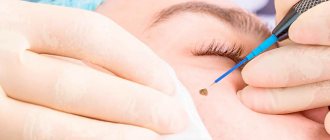
Before starting the procedure, the face is treated with antiseptic wipes. This is necessary to prevent the risk of infection getting into the wounds and the development of inflammation. To protect the eyes from the laser beam, the patient wears special glasses.
After completing the preparatory measures, the skin is exposed to a laser. The affected nodule heats up and its contents begin to evaporate. Particular attention is paid to removing millet from the eyelid. No technique other than laser can be used here. You must first consult an ophthalmologist.
How to care for your skin after the procedure
Immediately after the procedure, the patient will be able to see a mark on the skin that is similar to the healing site of a pimple. This area is covered with a dark thin crust on top; it cannot be torn off. It should fall off naturally. If it is accidentally picked off, an infection may develop. This will lead to rehabilitation lasting more than one month.
The crust will fall off on its own after 7 days. There will be no visible traces in its place. After some time, the skin will be completely renewed and the tone will even out. After 2-3 months it will be difficult to find the place where the milium was.
The final healing time for wounds after the procedure is 10 days. During recovery, you should avoid going to the pool, sauna, and swimming in open water. During the rehabilitation process, you cannot peel or expose the skin to any mechanical stress. Exposure to sunlight is contraindicated.
To make the recovery period go faster, the cosmetologist prescribes a set of ointments.
Characteristics of milia
Milia form gradually and painlessly on intact skin. Whiteheads can occur as individual elements, but more often appear in groups with typical localization on the face - in the paraorbital area, on the eyelids, temples and upper cheekbones, on the nose and cheeks; very rarely on other parts of the body - torso, genitals. Milia have the appearance of small rounded dense nodules of a white-yellowish hue, 0.5-3 mm in size with clear boundaries, protruding above the surface of the skin and resembling millet grains (popular name - “millet”). Milium is characterized by an uninflamed head, since the whitehead does not have a natural outlet and contact with the environment. Milia do not change in size for a long time. When microorganisms enter the milium, inflammation may develop with the formation of an abscess. Milia (milia) can often be found on the wings of the nose in recently born babies; they are considered borderline conditions in newborns and soon disappear spontaneously without any treatment.
Milia are recognized based on an examination of the skin by a dermatologist-cosmetologist based on characteristic external signs.
Milium must be differentiated from fibroma, fibrofolliculoma, trichodiscoma; as well as syringoma and xanthelasma of the eyelids. Clinical picture
From the point of view of dermatology, milium is a retention or follicular cyst, which is formed as a result of blockage of a pore by horny masses. There are two types of whiteheads:
- Primary milia, formed spontaneously.
- Secondary milia, called pseudomilium of Balser, form in scars at the site of traumatic or inflammatory skin lesions.
The wall of a whitehead is represented by the epidermis with all layers preserved; the contents of the cyst are dense horny masses mixed with fat.
Milia look like dense, yellowish or white nodules protruding above the surface of the skin. The size of whiteheads ranges from 0.5 to 3 mm. Most often, milia appear on the face, especially around the eyes, but sometimes whiteheads can be seen on other parts of the body, as well as on the genitals. Whiteheads can exist for years without changing. They do not cause any discomfort to a person, but create a cosmetic defect. Sometimes milia spontaneously open and disappear without a trace. When infected, whiteheads can become inflamed and fester. Establishing diagnosis
Diagnosis of milia is not difficult, since whiteheads have specific external signs. By making an incision into the wall of the cyst, the dense core can be easily removed. Histological examination of the contents of the milium reveals layered horny mass and fat impurities. It can be distinguished between a dense core and a loose outer mass. Milia should be distinguished from microretention cysts of the sebaceous gland, which are found in patients with seborrhea.
Contraindications to the procedure
Laser technology is considered a universal method of combating millet grass. But it has contraindications. Laser therapy should not be used:
- if a person develops an inflammatory process;
- for oncology (the procedure is not performed even if the patient has a benign tumor);
- during pregnancy;
- during exacerbation of skin diseases;
- for hypertension and endocrine disorders.
Laser treatment replaces mechanical cleaning, as it is characterized by high efficiency with minimal risk of skin injury.
How to get rid of milia?
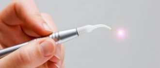
Today, the “gold standard” is CO-2 laser removal of milia
Benefits of laser milia removal:
- no direct contact with skin
- bloodless aseptic method
- high-precision impact
- does not affect healthy tissue
- effectiveness of the method
- excellent cosmetic result
At the Gradient clinic, milia removal is performed by experienced dermato-oncologist surgeons. The doctor will carefully examine the formations, make sure of their origin, carefully remove them and prescribe subsequent skin care at the sites of removal.
Structure of milia
Due to the fact that ordinary people do not fully understand the structural component of a white pimple, they try to get rid of the hated defect on their own. But this can only aggravate the situation, even if at home you can still squeeze out the contents of an unusual wen.
Some dermatologists say that the histological structure of the formation is based on blockage of the excretory duct of the sebaceous gland. This causes subcutaneous fat to accumulate and the gland to expand. This statement is only partly true, more suitable to the classic clinical picture characteristic of closed-type Camendons.
The latter differ from milia in their location, choosing for their growth the cheeks, as well as the chest, shoulders and all those areas that become a field for acne activity. People try to squeeze them out on their own, despite doctors’ warnings, especially in situations where there is no inflammation. Then, through the thinnest part of the skin, a dense fat clot breaks through, which has been accumulating for a long time, or an immediately hardened piece of sebum.
But with traditional white pimples, things happen a little differently. During histological examination, it became clear that formations of this type are located separately from the hair follicles. A retention cyst is an autonomous cavity, which does not depend at all on the location of the sebaceous or sweat glands, or their nearest ducts.
Instead, a whitehead consists of a cystic wall that is lined from the inside with epithelial cells. Inside, the cystic capsule hides layered, bulbous keratin-type accumulations. In practice, this means that inside the formation hides keratinized epithelial cells, and not the secretion of the sebaceous gland that has accumulated over a long period of time. Moreover, histology does not exclude a small fat content in the total mass.
Against this background, we can conclude that milium is an epidermal vesicle that is located in the superficial layer of the skin, containing keratitis inside. Scientists have never found the exact and only reason for their occurrence.
Among professional dermatologists, it is generally accepted that the catalyst for their occurrence is hyperkeratosis.
It involves accelerated cell division, which is localized in the upper layer of the skin. At the same time, the exfoliation process slows down. Because of such a contradictory process, it becomes clear why getting rid of “grains” with acne and acne remedies will not work.
No medicines, compresses or other traditional medicine recipes that provide external influence on the problem area guarantee a long-lasting and completely safe effect.
It is worth giving up even attempts to perform mechanical extrusion at home, even if you apply great force.
At best, you can only achieve the isolation of the specific contents of the cavity, which is characterized by:
- gelatinous texture;
- soft consistency;
- white.
But this will get rid of the defect only for a short period of time.
And in the worst case scenario, the victim will face an extensive inflammatory process. It occurs due to the contact of pathogenic microorganisms on the open wound surface. Pathogenic bacteria can also easily enter the body if there is insufficient care after mechanical intervention is completed.
How to prepare for removal
Although sometimes milia can be removed from the face without assistance, most often you need to take action to get rid of them. First, it is advisable to identify the cause of facial skin problems. Eliminating it will be an important step for the procedure to be effective. When planning to remove milia surgically, you need to start preparing about a month in advance. To do this you need:
- refuse to visit baths and saunas;
- Avoid damaging the skin with peeling or other methods.
During this time, you must visit a dermatologist or cosmetologist. Having assessed the condition of the skin, the number, location, and depth of nodules, they will determine the course of treatment. If there are few formations on the face, one session will be enough. A large number of milia must be eliminated in several stages. The location of the nodules will largely determine the method of their removal. These conditions will determine how much the service will cost.
Medical classification
In medical terminology, special skin formations are called milia. Externally, they look like a small wen that has already become keratinized, containing sebum inside its cavity. Their main problem is the complete absence of a natural hole that would allow them to get rid of the defect mechanically without the help of a professional.
Located above the epidermis, their upper part is completely clogged, which complicates their subsequent neutralization, even with specialized equipment. White pimples are formed as a natural response of the body to the inability to get rid of excess sebum with horny residues.
The patient feels as if a tubercle has appeared under a thin layer of skin, with a white or slightly yellowish head. Initially, milia can behave quite normally, without causing any significant discomfort to the victim, except for aesthetic unattractiveness. But this will continue only until the inflammatory process starts. It occurs when the ducts are no longer tightly clogged.
This outcome can be promoted by the victim’s desire to squeeze out whiteheads on their own, which often leads to infection. First, pronounced redness appears on the eyelids or in any other place with the former localization of the formation. Then it quickly degenerates into suppuration. In the most dangerous scenario, the patient may experience damage to the cells surrounding the problem area, triggering irreversible destructive processes.
The characteristic features that are inherent in classic milia are:
- the average size is a diameter of 2 to 3 mm;
- single or group arrangement;
- no pain or burning when touched.
Despite the fact that most patients seek help in eliminating such unsightly bumps, noticing their appearance under the eyes, this is not the only place where they usually grow. The risk group includes temples, cheeks, and nasal wings.
Dermatologists have a separate sorting according to the format of the manifestation of milia, which is also popularly called simply millet because of its resemblance to millet scattered on a flat surface. The primary type of formation makes itself felt spontaneously. It arises as a result of some strictly defined negative environmental factors or internal failures of the body.
The secondary version, which professionals call clinical, does not depend on skincare products or ultraviolet irradiation at all. It appears only in places of scars that occur after even the slightest injury to the skin. Sometimes the secondary course of the disease is a consequence of the attenuation of the inflammatory process.
Primary sources of problems
Before going to the beauty salon, potential patients are asked to understand the reasons for the prosyanka. There are many factors for its occurrence, so several aspects often provoke a surge in a peculiar release of sebum.
Among the most common sources of the disease is excessive use of decorative cosmetics.
Moreover, it is not even so important how high quality the product turned out to be. It’s just that when the sebaceous glands are clogged with a thick layer of powder, foundation or any other tinting agent, the pores of the skin stop working normally.
Also, the reasons for unsightly covering the face with small wen are:
- incorrect functioning of some internal organs;
- improper diet;
- excessive use of thermal procedures.
The first point of the program affects malfunctions not only of the gastrointestinal tract, but also of the pancreas and liver. Either one body or all at once may cease to perform its duties. In this situation, trying to excise the tubercles with a laser will be a futile exercise. You will have to start treatment from the inside to remove the barrier to glowing, beautiful skin.
Abuse of junk food becomes a direct path to obesity. The deposition of excess sebum in milia works on an identical principle. When the daily menu contains an abundance of excessively salty, sweet, fried or smoked food, this will quickly affect a person’s appearance.
Tanning is no less dangerous. Moreover, we are talking not only about visiting a solarium, but also about a long stay on the beach during the hottest hours.
In adolescents and middle-aged women, hormonal changes become the primary source of difficulties. An equally common cause is toxic poisoning of the body from within, which occurs with prolonged methodical accumulation of toxins. Bad habits such as drinking alcoholic beverages and smoking also add to the risk of being affected by fat “grains”. People who suffer from increased dryness of the skin of the face should be especially careful. But the most important pillar for the development of lesions is insufficient skin hygiene. This applies to both the inability to choose normal skincare products and the habit of not washing off makeup before bed.
Why should milia be classified as a separate group?
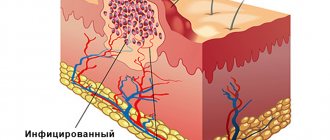
Milia are intradermal cysts filled with keratin masses.
Keratin is a protein that is part of the upper layers of skin, hair and nail plates.
If you think that you have milia, then specialists from the ATLANTIC Laser Surgery Center will help you confirm or refute your assumption.
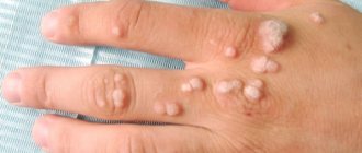
Rice. 2. Multiple milia on the skin
Milia can affect anyone, any age, any gender, and any skin type. It should be noted that children are more prone to developing milia, but removal is usually not necessary.
Ways to remove milia
| Removal method | Impact depth control | Application of anesthesia | Rehabilitation period | Probability of relapse | Consequences (scars, scars) |
| Laser removal | Yes | Painless | 1-2 days | low | No |
| Curettage | No | No | 1 Week | high | scarring |
| Electrocoagulation | Yes | No | 1-2 days | low | No |