Angioma is a neoplasm that is classified by doctors as a benign tumor. This pathology is a collection of blood and lymphatic vessels. Outwardly, most neoplasms resemble moles, and therefore are popularly called “red moles” or “port-wine stains.” In most cases, angiomas occur during fetal development, but sometimes they appear in older people or can be an external manifestation of a number of diseases.
What is angioma
Venous angiomas are localized in organs and tissues and can be multiple or single.
They are located in most cases on the upper half of the body. Their morphological basis is highly dilated lymphatic/blood vessels. The shape and size of pathological areas can vary, color - from colorless to red-blue. The lesions tend to progress rapidly. There are angiomas of the brain, liver, external genitalia, and bones. Angiomas are classified according to their structure:
- Simple (hypertrophic, capillary). Small venous and arterial vessels grow. If you study a photo of such an angioma, it will become clear that it is a bright red or bluish-purple spot. Its sizes can be different, even gigantic. If you press on the lesion, it will immediately turn pale due to the outflow of blood. Capillary angiomas of the skin extremely rarely transform into malignant formations.
- Branched (racellose). They are represented by plexuses of tortuous dilated vascular trunks. They are characterized by the presence of constant pulsation. The limbs and face are most often affected. Traumatization of venous branched angiomas is fraught with severe bleeding, which cannot always be stopped on its own.
- Cavernous (cavernous). They are formed by wide spongy cavities that are filled with blood. Cavernous angioma looks like a purple-bluish bumpy spot. Most often located under the skin. It feels hotter than the surrounding tissue. When stressed, it increases in size. A complex form is cavernous angioma of the brain, the treatment of which is very long and involves restoration of impaired blood flow.
- Combined. Combine subcutaneous and superficial location. Formed by overgrown vessels and other tissues (depending on location on the body).
The shape of angiomas is:
- stellate (point angiomatous formations, from which dilated small blood vessels extend in all directions);
- flat (vascular spots of pink-blue color located in the upper layers of the skin);
- nodular (seals protruding above the surface of the skin);
- serpiginous (rashes on the skin in the form of small burgundy nodules).
Types of neoplasms
There are several classifications of angiomas. They are distinguished by shape, cause of appearance, tissue composition and type of vessels that provoked the problem.
Thus, according to their shape, neoplasms are divided into pineal, nodular, branched (arachnid) and flat. Pineal angiomas are characterized by a convex shape; they rise noticeably above the skin. Nodular angiomas are usually small in size. Outwardly, they resemble small red dots. However, they do not have branches from the capillaries - a characteristic vascular network. It is characteristic of branched (arachnid) angiomas, which in appearance can actually resemble a spider with many legs. Finally, flat angiomas resemble ordinary moles in appearance.
Content:
- Types of neoplasms
- Reasons for appearance
- Methods for removing angiomas
Based on the reasons for their appearance, formations are divided into hemangiomas and lymphangiomas. Hemangiomas are a manifestation of blood vessel pathology, and the cause of lymphangiomas is a disruption in the functioning of the lymphatic system. They are diagnosed less frequently than hemangiomas and are point formations.
As for hemangiomas, they are divided into capillary, which are bluish-purple or bright red spots, as well as cavernous (cavernous) and punctate. Cavernous hemangiomas can acquire very significant sizes. These are soft tubercles that change shape when pressed. As a rule, they appear on the skin of the face, in the groin, and in the armpit area. Punctate hemangiomas are characterized by miniature sizes, and in appearance they resemble tiny red dots, similar to an injection mark.
Causes of angioma formation
Angioma in newborns is a standard clinical picture, since this disease in most cases is congenital. The source of the development of neoplasms in adulthood are persistent fetal anastomoses located between the veins and arteries. The proliferation of blood vessels in a benign tumor leads to the growth of the tumor itself.
The causes of angioma are as follows:
- hereditary predisposition;
- changes in hormonal levels;
- diseases of the gastrointestinal tract;
- dysfunction of lipid metabolism;
- skin pigmentation disorders.
In rare cases, venous angiomas of the skin occur after trauma to the skin area (bruises, cuts). They can also accompany malignant neoplasms of internal organs and cirrhosis of the liver.
Symptoms of angioma
Symptoms of angioma depend on the type of vascular tumor, its size and location. In newborns, it is diagnosed immediately or several months after birth, and can grow very quickly, covering large areas of the skin.
Vascular angiomas can affect:
- Integumentary tissues - skin, mucous membranes of the genitals, subcutaneous tissue. Angiomas are also found in the mouth.
- Musculoskeletal system - muscles, bones. A severe form of the disease includes hemangioma of the vertebral body.
- Internal organs.
If integumentary hemangiomas cause cosmetic defects, then hemangiomas of internal organs provoke the occurrence of disturbances of important functions - urination, breathing, vision, defecation.
Angiomas of muscles and bones are accompanied by:
- skeletal deformation;
- severe pain in the joints;
- frequent fractures;
- radicular syndrome (compression of the spinal nerves).
Venous angiomas of the brain (cerebellum, left/right frontal lobe, etc.) are very dangerous. They lead to subarachnoid hemorrhage and epilepsy. Also, due to compression of the brain vessels by the tumor, the following symptoms may occur:
- convulsions;
- headache;
- nausea, vomiting;
- dizziness;
- noise in the head;
- taste disturbances;
- speech problems.
During the growth process, angiomas can become inflamed, causing phlebitis and thrombosis. A common complication is bleeding due to trauma.
If you notice similar symptoms, consult a doctor immediately. It is easier to prevent a disease than to deal with the consequences.
After the procedure
The first result of treatment can be noted within a couple of minutes - the formation decreases in size or disappears, which depends on its initial size. The skin in the area of the angioma becomes lighter and becomes covered with a crust that cannot be peeled off or combed. As it heals, it goes away on its own.
During the healing period, it is not advisable to take long baths; it is better to cover the area with an adhesive plaster with a soft backing while taking a shower. It is necessary to avoid being in direct sunlight, avoid solariums, baths, and saunas. For about a month it is forbidden to scrub or rub the area where the formation was.
The multidisciplinary medical clinic "Baltmed" carries out safe and effective removal of skin tumors after preliminary diagnosis. You can make an appointment with a doctor by calling the specified phone number or using an application on the website. The price for removing angioma with a laser is indicated in the corresponding section.
Diagnostics
Angiomas located on the surface of the skin are diagnosed during examination and palpation. The characteristic coloring, ability to contract and increase in size when pressed and strained allow the doctor to easily make the correct diagnosis.
If the localization of the lesion is more complex (internal organs, bones, brain), a number of diagnostic measures are required:
Skeletal hemangiomas are identified using x-rays of the spine, ribs, bones, skull and pelvis.
To identify angiomas of internal organs, antiography (contrast x-ray examination of blood vessels) of the kidneys, brain, lungs, etc. is performed.
Pharyngeal angiomas are examined by an otolaryngologist.
Ultrasound diagnostics allows you to determine the exact depth of growth of the angioma, its structure and location features. If a vascular angioma is suspected, the doctor may additionally prescribe a puncture. The resulting yellowish liquid is examined in the laboratory. This makes it possible to differentiate angioma from lymphadenitis, lipoma, cyst, hernia.
Advantages of laser exposure
The undeniable advantages of laser treatment of angioma include:
- The versatility of the method. Laser can eliminate formations on any part of the body.
- Maximum aesthetics. After the procedure there are no rough scars left.
- High level of efficiency, minimal tissue trauma.
- Safety. There is no risk of bleeding or infection, since the beam seals blood vessels and disinfects the skin.
- No preparation required. If there are no contraindications and there is no need to consult doctors of related specialties regarding existing diseases, the procedure can be performed on the day of treatment.
- Short rehabilitation period. The skin is restored quickly and actively.
To completely remove the formation, one session is enough; only in cases of large angioma, repeated procedures may be required.
Treatment of angioma
Treatment of angioma is aimed at stopping its growth, eliminating the pathological process and normalizing the functioning of the vascular network. Indications for urgent intervention are:
- extensive tissue damage;
- rapid growth of the tumor;
- localization in the neck, head;
- frequent bleeding;
- dysfunction of the organ in which the angioma is located.
Watchful waiting is justified only if regression of a vascular benign tumor is observed.
Among the most common treatments for angiomas:
- Diathermoelectrocoagulation. Cauterization with electric current is indicated if it is necessary to remove hard-to-reach punctate angiomas and angiofibromas. The method cannot be used if the angioma is deep and occupies a large area.
- Laser treatment. Using a laser, the doctor removes pathological tissue layer by layer. The advantage of laser treatment of angiomas is minimal bleeding.
- Radiation treatment. Allows you to achieve good results when removing angiomas of complex anatomical localization.
- Surgery. It is used if the vascular tumor is deep and it is not possible to remove it without affecting the surrounding healthy tissue.
- Cryotherapy. Makes it possible to remove simple small angiomas of any location. During the procedure, liquid nitrogen is applied to the tumor. The method is painless and does not cause bleeding.
- Sclerosing therapy. Seventy percent alcohol is injected into the tumor. The treatment is painful and is suitable if the angioma is located in the deep layers of the skin.
- Hormonal therapy. Relevant if the lesion is extensive, angiomas grow quickly.
- Excision of angiomatous areas followed by reconstruction of the vessel.
- Ligation of the arteries supplying the angioma. A ligature is applied to the end of the artery, as a result of which the neoplasm gradually dies.
It is possible to treat angioma with folk remedies:
- Apply the kombucha to the tumor site and fix it. After a day, replace the compress. The duration of such treatment is 2-3 weeks.
- Dilute a tablespoon of copper sulfate with 100 ml of clean water. Wipe the neoplasm with the resulting liquid 4-5 times a day for 10 days.
- Apply a compress of onion pulp to the affected area of skin for 10 days. Change the bandage every 12 hours.
- Cover the angioma with fresh grated carrots and tie gauze on top. Change 3 times a day.
- Mix fresh celandine juice in a ratio of 1:4 with petroleum jelly and add a drop of 0.25% carbolic acid. Use the ointment daily in the morning and evening.
- If the angioma has affected the internal organs, you can use the following recipe: pour 3 tablespoons of potato flowers into 300 ml of boiling water. Leave in a thermos. Drink half a glass 3 times a day half an hour before meals. The treatment course is 2 weeks.
It should be remembered that any folk recipe can be used only after consultation with your doctor. Some medicinal plants can provoke the growth of angiomas, so preparing decoctions and infusions from them yourself is not recommended.
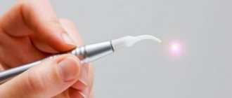
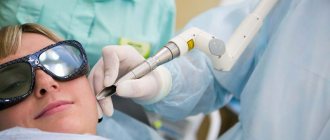
Removal of superficial angiomas with laser
Laser removal of angioma is carried out if it is superficial and located on the skin. The device generates radiation that penetrates several millimeters into tissues. Blood vessels absorb heat and stick together. As a result, the growths lighten, decrease in size and disappear.
Laser therapy can treat any angioma. On modern installations, you can choose the size of the light spot and power. The beam acts in a targeted manner and does not affect healthy surrounding tissues; the procedure does not cause bleeding.
During therapy, the beam destroys melanin cells, so after the session a pink spot appears at the site of the growth. For some time, the area will be slightly lighter than the rest of the skin. But after a few weeks the pigmentation will be restored.
The laser removes the angioma layer by layer. But he may not be able to cope in one go if the formation is large and deep.
The beam triggers tissue regeneration, so the skin is completely restored in a maximum of a month. After laser removal of a tumor, the risk of recurrence is minimal. The procedure takes no more than half an hour including examination and anesthesia.
You don’t have to apply for sick leave or go on vacation. During rehabilitation, you can lead your usual lifestyle, but you will have to comply with some restrictions.
Danger
A small spot formed on the skin can go unnoticed or be ignored by the patient for a long time. Since it can transform into a malignant formation, the lack of necessary treatment can lead to dire consequences.
Particular attention should be paid to angiomas located in places of increased friction with clothing (neck, chest, abdomen, shoulders), on the scalp. Frequent traumatization can lead to inflammation of a benign tumor and its rapid growth and bleeding.
The greatest danger to life is posed by angiomas of the brain and internal organs. As practice shows, if you do not treat a cerebral angioma, the prognosis is unfavorable - the area of accumulation of blood vessels will increase, which will lead to their rupture, hemorrhage in the brain and death.
Indications and contraindications
It is not necessary to remove angioma with a laser if it does not cause concern. Most often, tumors are removed for cosmetic purposes. If they do not spoil the appearance, do not grow and do not constantly rub against clothing, therapy can be postponed.
Doctors recommend removing even small growths from the mucous membrane of the lips, scalp, and eyelids. They are constantly exposed to mechanical stress, which can provoke the growth of a vascular tumor.
You should immediately make an appointment with a doctor if the angioma begins to peel off and constantly itches or bleeds due to friction with the bra straps or shirt collar. Also, indications for removal are the rapid growth of the tumor in length and swelling.
But the dermatologist does not always allow therapy. The laser is not recommended for use in the following cases:
- reduced immunity;
- blood diseases;
- inflammatory process in the body;
- the presence of infectious pathology;
- tendency to form keloid scars;
- oncology (even cured);
- diabetes.
The doctor may also prohibit the procedure in case of cardiovascular diseases, cerebral circulatory disorders, elevated temperature, exhaustion, or nervous disorders (psychosis, epilepsy). Each case is considered individually.
Pregnant and breastfeeding women will need to first consult an obstetrician-gynecologist. Due to hormonal surges, the response to therapy can be unpredictable.
The laser is used to remove angioma in children over 3 years old. Younger children require permission from a pediatrician.