When there is a lack of oxygen in the tissues, the skin turns blue. When a tight bandage is applied to any part of the body, such as a hand or foot, due to decreased blood flow, you may observe bluish skin. After exposure to the cold, you may see blue fingers and lips. Skin color will return after warming up. Often during feeding, newborns will develop a blue and white triangle around their mouth. After finishing feeding, it disappears. If there is blue discoloration in other areas of the skin, this may mean oxygen starvation.
URGENT CARE
Call an ambulance if:
- if the child has a fever, difficulty breathing and blue lips and tongue
- The child's whole body begins to turn blue
ATTENTION!
Call “emergency help” if the child cannot breathe, speak, or turns blue. At this time, begin first aid measures.
| ASK YOURSELF A QUESTION | POSSIBLE REASON | WHAT TO DO |
| The child is trembling, has been in water or cold for a long time | Hypothermia | Try to dress your baby according to the weather. If he is very cold, you can dry the child and take him to a warm room or let him take a warm bath. |
| The child has difficulty and noisy breathing, hoarseness, barking cough | Croup | Call the pediatrician |
| The child has difficulty exhaling, strained breathing, wheezing and wheezing can be heard. The skin around the mouth and lips turns blue | An attack of obstruction or asthma | Call "emergency" |
| The skin around the mouth and lips turn blue after a quarrel or outburst of anger | Holding your breath | Consult your pediatrician as planned ; he will recommend an examination to rule out organic causes of the disease and give advice on what to do if the attack recurs. |
| A child under one year old has been sick with a viral infection for a couple of days, breathing has become difficult and noisy, irritation is present, and appetite is reduced (does not eat at all). | Bronchitis, bronchiolitis | Call a pediatrician for an examination , “emergency care” . Once the diagnosis is confirmed, the issue of hospitalization may be decided. |
| The child suffered a viral infection. He has rapid breathing, wheezing, increased temperature, blue lips and nasolabial triangle, lethargy and poor health. | Pneumonia | Call the pediatrician ; if the diagnosis is confirmed, hospitalization is possible |
| A child's fingers, tongue, mucous membranes and lips turn blue after physical activity or during active play | Lung or heart disease | Avoid physical activity until the cause is determined. Make an appointment with your pediatrician , and after the examination, treatment will be prescribed. You may need to consult specialists. |
| The child has seizures and involuntary urination | Seizure | Call "emergency" |
FOR INFORMATION
What to do if you hold your breath?
When a child screams or cries, is close to hysteria, he cannot take another breath. His face may become pale or blue. There may be convulsions, and the child falls unconscious. This situation scares more parents than the baby. Parents try to get the child to take another breath. But even if the baby is very angry, he will not be able to hold his breath for a long time without harming himself.
After convulsions or loss of consciousness, breathing will restore itself. But be sure to consult a doctor after an episode of loss of consciousness or convulsions, to rule out a serious illness in your baby. You may need to consult a psychologist if your child has an emotional disorder. This condition does not threaten the child's health. Do not show your child your concern by holding your breath against an emotional background. Over time, he himself will find another way of emotional relief. Holding his breath can become a habit if he sees that he is achieving his goal in this way. Your task is not to show your concern. If after loss of consciousness the child does not begin to breathe immediately, then perform artificial respiration immediately. And call “emergency help.”
Classification of the disease
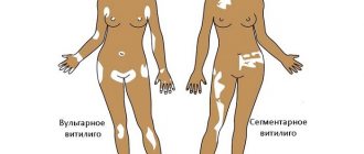
Vasculitis is classified based on etiology, pathogenesis, type of vessels affected and the nature of the rash. Genetic and demographic factors, clinical manifestations and predominant lesions of internal organs are also taken into account.
Based on the factors preceding the disease, primary and secondary vasculitis are distinguished. Primary - caused by inflammation of the vascular wall, secondary - in which vascular inflammation is a reaction to another disease (lupus erythematosus, rheumatism, diabetes mellitus, granulomatosis, sarcoidosis, hepatitis).
according to the diameter of the affected vessels :
- small vessels - affects capillaries and venules, arterioles and small arteries of internal organs;
- middle vessels - mainly affects the abdominal arteries and their branches. Often cause stenoses and aneurysms;
- large vessels - affect the aorta and its main branches.
In addition, there are groups of vasculitis :
- small vessels with immune complexes;
- with trigger connective tissue diseases;
- with an established etiology of a metabolic or infectious nature;
- with damage to one organ.
What is infantile hemangioma?
This is a tumor-like formation of a qualitative nature, which is formed by fragments of blood vessels of varying thickness. The nature of the origin is as follows: during the process of intrauterine development, the formation of blood vessels is disrupted. As a result, almost immediately after the birth of the child, or some time (about a month) later, a hemangioma appears on his body.
Cavernous hemangioma
By the way. Most often, this formation appears on the skin, at any point. But in 20% of cases, the tumor can localize itself to internal organs, muscle and even bone tissue.
The neoplasm looks like this:
- a thickened tumor-like spot of various sizes and degrees of elevation above the surface of the skin or depression;
- the color is either pale burgundy, closer to pink, or bright burgundy;
- the edges are uneven;
- the structure is dense;
- composition – atypical and normal cells.
New growth on a child's cheek
Important! The key point is that although this is a full-fledged tumor, it is always benign. Therefore, there is no need to do anything. Over time, in most cases, the hemangioma subsequently resolves on its own, without a trace.
In children, quite often hemangioma simply resolves over the years.
Of course, exceptions, deviations and anomalies occur, since each person’s body is individual, including a newborn baby. The most important thing for parents when a neoplasm is detected is not to panic and carefully study the issue of how the local pediatrician and this material can help them.
Symptoms of vasculitis in a child
There are dozens of vasculitis - each of them affects one or another type of vessel and is accompanied by specific symptoms. They appear when damaged:
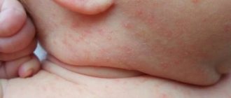
- skin - spotty hemorrhages;
- nerves - partial or complete loss of sensitivity;
- joints - joint pain;
- visceral vessels – abdominal pain;
- kidney – glomerulonephritis;
- brain - strokes.
General symptoms:
- weakness and increased fatigue,
- temperature increase,
- loss of appetite,
- pallor.
The most common vasculitis:
- hemorrhagic,
- allergic,
- urticarial,
- Wegener's granulomatosis,
- Kawasaki disease.
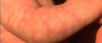
Hemorrhagic vasculitis is manifested by a palpable hemorrhagic skin rash, pain in the ankle and knee joints, sometimes in smaller ones. Cramping abdominal pain lasting from 3 to 10 days is not uncommon, and its clinical manifestations are similar to appendicitis, ulcer perforation and intestinal obstruction. Sometimes they are accompanied by nausea and vomiting, blood in the vomit and stool, and kidney damage - homerulonephritis. The diameter of the rash is 1-3 mm. The rash is symmetrical and initially appears on the legs and feet. Erythema and areas of necrosis are possible. Source: E.V. Borisova Hemorrhagic vasculitis in children // Pediatrics, No. 6, 2004, pp. 51-56
Symptoms of allergic vasculitis:
- erythematous and hemorrhagic spots and nodules;
- hemorrhages under the toenails;
- itching and pain in the rash area;
- necrosis – necrosis and blackening of the skin in the area of the rash;
- pain in muscles and joints.
The disease affects the lower extremities, and in generalized forms it also affects the trunk and forearms. In Behcet's disease, the mucous membranes of the mouth and eyes are affected, ulcers and erosions form.
With urticarial vasuclititis, dense blisters appear. Fever and muscle weakness, joint pain and kidney failure may occur. Diarrhea, conjunctivitis, laryngeal edema, increased intracranial pressure and cardiac arrhythmias are common.
Wegener's granulomatosis is manifested by damage to the kidneys and lungs, laryngeal stenosis, and nodular rashes on the skin.
Kawasaki syndrome - damage to the blood vessels of the heart, respiratory tract and lymph nodes. It begins rapidly with an increase in temperature to 38-41 oC. Other symptoms:
- debilitating fever;
- a rash with plaques that resembles scarlet fever;
- damage to the eyes and respiratory system;
- thickening and redness of the skin on the palms and soles;
- enlarged cervical lymph nodes;
- redness of the tongue;
- peeling of the skin of the fingers;
- aneurysm and heart pain. Source: I.Ya. Lutfullin Kawasaki syndrome: clinical algorithms and the problem of underdiagnosis of the disease // Bulletin of modern clinical medicine, 2016, vol. 9, issue 2, 52-60
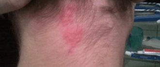
Soon after the birth of a child, parents may notice a formation on the skin of blue, red, pink color, which can quickly grow, capturing new territories on the baby’s tiny body. Most likely, this is a benign vascular tumor, a hemangioma, with all the inherent signs of a benign tumor.
A feature of the course of vascular neoplasms - hemangiomas in young children is the unpredictability of their “behavior”. A punctate hemangioma of the cheek in a one-month-old baby can turn into an extensive and deep tumor with a complex anatomical localization and without a tendency to stop growth within 2-3 weeks. This leads to disruption of the functions of various important organs responsible for vision, hearing, breathing, etc. Due to the unpredictability of the development of hemangiomas in children under one year of age, hoping for independent resorption of large and deep hemangiomas using expectant management means voluntarily taking a conscious risk of the health and life of the baby. It is necessary to distinguish between a vascular tumor and other formations: medial spot, nevi Unna, rosacea, etc.
Therefore, if you find any spots, spots, or blood vessels on the baby’s body, immediately contact pediatric surgeons and oncologists.
Why does hemangioma appear?
It is believed that a tumor as a disease occurs as a result of the body’s reaction to harmful external and internal influences. Therefore, to prevent vascular tumors during pregnancy, simple rules must be followed: contact with harmful carcinogenic substances should be completely avoided; You should not smoke, you should not be in a room where people smoke or have smoked; Uncontrolled use of any medications is strictly prohibited. There is an opinion that the appearance of hemangiomas is associated with complex disorders of intrauterine development of the fetus, especially in the first trimester of pregnancy, when the vascular system of the unborn baby is formed. During this period, emotional and chemical stress, low-frequency electromagnetic influences, injuries, infections, hormonal disorders and any unfavorable factors of a physical or chemical nature that have embryotoxic properties are especially dangerous. Particularly dangerous factors of a physical nature include ultraviolet radiation (solariums!), ionizing radiation, mechanical injuries and elevated temperature in a pregnant woman with long-term exposure. Among the biological factors include viral and bacterial diseases, decreased immunity, and parasitic infections of the mother during pregnancy. Sometimes it is enough to dye your hair in the early stages of pregnancy and the child develops a “stork’s kiss,” as hemangiomas are often called.
Experts are still debating whether hemangiomas are true tumors or a congenital malformation of the vascular system. These tumors have the ability to self-resorb and malignant degeneration. Nowadays, hemangiomas occur in every tenth newborn. In girls it is 2-3 times more common than in boys. If a child already has one hemangioma, then during the first six months of life, 75% of such children may develop hemangiomas in other parts of the body. Recently, the number of children with multiple hemangiomas has increased to 43%. Hemangiomas can grow unevenly. Along with rapid growth, often leading to severe irreversible damage, sometimes there is a complete lack of growth, and in some rare cases even reverse development.
What complications can occur with hemangiomas?
The vast majority of hemangiomas do not have complications. However, in some cases, ulcers and ulcerations occur on them; some hemangiomas are complicated by bleeding. A feature of the course of hemangiomas in the perineum and external genitalia is their tendency to frequent ulceration. Contamination of these areas with urine and feces, friction with diapers or clothing can lead to the formation of ulcers, drip bleeding, and infection. In cases of internal hemangiomas, for example, the larynx or the inside of the nasal passage, the breathing process is complicated. Vascular tumors of the eye can cause occlusion and amblyopia. Hemangiomas of the oral mucosa and lip area disrupt the processes of sucking and digestion. With very large tumors, cardiac dysfunction may occur due to the large amount of blood that must be pumped into the excess blood vessels of the hemangioma. Angiomas adjacent to the bone can also lead to bone erosion. Children with hemangiomas may develop thrombocytopenia due to the death of platelets in the vascular tumor. There are gigantic hemangiomas of skeletal muscles, consisting of thin-walled capillaries infiltrating the entire limb.
How to assess the condition of hemangioma in a child yourself?
Signs of growth, stabilization and regression:
1.Color - a fast-growing tumor is bright red, tense, bulging, the surface is shiny or peeling
2.rapid increase in tumor size: dynamic measurement of the tumor is necessary. You can transfer the contours to cellophane and get a stencil that reflects its size and area. Mark on the stencil the date and overall height of the child at the time of measuring the tumor or the length and circumference of the body part at the measurement site. After 3-4 weeks, take a repeat measurement to create a repeat stencil, observing the same position of the child’s body (standing, lying, sitting). Re-enter the date and information about the child’s height. The assessment is carried out by simple mathematical comparison. If the increase in the size of the hemangioma area in millimeters is equal to or exceeds in digital value the number of centimeters by which the child has grown, then this is an indicator of the progressive development of the neoplasm itself. Such a growing hemangioma requires immediate treatment.
A sign of stabilization of growth or the beginning of regression is its pallor, a decrease in volume, the appearance of areas of whitish coloring in the mass of the tumor - a symptom of “fly agaric”.
If there is a hemangioma, in the interests of the child, taking into account the unpredictability of its development, it is necessary to carry out therapeutic or preventive measures, and the sooner the better.
What treatment methods for hemangiomas exist?
Currently, more than 60 methods of treating hemangiomas are known: surgical, sclerosing, electrocoagulation, radiation, hormonal, laser, cryogenic, medicinal, etc. Each of the methods has certain advantages and disadvantages, and the question is which one is the most rational with in terms of simplicity, accessibility, convenience for the patient, effectiveness in terms of cosmetic and functional results. The choice of treatment method should be based on a comprehensive assessment of each case, taking into account the type, location, clinical form, size, nature of the hemangioma, and the age of the patient. Conservative and invasive methods are used to treat hemangiomas. Of the conservative methods, the most commonly used are hormone therapy, treatment with propranolol, radiation methods, and photochromotherapy. Among the invasive methods, the most popular are surgical excision, cryotherapy (cold treatment), electrocoagulation, laser therapy, sclerotherapy, and embolization of regional vessels. Unfortunately, each of these methods does not provide a guaranteed cure, but has a number of side effects. At the Oncology Research Institute in Rostov-on-Don, a non-invasive, painless method of treating hemangiomas in children was developed and patented: photochromotherapy (a treatment method in which a certain modified red light). This medical technology is protected by Russian Patent No. 2240159 for the invention “Method for treating skin tumors in children under one year of age” and has been widely and successfully used for more than 10 years.
Photochromotherapy - the use of the method is based on the ability of monochrome red light to induce photobioadaptive processes in the child’s body, leading to the activation of humoral factors regulating local blood flow and induction of reparative tissue reactions, which contributes to the stabilization of growth and regression of vascular neoplasms of the skin. This technique is non-invasive and allows it to be used for both therapeutic and preventive purposes. The developed method for treating skin tumors is suitable for children of any age - both up to one year of age and older.
Contraindications:
individual intolerance to light therapy, severe neurological symptoms, acute infectious diseases of unknown origin. Considering the simplicity, harmlessness, painlessness and high efficiency of this method, it can be actively recommended for the treatment of hemangiomas in children.
Errors in the treatment of hemangiomas
The traditional mistake is to wait and see
in the hope of spontaneous resorption. It is not possible to hope for spontaneous regression of large and deep hemangiomas due to their rapid growth or other complications.
Another common mistake is prescribing troxevasin
to the area of hemangiomas. Monitoring of children after the administration of troxevasin in the early stages showed stimulation of tumor growth in 98% of children. This also applies to Lyoton and other heparin-containing drugs. One of the erroneous prescriptions is to apply ice; after such an effect, a phase of sharp vasodilation begins, and the hemangioma grows. There is no point in using Vishnevsky's liniment, and the use of decoctions of St. John's wort, primrose, oats, bee products, acetic and sulfuric acids can significantly worsen the process, lead to dangerous bleeding and tumor growth. The obvious harm is lost time, a neglected process.
What should children with hemangiomas be wary of?
1. Injuries . Danger of bleeding in case of injury - call an ambulance if there is bleeding. It is necessary to apply a hemostatic sponge and press firmly. Cold compress - ice cubes, preferably made from a decoction of oak bark, but you can keep it for no more than 10-15 minutes, then remove it for 15 minutes and repeat the procedure. Hydrogen peroxide 1-2% and antiseptic disinfectants (miramistin or chlorhexidine) are used.
2. Sun. _ You should be wary of direct sunlight. You should take a walk in the summer until 10 a.m. and from 6 p.m. From 11 a.m. to 5 p.m., direct sunlight causes irreparable damage to the skin. Before a walk in the sun, give your baby salted water or tea with lemon. Use sunscreen.
Sunscreens for babies: AVEN: sunscreen for babies spf 50+, Garnier AMBRE SOLER: sunscreen milk-screen baby spf 50, spray, sunscreen color, control for babies, spf30, Vichy Capital Soleil, baby sunscreen spray, spf 50, sunscreen hypoallergenic baby cream, spf 30. La Roche Pose antiheliosdermoKIDs: milk (for babies), spf 50. Mustella sunscreen milk for babies spf 50+, sunscreen spray for children, spf 30. Uriage baby sunscreen milk for hypersensitive skin spf50 spf 30. Floresan, sunscreen milk for babies spf25. Bübchen sunscreen spray for children spf 30, sunscreen milk for children spf 20, sunscreen foam for children spf 15 and 25. Nivea sun sunscreen spray color for children spf 30, children's sunscreen lotion spf 50+. Oriflame children's sunscreen spf 40, spray spf30."Our Mother" Russia children's sunscreen cream spf 15, children's sunscreen mousse with a water-repellent effect, spf 20, children's sunscreen milk spf15."Our Sun", Russia children's sunscreen cream spf 20.1. You need to choose the right protection product: creams are useful for children For those with dry skin, sprays are most convenient.2. Check safety: apply the cream behind the ear, on the elbow and knee bends, observe the skin and the child’s reaction to the smell for 24 hours. 3. Apply the product half an hour before going outside. 4. Distribute correctly over the entire surface of the skin - colored sprays are convenient. 5.Storage sunscreen in the refrigerator for a maximum of six months.
3. Bathing . It is not recommended to bathe in hot water up to 37 degrees, do not bathe in a manganese solution, use neutral soap on the hemangioma area once every 10 days.
4. Before recovery, it is not recommended to do routine preventive vaccinations, since the possibility of provoking the rapid growth of hemangiomas in children after vaccination cannot be ruled out.
Photochromotherapy.
This is a type of treatment with visible electromagnetic radiation (light quanta) with a certain wavelength. The clinical use of red light with a wavelength of 0.67 microns allows for a therapeutic effect due to the photolytic breakdown of biomolecules, which leads to the growth of free forms with high biological activity. This medical technology is protected by RF patent No. 2240159 for the invention “Method for treating skin tumors in children under one year of age.” The technique is non-invasive, which allows it to be used in the treatment of early infants with hemangiomas of critical locations (head and neck, genitals, mammary glands, places of natural skin folds, articular surfaces. The high efficiency of the FCT method lies not only in achieving a local therapeutic result (photo-induced regression of the vascular tumor itself), but also in increasing the overall protective and compensatory potential of the child, achieving an immunostimulating and anti-stress effect on his body.
Treatment of vasculitis
Conservative therapy for vasculitis is aimed at suppressing pathological autoimmune reactions with the production of antibodies. It is carried out in stages:
- relief of symptoms and achievement of remission;
- maintaining remission for 0.5–2 years;
- treatment of relapses.
In addition, prevention or elimination of concomitant diseases and complications is carried out.
The treatment regimen is based on the drugs of choice:
- hormones – for giant cell arteritis of GCA, polymyalgia rheumatica and Takayasu’s disease;
- hormones and cytostatics - for systemic, hemorrhagic and cryoglobulinemic vasculitis, polyarteritis nodosa and a number of others;
- immunosuppressants with hormones - for hemorrhagic vasculitis, if combination therapy with cytostatics is not effective;
- monoclonal antibodies - for systemic vasculitis, if combination therapy is not effective;
- basic antirheumatic drugs – in case of contraindications to the drugs of choice;
- immunoglobulins - for severe complications.
Diet therapy is also indicated . For functional kidney damage, plasmapheresis or hemosorption - hardware blood purification - are prescribed. In addition, according to indications, antiviral and symptomatic agents, antibiotics, drugs to maintain cardiac activity and prevent blood clotting are prescribed.
Conservative treatment of vasculitis is complex and long-term. In some forms, against the background of overgrowth, spasms and thrombosis of large vessels, surgical treatment is indicated. Without timely and correct treatment of vasculitis in children, the prognosis is unfavorable. The nature of the consequences depends on the type of disease.
Sources:
- https://academic.oup.com/ndt/article/30/suppl_1/i94/2324852 Despina Eleftheriou, Ezgi Deniz Batu, Seza Ozen, Paul A. Brogan. Vasculitis in children // Nephrology Dialysis Transplantation, Volume 30, Issue suppl_1, 1 April 2015, Pages i94–i103.
- E.V. Borisova. Hemorrhagic vasculitis in children // Pediatrics, No. 6, 2004, pp. 51-56.
- AND I. Lutfullin. Kawasaki syndrome: clinical algorithms and the problem of underdiagnosis of the disease // Bulletin of modern clinical medicine, 2016, vol. 9, issue 2, 52-60/
The information in this article is provided for reference purposes and does not replace advice from a qualified professional. Don't self-medicate! At the first signs of illness, you should consult a doctor.