| A healthy 25-year-old brunette went to get medical help for skin depigmentation for 12 months. She first noticed the white skin on the stitches; The doctor suspected a fungal infection and prescribed imidazolic cream, but there was no relief. After being released on the seashore, the women experienced additional signs of depigmentation on the elbows, foreheads, upper eyelids and lower part of the chest. The dermatologist made a diagnosis of vitiligo and prescribed a sore throat cream, but did not hope for effective treatment. The illness senses the power of inferiority, as reflected by its external appearance. How to cover and treat the patient? |
CLINICAL PROBLEM
Vitiligo is the most widespread depigmentation disease, affecting approximately 0.5% of the world population. In approximately half of patients, vitiligo develops before the age of 20. Men and women get sick at the same frequency; illness is not affected by skin type or race.
Non-segmental (or generalized) and segmental vitiligo are characterized by a different clinical picture and medical history (div. Table 1 and Fig. 1).
The most common type of vitiligo is non-segmental vitiligo (85–90% of cases), prosegmental vitiligo, which tends to develop earlier in life, can account for up to 30% of cases in children (Hann SK et al. , 1996). Non-segmental and segmental vitiligo can be differentiated from the focal form, which is characterized by a small area of distribution (< 15 cm2). Rarely do non-segmental and segmental vitiligo develop simultaneously; in such cases, segmental disorders are less amenable to treatment. Table 1. Characteristic signs of segmental and non-segmental vitiligo
| Segmental vitiligo | Non-segmental vitiligo |
| Often begins in childhood | They may begin in childhood, but more often in adulthood |
| Characteristic soft cob, after which the illness stabilizes | Progressive recovery from illness due to illness |
| Nezabar after the onset of illness affects the hairline of the skin | Affects the hairline of the skin in the later stages |
| Definitely not associated with other autoimmune diseases | Often accompanied by autoimmune diseases or a similar complex family history |
| Often blames accusations | Often occurs on plots that are sensitive to pressure and rubbing and susceptible to injury |
| Often responds to autologous skin transplantation with stable repigmentation | In situ relapses often occur after autologous skin transplantation |
| It is important to differentiate between nevus depigmentosus , especially with early onset of illness |
Rice. 1.
Nonsegmental and segmental vitiligo
.
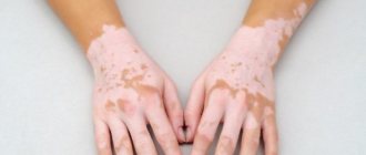
Non-segmental vitiligo (Fig. 1A) is characterized by white lesions that are often symmetrically distributed and increase in size over time, with a significant loss of functional epidermal melanocytes and other melanocytes in hair follicles. Segmental vitiligo (Fig. 1B) mainly expands on one side of the body and may resemble a dermatoma in some areas. In most patients, only one segment of depigmentation is visible (Fig. 1B), although in rare cases it may develop into two or more segments on one or both sides of the body.
From a pathohistological point of view, the criterion for the diagnosis of vitiligo is the loss of epidermal melanocytes (Dell'anna ML et al., 2006). In plots with constant loss of pigment, inflammation is often absent, but with non-segmental vitiligo, especially with rapidly progressing migration, mononuclear cells can be detected at the border of depigmented plots. The underlying etiology of nonsegmental vitiligo is still debated, but may include immunological factors, oxidative stress, or impaired functioning of the sympathetic nervous system. In the pathogenesis of non-segmental vitiligo, the migration of melanocytes 24 years after mechanical trauma in the tissue, which has been torn near the site of injury, indicates an important role for the Koebner phenomenon (skin tissue on non-specific , extremely traumatic stimuli, such as pressure or rubbing). In segmental vitiligo, the main contributing factor is impaired functioning of the sympathetic nervous system, although genetic abnormalities (mosaicism) also play a role.
CUTTING
Causes of vitiligo
There are several causes of vitiligo:
- autoimmune diseases (systemic lupus, rheumatoid arthritis, diabetes mellitus, thyroid diseases, etc.). These pathologies cause a malfunction in the immune defense, in which pathological antibodies begin to be produced against healthy (unaffected) tissues;
- genetic predisposition. In approximately 15-20% of cases, the disease is hereditary;
- endocrine disorders. It has been proven that hormonal disruptions caused by disruption of the endocrine glands (thyroid, ovaries, pituitary gland, adrenal glands, pancreas) can trigger the onset of the disease;
- trophic changes in the skin. This can be various types of skin damage: cuts, scars, microtraumas, burns (both thermal and solar), skin contact with chemicals, as well as prolonged exposure to the sun, especially without sunscreen. All these factors trigger inflammatory reactions, as a result of which melanocytic cells begin to destroy;
- diseases of the liver and gastrointestinal tract, vitamin deficiency, and taking certain medications can also give impetus to the development of vitiligo.
Repigmentation stage
This form is observed quite rarely in patients. Most often, it is discovered when the appearance of spots is the result of medications or other medical procedures.
If treatment is started at an early stage of vitiligo, it is possible to achieve normal melanin production in the skin. This is most often observed in children under 4 years of age: when melanocyte function is restored, skin repigmentation occurs.
If, nevertheless, it is impossible to resume normal pigment production in the affected areas, then therapeutic measures are taken to prevent further progression of the pathology, the goal is to stop the growth of depigmented areas.
Symptoms of vitiligo
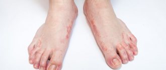
The manifestation of the disease begins with the appearance of a primary white spot. The spot can appear both on smooth skin and on mucous membranes, the spots can increase in size and begin to merge with each other, most often the lesions are symmetrical in nature.
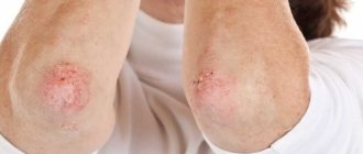
The lesions are localized most often on the extremities, mainly on the flexor and extension surfaces, face, skin folds, in places of constant trauma and friction. It should be noted that vitiligo does not appear in the area of the palms and soles
Increased pigmentation around the lesion
One of the characteristic signs of vitiligo is a rim of hyperpigmentation around the lesion; it may be only slightly darker than healthy skin in this area.
Hair bleaching within the lesion
The hair on the body, scalp, eyebrows, beard, etc. is bleached.
Introduction
Vitiligo is an acquired autoimmune disease characterized by depigmented patches on the skin that occur due to the destruction of melanocytes.
Focal and segmental forms of vitiligo affect no more than 10% of the body surface area and are considered stable. Generalized forms of vitiligo usually include lesions covering more than 10% of the body surface area, located symmetrically on both sides and, as a rule, developing during a relapsing and remitting course of the disease. Treatment can be complex, although available treatments include medications, surgery, and phototherapy. Proper characterization of the type of vitiligo, consideration of the extent and duration of the disease, and the effectiveness of previous treatments can guide treatment and maximize its effectiveness.
Drug treatment
For early, stable, localized disease (up to 12 months from onset), topical corticosteroids, topical immunomodulators, topical narrow-band ultraviolet B (NB-UVB) phototherapy, or a combination of these may be effective..
For generalized, stable disease, the mainstay of therapy is topical corticosteroids or immunomodulators in combination with NB-UVB phototherapy. In patients with generalized, rapidly progressive vitiligo, pulse therapy with oral corticosteroids may help induce stabilization, a three-month study showed.
Topical therapy
Topical corticosteroids are effective medications for vitiligo due to their immunosuppressive and anti-inflammatory properties. Superpotent or high-potency corticosteroids are suitable for the trunk and extremities, while intermediate-potency topical corticosteroids (or topical calcineurin inhibitors) are better suited for the face, neck or intervertebral areas, and in children.
Cyclic use (1-week cycles on 1-week intervals for 6 months or 5-day cycles on 2-day intervals) may help avoid the cumulative side effects of topical corticosteroids, including skin atrophy, telangiectasias, and steroid acne. Treatment should not exceed 14 days per month per package.
Topical calcineurin inhibitors, such as tacrolimus (0.1%) or pimecrolimus (1%), are immunomodulatory, steroid-sparing agents that can be used at any site except the mucous membranes and are almost as effective as topical corticosteroids, but more effective in combination with them, providing a gentle effect. Twice daily use may help stabilize the disease, and twice weekly use may be used for maintenance therapy.
Topical calcineurin inhibitors can be used in combination with topical corticosteroids on off-days. Due to its immunosuppressive properties, tacrolimus may theoretically increase the risk of carcinogenicity with long-term use, but no cases have been reported in humans. In a murine model examining skin photocarcinogenicity, topical 0.1% tacrolimus was associated with the development of lymphoma.
In a single-arm comparison study of 9 patients with generalized vitiligo, Ostovari et al found that repigmentation was optimized in all patients when topical calcineurin inhibitors were combined with UVB (excimersq laser 308 nm), in while on the side treated with 0.1% topical tacrolimus alone, no repigmentation was observed.
Vitamin D analogues, such as topical calcipotriol, used in combination with phototherapy, can reduce the time to repigmentation and the total accumulated photodose.
Oral therapy
For advanced vitiligo, oral minipulse corticosteroid therapy may be used to promote disease stabilization through immunosuppression. A typical regimen involves a low dose of oral betamethasone or dexamethasone taken 2 consecutive days per week for 3 to 6 months. Failure to stabilize the disease after 3 months of combination therapy (minipulse corticosteroid therapy + NB-UVB phototherapy) requires discontinuation of minipulse corticosteroid therapy alone and re-evaluation at 3-month intervals.
Alternatively, if minipulse corticosteroid therapy is avoided, daily dosing of 20 mg oral prednisolone may be considered until disease progression stops. In an early study published in 1993, 40 patients with extensive or rapidly progressive vitiligo were treated with 5 mg betamethasone/dexamethasone as a single daily oral dose on 2 consecutive days per week for 1 to 3 months, which resulted in reversal of disease progression in 89% patients. To stop the progression of the disease in 2 patients, it was necessary to increase the dose to 7.5 mg per day. During 2–4 months of treatment, 80% of patients noted an increase in spontaneous repigmentation, which was maintained with continued therapy.
Side effects from oral corticosteroids were observed in 42.5% of patients. In a study published in 2001, 29 patients with progressive (n = 25) or stable (n = 4) vitiligo received 10 mg of oral dexamethasone for 2 consecutive days per week for 24 weeks. After an average of 18.2 ± 5.2 weeks, 88% (22 of 25) of patients with progressive vitiligo experienced cessation of disease activity.
Side effects associated with oral corticosteroids were observed in 69% of patients studied. In a more recent study published in 2013, 444 patients with advanced vitiligo were treated with 2.5 mg dexamethasone orally for several days a week. With this regimen, arrest of disease progression was observed in 91.8% of these patients, with some repigmentation observed for an average of 16.1 ± 5.9 weeks. Disease relapse occurred in 12.25% of patients, although the mean period to first relapse was 55.7 ± 26.7 weeks.
Adverse effects from oral corticosteroids were observed in 9.2% of patients. Patients should be warned about the possible side effects associated with taking any oral corticosteroid, incl. such as acne, weight gain, headache, nausea and lethargy. There is reason to believe that confetti-like spots and köbnerization are good markers of instability. These markers can be used to decide whether to initiate oral corticosteroid therapy.
To stop the progression of vitiligo, as well as to stimulate repigmentation, minocycline, which has anti-inflammatory, immunomodulatory, antioxidant and anti-apoptotic properties, as well as direct free radical scavenging properties, may be effective. In a study of patients with gradually progressive vitiligo (most cases were generalized vitiligo), patients were prescribed 100 mg of oral minocycline per day. Of the 32 patients included in the study, 29 experienced cessation of disease progression, incl. in 10 out of 29 patients - already 4 weeks after the start of treatment. 7 of 32 patients showed moderate or severe repigmentation.
A randomized trial comparing 6 months of oral dexamethasone (5 mg per week, 2.5 mg/day for 2 consecutive days per week) with oral minocycline (100 mg per day) in 50 patients with unstable generalized vitiligo , showed no statistically significant differences in vitiligo activity scores and vitiligo score index between groups. Another study compared 3 months of oral minocycline (100 mg daily) with NB-UVB phototherapy (twice weekly) in 42 patients with unstable vitiligo and found a statistically significant improvement in repigmentation in the NB-UVB group.
A recently published case series showed that the use of methotrexate, an immunomodulatory systemic agent, improves vitiligo in a third of patients with rapidly progressive generalized vitiligo when administered at an escalating dose of 12.5–25 mg per week. Repigmentation in these previously unresponsive patients to topical calcineurin inhibitors and phototherapy was noted within 6–14 months.
A prospective randomized trial was conducted comparing 24 weeks of oral methotrexate (10 mg weekly) with minipulse corticosteroid therapy (dexamethasone 5 mg weekly, 2.5 mg administered on 2 consecutive days weekly) in 50 patients with unstable vitiligo. New lesions developed in 6 of 25 patients in the methotrexate group and in 7 of 25 patients in the minipulse corticosteroid therapy group, although the difference was not statistically significant. Both groups showed similar reductions in vitiligo disease activity scores.
Due to the limited availability of studies demonstrating its superior efficacy compared to other available treatments, the use of methotrexate may be considered as a steroid-sparing agent in patients with vitiligo refractory to topical treatment and phototherapy.
The role of antioxidants in the treatment of vitiligo remains controversial. Based on a systematic review of the literature by Speeckaert et al., it was found that patients with vitiligo have impaired oxidative processes, which is also characteristic of other immune-mediated skin diseases. In addition, it has been noted that increased oxidative stress in unlesioned skin may be specific to vitiligo.
Among antioxidant therapies, oral Polypodium leucotomos extract has been studied in patients with generalized vitiligo due to its known antioxidant and immunomodulatory properties.
In a 26-week randomized clinical trial, patients received NB-UVB phototherapy (twice weekly) in combination with oral Polypodium leucotomos (250 mg three times daily) (n = 25) or placebo (n = 24). The improvement in repigmentation was greatest in the group receiving oral phototherapy Polypodium leucotomos + NB-UVB and was most noticeable on the head and neck, which was statistically significant.
A pilot randomized, placebo-controlled clinical trial assessed the effects of oral Polypodium leucotomos + PUVA compared with placebo + PUVA in 19 patients. Repigmentation greater than 50% was observed more frequently in the Polypodium leucotomos + PUVA group compared to the placebo + Polypodium leucotomos group.
Ginkgo biloba extract has been shown to have some antioxidant and anti-inflammatory properties and may help stabilize vitiligo. In a placebo-controlled, double-blind study evaluating the effectiveness of ginkgo biloba extract in patients with gradually progressive vitiligo, 40 mg of oral ginkgo biloba extract was given to 24 patients 3 times daily for 6 months, while 18 patients received placebo. 20 patients in the active treatment group and 8 patients in the placebo-controlled group experienced a statistically significant cessation of disease progression. 10 patients in the active treatment group showed significant repigmentation compared with 2 patients in the placebo-controlled group.
A separate 12-week pilot clinical study of 11 patients found that taking 60 mg of oral ginkgo biloba extract twice daily resulted in improvements in vitiligo total area index scores. Additional clinical trials with larger cohorts are needed to better evaluate the effectiveness of antioxidants, including Polypodium leucotomos and Ginkgo biloba extract.
Phototherapy
Narrow band phototherapy (NB-UVB) is recommended for rapidly spreading or widespread generalized vitiligo involving more than 5-10% of the body surface. The immunosuppressive effects and ability to induce melanocyte differentiation and melanin production make NB-UVB an effective therapy. A starting regimen of 200 mJ two or three times a week is suitable for all phototypes as it prevents the risk of phototoxic reactions. In the absence of symptoms, when light pink erythema lasting less than 24 hours (the treatment end point) is no longer observed, the dose can be increased in increments of 10% to 20% until this end point is restored.
Patients should be warned of the risk of phototoxicity, and if this occurs, the next dose should be reduced or skipped, depending on the severity of the reaction. Alternatively, a fixed dosing regimen can be used that accounts for differences in skin phototype in the minimum erythemal dose, but this is only recommended for dark skin phototypes due to the increased risk of phototoxic reactions.
In patients with a limited area of vitiligo (less than 10% of the body surface) or with early segmental diseases, local phototherapy (excimer lasers and excimer lamps) is advisable. This approach avoids the overall tanning effect of NB-UVB, although local phototherapy does not stabilize the disease because does not have a therapeutic effect on clinically unaffected skin.
You can prescribe phototherapy twice or thrice a week, because... Available comparative studies have not demonstrated superiority in achieving repigmentation of any of these treatment regimens. In studies using the excimer laser, the onset of repigmentation occurred earlier with three times weekly dosing, and repigmentation had a greater relationship with the total number of treatments, which can likely be extrapolated to NB-UVB phototherapy.
The Vitiligo Working Group recently released guidelines for the use of NB-UVB, which recommend the maximum tolerable dose of NB-UVB for this treatment as 1500 mJ/cm2 and 3000 mJ/cm2 for the face and body, respectively. In patients with skin phototypes IV-VI, the Vitiligo Working Group recommends no upper limit for NB-UVB dosage. In patients with skin phototypes I-III, an upper dosage limit for NB-UVB is not proposed because there are limited long-term studies to examine the relationship between cumulative exposure to NB-UVB and the potential for cutaneous malignancy, which remains a concern. Skin phototype and photosensitivity may also be used to determine upper dosage limits for NB-UVB.
NB-UVB has become the preferred route of phototherapy because it has been demonstrated that this type of phototherapy is more effective, does not require a photosensitizer, requires a lower cumulative dose, and has fewer side effects compared to PUVA. NB-UVB is also safe for children and pregnant or breastfeeding women. PUVA may still be used for dark skin phototypes and treatment of resistant vitiligo. Despite the concerns, there are no studies demonstrating an increased risk of cutaneous malignancies with the use of PUVA or NB-UVB. Recent literature suggests that vitiligo may protect the body from nonmelanoma skin cancer and melanoma.
Surgery
Surgery may be considered for localized or generalized vitiligo that is stable or untreated for more than a year from onset. It is also suitable for patients who have completed a 6-month course of topical therapy with or without phototherapy without response. In generalized vitiligo, the results are less promising due to the unstable course of the disease.
Disease stability is an important variable when selecting candidates for surgical treatment. Published studies define stability as the absence of new or enlarging depigmented spots over a period of 6 months to 2 years. Other cutaneous markers of instability include köbnerization, confetti-like patches of depigmentation, trichrome pattern, or inflammatory borders.
Patient interview, serial photography, and validated scoring systems such as the Vitiligo Area Severity Index, the European Vitiligo Task Force score, and the Vitiligo Disease Activity Score can be used to confirm stability. In uncertain cases, a trial graft may be performed in a stable depigmented lesion.
The most favorable results of repigmentation in skin recipients are observed on the face and neck, followed by the extremities (except fingers) and then the torso. Acrofacial vitiligo (including the distal fingertips, periungal and perioral areas) usually shows poor repigmentation. The skin over the joints also does not respond well to treatment due to its susceptibility to injury. Other contraindications to surgery include a history of keloid formations, coagulopathies, and blood-borne infections. Surgical therapy can be divided into tissue-based and cell-based transplantation methods.
Tissue transplantation methods
Tissue grafting involves transferring intact tissue from an area of normal, intact skin to an area of depigmented skin. Many tissue grafting techniques are available, but the most widely used are aspiration-assisted epidermal grafting and mini-perforation.
Epidermal grafting is useful for difficult-to-treat areas such as the eyelids. Complications such as peripheral halo depigmentation, milia, hypertrophy and hyperpigmentation (especially in darker skin phototypes), including infection (rarely), may occur.
When using mini-perforation, grafts are transferred from the donor zone to cells in the depigmentation zone at a distance of approximately 5-10 mm from each other. Complications such as unevenness, color discrepancy, hypertrophic scarring, keloid formation and graft rejection may occur in the skin at the graft site, and depigmentation and scarring at the donor site.
Follicular unit transplantation (FUT) is based on the concept that pigmentation can be restored to damaged areas of vitiligo by using undifferentiated stem cells located in hair follicles. Removal of the epidermis (or ultraviolet radiation) may induce inactive melanocytes to become active, which can then migrate upward into the epidermis from the outer root sheath, causing perifollicular repigmentation. Donor grafts are typically harvested from the back of the ear or the back of the head using the "strip method" (also known as the FUT method), then dissected into follicular units and inserted into slits made in the depigmented area using either a follicle machine. hair transplant or 18 gauge needle. The FUT method can be beneficial for hairy areas, including hard-to-reach areas such as eyelashes.
Follicular unit extraction isolates the hair follicle using a 1mm punch biopsy, which is then transferred into pits created using a 1mm punch biopsy approximately 3-10mm apart. In contrast to FUT, follicular unit extraction is considered a simpler procedure and is preferred when limited area is available for grafting and is also preferred as a spot treatment for depigmented areas that have not repigmented after FUT.
Cell transplantation methods
Cell transplantation involves the transfer of melanocytes and keratinocytes from the donor to the depigmented area in the form of a suspension, with or without the use of cell culture. Non-culture epidermal suspensions (also called melanocyte keratinocyte transplantation procedure; NCES) have gained worldwide acceptance as the standard for vitiligo transplantation because they can be performed in a single office visit and do not require the use of a laboratory.
The main advantage of NCES is the small area of the donor site required to cover a large area of the depigmentation zone, in a ratio of 1:10, respectively. An ultra-thin skin graft is taken from the donor site and after processing the cells, a cell suspension is formed and applied to the depigmented area denuded to the dermal-epidermal junction using dermabrasion or carbon dioxide ablative laser.
Suspensions of cells from the outer root sheath of the hair follicle were introduced not so long ago, in 2009 by Vanscheidt and Hunziker. This method involves collecting hair follicles using the follicular unit extraction method, inducing cell separation using trypsin, incubation, centrifugation and ultimately transplanting the collected cells into the vitiligo area. A notable disadvantage of hair follicle outer root sheath cell suspension is the low cell yield.
Treatment options for resistant forms
Lack of response to treatment, as evidenced by lack of stability or repigmentation after 3 and 6 months of combination therapy, respectively, should warrant consideration of other treatments to improve dyschromia. The choice of treatment depends on the severity of the disease and its impact on quality of life.
Masking agents
For vitiligo, camouflage agents are classified into temporary and permanent. Temporary options include foundation and sunblock, while permanent methods include micropigmentation (tattooing). The ideal camouflage product is one that is waterproof, sweat-resistant, opaque and shows good skin color matching.
Camouflage makeup foundations contain 25% more pigment than traditional makeup and are typically waterproof, reducing the need for reapplication. Dihydroxyacetone is a sunless tanning product that causes temporary skin coloration by reacting with proteins in the stratum corneum to form brown chromophores called melanoidins. Dihydroxyacetone provides camouflage effects over a longer period (approximately 5-7 days), although its use may be limited due to poor skin color matching, allergic contact dermatitis, and potential cytotoxic effects on keratinocytes in a recent in vitro study.
Micropigmentation (skin tattooing) is generally not recommended due to the risk of köbnerization and discoloration over time due to improper pigment depth. This leads to the need to re-apply the tattoo. Oxidation of tattoos containing metal oxides can also cause black discoloration that is difficult to remove. Summer tanning can also lead to undesirable contrasts in skin color. Other complications include contact dermatitis, granulomatous reactions, and infections with herpes simplex virus, human immunodeficiency virus, hepatitis B and C, and secondary infections.
Depigmentation
In patients with extensive, treatment-resistant vitiligo involving more than 50% of the body surface, depigmentation techniques may be considered. Hydroquinone monobenzyl ether (MBEG) is the only drug approved by the FDA for depigmentation. In areas of residual pigmentation, 20% MBEG can be applied twice daily.
Lack of effect after 4 months is an indication to increase the concentration of MBEG to 30%, and if no effect is observed after 6 months, MBEG should be discontinued. Once complete depigmentation is achieved, use can be reduced to several times a week as a maintenance treatment. Complete depigmentation usually takes 4 to 12 months, although dark skin phototypes may require longer exposure. Side effects include irritant contact dermatitis and rarely conjunctival melanosis. Mequinol, a phenol derivative, can also be used to achieve depigmentation, although the onset of depigmentation is usually slower.
Laser-mediated depigmentation can be used to destroy remaining melanosomes in melanocytes through photothermolysis and photoacoustic effects. The most commonly used for this purpose are ruby (694 nm), alexandrite (755 nm) and neodymium (532 nm) Q-switched lasers. This method is usually reserved for stubborn areas on the hands and face. Koebnerization that occurs with laser therapy may also be useful in improving depigmentation.
New treatments for vitiligo
Recent studies have shown that the interferon gamma-CXCL10 gene axis may be an effective target for the treatment of vitiligo, leading to the emergence of a new class of targeted immunotherapies, Janus kinase inhibitors. Significant repigmentation has been reported following treatment with two oral Janus kinase inhibitors, tofacitinib and ruxolitinib. Topical ruxolitinib has also been shown to be effective, especially for facial repigmentation. Combination with natural sunlight or NB-UVB may result in increased efficacy, as observed with oral tofacitinib. Although these treatments appear promising, studies in larger groups are needed to confirm their effectiveness. A multicenter phase II clinical trial (ClinicalTrials.gov Identifier: NCT03099304) involving topical tofacitinib for vitiligo is currently underway.
Afamelanotide, a synthetic alpha-melanocyte-stimulating hormone, requires the presence of the melanocortin 1 receptor and NB-UVB engagement to stimulate melanoblast differentiation, and may be a new effective therapy for vitiligo.
Additional recommendations
To ensure appropriate treatment and early detection of treatment failure, frequent monitoring of the effectiveness of treatment is necessary, allowing for switching to alternative, potentially more effective treatments if necessary. It is generally recommended that patients be re-evaluated every 3 to 6 months until an effective and stable treatment regimen is established. Because of the significant psychosocial burden experienced by patients with vitiligo, healthcare providers should be mindful of the importance of conversations with the patient aimed at promoting mental health.
Conclusion
Vitiligo is a debilitating psychosocial disease that requires a multidisciplinary approach to treatment. Before formulating a treatment plan, patients should be aware of all available repigmentation and depigmentation options. Frequent follow-up of the disease and interviews with the patient to help determine the extent to which the disease is affecting quality of life can help guide the selection of optimal treatment and provide individualized care.
Methods for diagnosing vitiligo
Typically, the diagnosis of vitiligo is made based on:
- clinical picture (symptoms characteristic of the disease);
- collecting anamnesis (clarification of the hereditary nature of the disease, the presence of autoimmune diseases, etc.);
- laboratory and instrumental research data.
Laboratory research
Indicators of general and biochemical blood tests, general urinalysis, and analysis of thyroid hormones are assessed. Additional blood tests for vitamins and minerals may also be required.
Instrumental studies
Studies are being carried out in the rays of a Wood's lamp: vitiligo is characterized by a bluish-white glow. In very rare cases, a skin biopsy is used.
Sign up for diagnostics To accurately diagnose the disease, make an appointment with specialists from the Family Doctor network.
Why you need to contact the Mama Papa Ya clinic
Our clinic offers quality services at affordable prices:
- consultation with a qualified dermatologist familiar with modern methods of treating the disease;
- the opportunity to attend physical therapy;
- the use of non-traditional methods of treatment, such as acupuncture;
- the ability to treat family members of different ages;
- careful observation of a specialist for the effectiveness of treatment, if necessary, the use of additional methods.
Call our clinic by phone or make an appointment at a time convenient for you through the form on our website.
Reviews
Good clinic, good doctor!
Raisa Vasilievna can clearly and clearly explain what the problem is. If something is wrong, she speaks about everything directly, not in a veiled way, as other doctors sometimes do. I don’t regret that I ended up with her. Anna
I would like to express my gratitude to the staff of the clinic: Mom, Dad, and me. The clinic has a very friendly atmosphere, a very friendly and cheerful team and highly qualified specialists. Thank you very much! I wish your clinic prosperity.
Anonymous user
Today I had a mole removed on my face from dermatologist I.A. Kodareva. The doctor is very neat! Correct! Thanks a lot! Administrator Yulia Borshchevskaya is friendly and accurately fulfills her duties.
Belova E.M.
Today I was treated at the clinic, I was satisfied with the staff, as well as the gynecologist. Everyone treats patients with respect and attention. Many thanks to them and continued prosperity.
Anonymous user
The Mama Papa Ya clinic in Lyubertsy is very good. The team is friendly and responsive. I recommend this clinic to all my friends. Thanks to all doctors and administrators. I wish the clinic prosperity and many adequate clients.
Iratyev V.V.
We visited the “Mama Papa Ya” Clinic with our child. A consultation with a pediatric cardiologist was needed. I liked the clinic. Good service, doctors. There was no queue, everything was the same price.
Evgeniya
I liked the first visit. They examined me carefully, prescribed additional examinations, and gave me good recommendations. I will continue treatment further; I liked the conditions at the clinic.
Christina
The doctor carefully examined my husband, prescribed an ECG and made a preliminary diagnosis. She gave recommendations on our situation and ordered additional examination. No comments so far. Financial agreements have been met.
Marina Petrovna
I really liked the clinic. Helpful staff. I had an appointment with gynecologist E.A. Mikhailova. I was satisfied, there are more such doctors. Thank you!!!
Olga
Features of treatment of leukoderma on the genitals
If you decide to treat vitiligo in the intimate area, this should only be done under medical supervision. Any careless action can cause problems with the reproductive organs; self-medication in most cases causes even more harm to the body. What will the doctor do?
First, in any case, the patient will be carefully examined. It is important to identify all diseases, chronic, latent, sluggish, and treat them - perhaps this will be enough to get rid of vitiligo. Then the doctor will decide which actions are more appropriate: to even out the skin tone by lightening healthy areas, or to restore pigmentation in areas affected by vitiligo. The following methods and means can be used:
- MBEG ointments are preparations for skin lightening.
- Transplantation of donor melanocytes or cells from a healthy area of the patient’s own skin.
- A course of taking immunomodulators and corticosteroids.
- Vitamin therapy.
- Photosensitizers are drugs that increase the skin's sensitivity to ultraviolet radiation and thereby stimulate its pigmentation.
Each product is designed to neutralize various factors that can trigger vitiligo. Therefore, it is important to find out what actually served as the impetus for the formation of pathology. But since this is difficult to do, doctors use a combination of several drugs and techniques with different effects.
There is no universal treatment regimen that would suit every patient. All drugs, procedures, dosages, and duration of therapy are selected individually and adjusted if necessary. The doctor constantly monitors the patient, monitors the treatment process and, if necessary, intervenes.
Differences from treatment on other parts of the body
Leukoderma on the genitals, if not complicated by infections and inflammations, cannot affect the reproductive functions of male or female organisms. But since important organs are in close proximity, a special approach is needed. There are two important points to remember:
- Products and procedures should not injure or irritate sensitive mucous membranes.
- Systemic drugs should not have side effects on the organs of the genitourinary system (and others too).
Those who suffer from leukoderma should be especially careful when sunbathing and carefully choose intimate hygiene products and underwear. The main task is not to provoke the progression of the disease and not to irritate vulnerable skin with aggressive cosmetic products and low-quality, uncomfortable underwear.
Special preparations and methods
There are a number of drugs designed specifically to even out the skin tone and mucous membranes of vitiligo in intimate places. These are a variety of foams, gels and ointments. The Sesderma brand line has proven itself well. You can also use other external products - for example, Vitasan cream.
As for the lamp, it is better not to use it, like a laser, to irradiate the mucous membrane in the intimate area - it is easy to burn delicate skin. Also not suitable for treating the genitals are tinctures based on red or black pepper, acetylsalicylic acid, or apple cider vinegar.
We must not forget about a diet rich in minerals and vitamins - this is a very important point. During the period of remission, to maintain the achieved result and prevent exacerbations, it is recommended to use folk remedies based on medicinal plants.
Therapeutic baths
Treatment of vitiligo at home should be combined with bathing. This procedure will not only relieve the signs of vitiligo, but also calm the nervous system.
Effective home bath recipes:
- 50 ml of valerian tincture is mixed with 40 ml of pine needle extract. The mixture is added to a bathtub filled with water. Taken daily for 25 minutes. The course of home therapy is 15 days.
- You need to buy Dead Sea salt at the pharmacy. Pour 1 cup of product into a filled bath. Take in the morning for 20 minutes for no more than 14 days.
- Grind the roots of pelargonium officinalis, pour a liter of boiling water and leave for 4 hours. After filtering, add the liquid to the bath. Take for 2 weeks no more than a quarter of an hour before bedtime.
- 4 tbsp. l. St. John's wort is poured into 1 liter of boiling water and left for an hour. After filtering, the product is added to the bath. The duration of the appointment is 20 minutes. The course of home treatment is 2 weeks.
Traditional medicine for vitiligo also recommends taking baths based on infusions of chamomile, sage, string, and calamus.