Most often, such seals do not pose a danger, since they are benign tumors. But nevertheless, the formation of a lump on the shoulder can lead to the development of a pathological process in the body, so the disease requires mandatory treatment. This is especially true for growths that have a high degree of oncogenicity.
As a rule, people go to a medical institution after a noticeable thickening and increase in the size of the resulting growth. In most cases, the growth of the lump is accompanied by redness of the skin and pain. Such neoplasms can appear on any part of the body - on the forearm, arms, neck, etc.
Review
Various formations under the skin: bumps, balls, compactions, tumors - this is a common problem that almost every person faces.
In most cases, these formations are harmless, but some of them require emergency treatment. Lumps and lumps under the skin can develop on any part of the body: face, arms and legs, back, abdomen, etc. Sometimes these formations are hidden in the folds of the skin, on the scalp, or grow so slowly that they remain unnoticed for a long time and are discovered reaching large sizes. Benign neoplasms of the skin and soft tissues usually proceed asymptomatically.
Lumps or lumps that cause pain or discomfort are often the result of infection. They may be accompanied by an increase in general or local temperature. The skin over them usually turns red. Associated disorders occur: general malaise, headache, weakness, etc. With timely treatment, such formations usually go away quickly.
Much less common are malignant neoplasms of the skin and underlying tissues, which can be palpated or noticed on your own. You need to be able to recognize these diseases in time and consult a doctor as soon as possible. Below we outline the most common skin lesions that may cause concern.
When to see a doctor
An appeal to a surgeon, oncologist, and also a dermatologist should take place in the first 1-2 days after a person discovers foreign tumors in the form of lumps on his hands. Regardless of whether they are single or multiple formations, they differ in pain syndrome or are completely painless.
Delay in diagnosis and initiation of treatment is fraught with the development of complications, the appearance of polycystic formations, the development of cancerous tumors and the degeneration of benign neoplasms. The average cost of diagnostics in a private clinic is 3000-5000 rubles. In government institutions, medical services are provided free of charge.
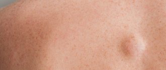
Lipoma (wen)
Lumps under the skin most often turn out to be lipomas. These are benign, completely safe tumors from fat cells. Lipoma can be felt under the skin as a soft formation with clear boundaries, sometimes with a bumpy surface. The skin over the lipoma is of normal color and density, easily folded.
Most often, lipomas appear on the scalp, neck, armpits, chest, back and thighs. When they reach large sizes, they can cause pain by squeezing neighboring organs or muscles. Find out more about how to get rid of lipoma.
Diagnostics
To make a correct diagnosis for tumors under the skin, the doctor can:
- Carry out a visual inspection. This allows you to make a preliminary diagnosis or outline the further direction of the search.
- Collect anamnesis. The patient must inform the doctor about all the features of the appearance of the tumor, previous injuries and cases of similar problems in relatives.
- Order general tests (blood, urine, etc.).
- Scrap the skin on the affected area.
- Collect tumor tissues and send them for histological examination.
- Prescribe an ultrasound, x-ray or CT scan (computed tomography).
Usually, carrying out the above actions is enough to make a correct diagnosis. It is worth recognizing that most often the doctor is able to determine the type of neoplasm already during the initial visit.
Atheroma
Atheroma is often confused with lipoma, also called a wen. In fact, it is a cyst, that is, a stretched sebaceous gland in which the excretory duct is blocked. The contents of the atheroma, sebum, gradually accumulates, stretching the capsule of the gland.
To the touch it is a dense, round formation with clear boundaries. The skin over the atheroma cannot be folded; sometimes the surface of the skin takes on a bluish color and you can see a point on it - a blocked duct. Atheroma can become inflamed and fester. If necessary, it can be removed by a surgeon.
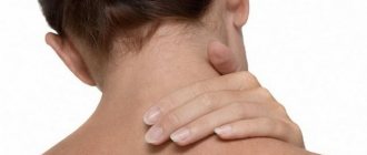
Nodules on the joints
Various joint diseases: arthritis and arthrosis are often accompanied by the appearance of small, hard, immobile nodules under the skin. Such formations in the elbow joint are called rheumatoid nodules and are characteristic of rheumatoid arthritis. Nodules on the extensor surface of the joints of the fingers - Heberden's and Bouchard's nodes accompany deforming osteoarthritis.
Gouty nodes - tophi, which are accumulations of uric acid salts and grow on the joints of people who have suffered from gout for many years, can reach a significant size.
Treatment methods
Methods for getting rid of tumors are selected individually, depending on what the tumors that have arisen on the body are:
- Sometimes the doctor advises only to observe the growths (lipoma or fibroma) if they do not cause any discomfort.
- To get rid of lipoma, fibroma, atheroma and hygroma, surgical treatment can be performed. Most often, tumors are removed using a scalpel, but modern surgeons also practice radio wave and laser treatment.
- Heberden's and Bouchard's nodes require targeted therapy for the underlying pathology. Patients are prescribed medications that stimulate the restoration of joint cartilage and slow down destructive processes.
- The appearance of a hernia most often becomes an indication for planned surgical intervention. If an acute strangulation of the hernia occurs by accident, the operation is performed immediately - as an emergency.
- When a wart is detected, doctors most often recommend surgical removal and targeted antiviral therapy to prevent relapse of the disease.
- Diagnosed cancer becomes an indication for removal of the disease focus and targeted chemotherapy or radiation therapy. With early diagnosis, the prognosis is usually favorable.
Thus, the approach to treating subcutaneous tumors on the body may differ. Only a doctor can select the most optimal method of therapy after conducting a comprehensive diagnosis.
Hernia
It feels like a soft protrusion under the skin, which can appear during exercise and disappear completely when lying down or at rest. A hernia forms in the navel, postoperative scar on the abdomen, in the groin, on the inner surface of the thigh. The hernia may be painful when palpated. Sometimes you can push it back in with your fingers.
A hernia is formed by the internal organs of the abdomen, which are squeezed out through weak spots in the abdominal wall during an increase in intra-abdominal pressure: when coughing, lifting heavy objects, etc. Find out whether a hernia can be cured using traditional methods, and why it is dangerous.
Seals in the form of subcutaneous balls
Tumors under the skin in the form of lumps and balls are a fairly common occurrence. As a rule, such neoplasms are of a completely benign nature - they do not pose a serious threat to human life, although they may cause some discomfort caused by their growth and compression of nearby structures. However, we should not exclude the possibility that the bumps under the skin are a symptom of some rather serious diseases, including cancer.
Only a dermatologist can determine what exactly the ball or lump under the skin is. To make a correct diagnosis, various additional studies may be necessary.
Such a neoplasm can be localized in different parts of the hand. Most often, lumps that look like soft lumps appear on the shoulders, but they can also form in other places. Such tumors can be essentially:
- Lipoma. This tumor occurs due to an abnormal accumulation of fat cells under the skin. Usually it develops gradually and can reach quite large sizes, but does not cause any discomfort to the patient. When palpated, such a tumor seems elastic, quite dense and mobile. Lipoma is not prone to malignancy, but can cause significant psychological discomfort.
- Hygroma. This benign cystic neoplasm is often the main cause of the appearance of a noticeable raised ball on the hand. Hygroma consists of a dense wall formed from connective tissue, as well as viscous contents. Hygromas are always associated with joints or tendon sheaths and are localized near them. Such a tumor can be completely soft or elastic, sometimes quite hard tumors are found. Pressure on the area of the hygroma may be accompanied by acute pain, as the tumor often presses on the nerve fibers.
- Atheroma. This is the name given to a tumor caused by a blockage of the sebaceous gland duct. Such a neoplasm can occur on any part of the body where there is hair. Atheroma is characterized by a superficial location, a dense-elastic structure, clear contours and mobility. This neoplasm is prone to inflammation and suppuration, which is manifested by redness, pain, swelling and local fever.
Rheumatoid node. This neoplasm has a fairly dense structure and can reach the size of a grain of rice to a walnut. A rheumatoid nodule occurs against the background of rheumatoid arthritis in the subcutaneous tissue. A typical location is the extensor surface of the elbow joint. Also, such neoplasms can be found on the forearms, the backs of the palms and other parts of the body. They don't hurt, but they can become infected.
The listed neoplasms are extremely rarely localized on the palms and, especially, on the fingers. Only a doctor can determine what exactly a particular subcutaneous lump is.
Subcutaneous tumors on the fingers are most often:
- Heberden's and Bouchard's knots. These are dense neoplasms that are localized on the nail phalanges of the index and middle fingers. At first, their size does not exceed a grain of rice, but subsequently the compactions grow to the volume of a large pea. Nodules appear on both hands at the same time; at first, this process may be accompanied by signs of inflammation - soreness, swelling and redness of the skin. The main reason for their appearance is arthrosis of the fingers.
- Fibroids. These are quite rare benign tumors, which are based on connective tissue and collagen fibers. Such neoplasms can arise at the site of insect bites and microtraumas, but more often they appear for unknown reasons. Fibroids start as a small nodule and slowly grow to become a noticeable lump. Such tumors usually do not cause any discomfort to the patient.
- Hygroma. Such a tumor, as we have already found out above, has a dense wall of connective tissue and viscous contents. It can be localized on the dorsum and palmar surface of the fingers. At rest, such neoplasms usually do not cause discomfort, but they hurt when moving or pressing.
- Banal calluses. Sometimes lumps under the skin occur due to prolonged friction and unusual stress. They may be painful, or they may not cause any particular discomfort if they form gradually.
Fingers are an atypical place for various lumps and lumps under the skin. Tumors in this area are extremely rare.
If an incomprehensible hard ball appears on your hand in the area of the hand, it is most likely a hygroma. As a rule, such a cystic tumor is quite elastic, but in medical practice there are also quite dense pathological neoplasms. In density they can resemble bone or cartilage.
As we said above, hygromas are in close relationship with the joint or tendon sheath, and accordingly, they are localized near them.
A hard lump on the wrist may also be a sign of:
- Deformations of bone tissue.
- Suffered and improperly treated trauma. In this case, the patient is concerned about a significant limitation in the mobility of the wrist.
Joint diseases, for example, arthrosis or arthritis.
As a rule, making a correct diagnosis for a lump on the wrist is not difficult. When faced with such a problem, you should first consult with a therapist, then you may need to visit a dermatologist, surgeon, rheumatologist and other specialized specialists.
The head quite often becomes the site of localization of various neoplasms. Here the following may arise:
- Lipomas or fatty tissues. This mobile, soft and painless neoplasm occurs due to the accumulation of fatty tissue. Lipomas are most often found under the hair, especially in the back of the head, but can actually be anywhere, including on the cheek or forehead.
- Atheromas. Such a neoplasm, as we have already found out above, occurs due to blockage of the sebaceous gland duct. Atheromas are most often localized behind the ears and on the neck, and can also occur on the face. Such tumors are more likely to occur in women with oily skin.
- Osteomas. This neoplasm is a benign bone tumor. It can be found on the bones of the skull and face (temporal, frontal), appearing as a hard lump in the shape of a hemisphere. Osteoma is considered a fairly rare pathology; it usually does not cause any discomfort to a person.
- Fibroids. This tumor often occurs on the head and neck and is less common on the face. The tumor usually has a round shape, with intact skin on top. Fibroma tends to grow slowly, usually does not cause discomfort and does not malignize.
- Trichoepitheliomas. This term is used to refer to a benign tumor-like formation of the hair follicle. Such a neoplasm most often occurs in women during adolescence; its location can be different parts of the body, but most often trichoepitheliomas are found on the face and scalp. A tumor on the forehead that does not hurt may well be trichoepithelioma. A single element may appear as a dense, dome-shaped neoplasm, about 2 cm in diameter. The tumor does not hurt, is covered with skin of natural color without ulceration. Over time, the growth may increase in size.
Warts. Such growths on the body can also resemble subcutaneous spherical seals, especially if they are localized under the hair, where it is quite difficult to see them independently. But most often, warts still stand out noticeably above the level of healthy skin. The reason for their appearance is the activity of the human papillomavirus. Such growths do not hurt, but they can become injured and become inflamed as a result.
In addition, a bump under the hair or on the face may be a trivial consequence of an insect bite that happened unnoticed by a person. It happens that the swelling from such damage lasts for a whole week or is even complicated by the addition of inflammation. But the bite site usually causes discomfort - it hurts and itches.
Subcutaneous spherical tumors on the legs, as well as in other places on the body, can be lipomas, atheromas or fibromas. But in addition, the cause of the appearance of spherical seals on the lower extremities may be:
- Phlebeurysm. Sometimes a vein swollen under the skin on the lower leg looks like a subcutaneous tumor. The patient may experience fatigue and heaviness in the legs, as well as some pain.
- Previously suffered trauma. A single bruise, surgery or bite on the leg can lead to the formation of a collection of connective tissue under the skin that will look like a lump. In some cases, a tumor occurs when a foreign body (for example, a piece of metal or a piece of glass) gets under the skin and becomes encapsulated.
- Rheumatoid nodes. Such tumors arise, as we said above, against the background of rheumatoid arthritis.
- Valgus foot deformity. In such a situation, a lump appears on the bone of the big toes. This is a very common problem, which is often diagnosed in the fair sex; its development can be caused by various joint diseases, previous injuries, genetic predisposition, wearing tight and uncomfortable shoes, etc. Most often, a bone requires surgical treatment; conservative therapy gives only a temporary effect.
- A wart. If there is a lump on your foot that makes it painful to step on, it could very well be a plantar wart. This new formation looks like a callus, but upon careful examination you can see one or more dark dots in the central part. The main cause of warts is the activity of the human papillomavirus.
If a formation on your leg causes significant discomfort, it is better not to hesitate to visit a doctor. Timely diagnosis will make treatment as fast and effective as possible.
The skin of the abdomen is a rather atypical place for the appearance of various benign neoplasms (lipomas, fibromas and atheromas). However, they may well occur in this area of the body. But in addition, a lump under the skin on the abdomen can be a sign of a hernia. In this case, it will be felt as a soft protrusion under the skin that occurs during exercise and completely disappears when lying down or simply at complete rest. A hernia is most often found in the navel area; such a protrusion can also be observed near a postoperative scar and in the groin. Its palpation may cause some discomfort and even pain.
A hernia is essentially the internal organs of the abdomen pushing outward under the skin through weak areas in the abdominal wall. This situation occurs when intra-abdominal pressure increases (during exercise, coughing, sneezing, etc.) and can be very dangerous. Hernias require surgical treatment.
Multiple lumps under the skin throughout the body are most likely multiple lipomas. When such symptoms develop, doctors talk about lipomatosis. This disease develops for reasons still unknown to science, but doctors suggest that its occurrence may be associated with impaired lipolysis and various endocrine ailments. The tendency to lipomatosis can be inherited. This disease usually makes itself felt not only by subcutaneous movable tumors, but also by:
- Soreness in the places where they are localized.
- Asthenia, adynamia, depression.
- Itchy skin.
- Increased blood pressure.
Lipomatosis lesions most often appear on the neck, chest and chin. They can compress peripheral nerves and other important structures in the body.
It is worth noting that subcutaneous spherical formations, which can be felt throughout the body, may be enlarged lymph nodes - important organs of the immune system. A change in their size can be observed in a variety of diseases, ranging from a common ARVI to cancer or HIV infection.
In fact, many serious diseases that can develop in humans can be triggered by stressful situations. And some ailments that manifest as bumps on the body are no exception. In particular, doctors note that they quite often have to deal with cases of lipomatosis and single lipomas, which arise against the background of severe emotional upheaval. And for patients with such tumors on the body, for successful therapy, it is extremely important to avoid both physical and psychological stress.
Exacerbation of HPV (which leads to the appearance of warts and papillomas) can also be triggered by severe emotional distress. And the lymphatic system can also respond to significant stress by enlarging lymph nodes throughout the body.
Even if bumps appear on the body after severe emotional stress, you should definitely seek medical help. Sometimes the consequences of mental suffering require completely physical treatment - the use of medications or even surgery.
Of course, when faced with any lump under the skin, many people first think about cancer. Indeed, some types of oncology may well manifest themselves as lumps and balls on the body. You can suspect the development of cancer by:
- Irregularities and unclear boundaries of education.
- Enlargement of nearby lymph nodes.
- The tendency of the tumor to actively grow.
- Adhesion of the neoplasm to the skin, lack of mobility during palpation.
- Bleeding and ulceration of the skin.
Despite the fact that cancerous tumors are very rare compared to benign skin tumors, the risk of their development cannot be excluded. It’s better to play it safe and show the lump that appears on your body to a doctor.
Enlarged lymph nodes (lymphadenopathy)
Most often accompanied by colds. Lymph nodes are small round formations that can be felt under the skin in the form of soft elastic balls the size of a pea to a plum, not fused to the surface of the skin.
Lymph nodes are located in groups in the neck, under the lower jaw, above and below the collarbones, in the armpits, in the elbows and knees, in the groin and other parts of the body. These are components of the immune system that, like a filter, pass interstitial fluid through themselves, clearing it of infection, foreign inclusions and damaged cells, including tumor cells.
An increase in the size of the lymph nodes (lymphadenopathy), which become painful when palpated, usually accompanies infectious diseases: sore throat, otitis media, flux, panaritium, as well as wounds and burns. Treatment of the underlying disease leads to a reduction in the node.
If the skin over the lymph node turns red, and palpation becomes sharply painful, the development of lymphadenitis is likely - a purulent lesion of the node itself. In this case, you need to contact a surgeon. Minor surgery may be required, and early treatment can sometimes clear up the infection with antibiotics.
If a dense, tuberous formation is felt under the skin, and the skin above it cannot be folded, the node is likely damaged by a malignant tumor. In this case, consult an oncologist as soon as possible. Read more about other causes of swollen lymph nodes.
Possible complications
In the event that a person who has discovered single or multiple lumps on his hands does not go to the hospital and does not treat the underlying disease, then the development of the following complications cannot be ruled out:
- degeneration of a benign cyst or tumor into a malignant neoplasm and its further growth with the transition to the final stages;
- the spread of cancer cells into the deeper layers of the dermis, damage to the bone tissue of the arm and muscle fibers, as well as the involvement of internal organs located in close proximity to the source of pathology in the oncological process;
- upper limb amputation;
- surgical removal of affected lymph nodes;
- inflammation of muscle fibers and temporary disability.
Regardless of what causes the appearance of lumps under the skin on the hands, the disease requires comprehensive treatment. In this case, you can count on a favorable prognosis and complete recovery of the patient without the risk of relapse.
Benign neoplasms are not dangerous to human health, but there is always a risk of their degeneration into a malignant tumor, which can lead to disability or, in the most severe cases, even death.
Warts, papillomas, condylomas, soft fibromas
All these terms refer to small growths on the skin of various shapes: in the form of a polyp, a mole on a thin stalk, growths in the shape of a cockscomb or cauliflower, a hard nodule or papilla protruding above the surface. These lesions may be yellowish, pale, brown, or flesh-colored and have a smooth or flaky surface. Read more and look at photos of warts and papillomas.
Their causes are different: most often it is a viral infection, mechanical injury, hormonal disorders. Sometimes warts and papillomas grow “out of the blue,” for no apparent reason, and can be located on any part of the body, including the mucous membranes of the genital organs. For the most part, these are harmless growths that cause only cosmetic discomfort or interfere with wearing clothes or underwear. However, the variety of their shapes, colors and sizes does not allow one to independently distinguish a benign wart, condyloma or soft fibroma from malignant skin diseases. Therefore, if a suspicious growth appears on the skin, it is advisable to show it to a dermatologist or oncologist.
Skin inflammation and ulcers
A whole group of skin lesions may be associated with infection. The most common cause of inflammation and suppuration is the bacterium staphylococcus. The skin in the affected area turns red, swelling and induration of varying sizes appear. The surface of the skin becomes hot and painful to the touch, and overall body temperature may also increase.
Sometimes inflammation quickly spreads across the skin, covering large areas. Such a diffuse lesion is characteristic of erysipelas (erysipelas). A more serious condition - phlegmon - is a purulent inflammation of the subcutaneous fatty tissue. Focal inflammatory diseases are common: carbuncle and furuncle, which are formed when hair follicles and sebaceous glands are damaged.
Surgeons treat purulent-inflammatory diseases of the skin and soft tissues. If redness, pain and swelling appear on the skin, accompanied by an increase in temperature, you should seek help as soon as possible. In the initial stages, the problem can be solved with the help of antibiotics; in more advanced cases, surgery must be resorted to.
Malignant tumors
Compared to other skin lesions, malignant tumors are very rare. As a rule, at first a focus of compaction or a nodule appears in the thickness of the skin, which gradually grows. Usually the tumor does not hurt or itch. The surface of the skin may be normal, flaky, crusty, or dark in color.
Signs of malignancy are:
- uneven and unclear boundaries of the tumor;
- enlargement of nearby lymph nodes;
- rapid growth in education;
- adhesion to the surface of the skin, inactivity when palpating;
- bleeding and ulceration on the surface of the lesion.
A tumor can develop at the site of a mole, like melanoma. It can be located under the skin, like a sarcoma, or at the site of a lymph node - lymphoma. If you suspect a malignant skin tumor, you should contact an oncologist as soon as possible.
Etiology of compactions under the skin
Lump on face under skin
The appearance of painful or painless lumps on the arm under the skin can be due to a number of reasons:
- long-term insolation;
- metabolic disorders;
- injuries;
- weak immunity;
- unfavorable environment;
- infections;
- poor body hygiene;
- exacerbation of any disease;
- emotional stress.
Advice. The etiology of subcutaneous neoplasms is quite extensive, so it is sometimes very difficult to establish the true cause that provoked the development of pathology. To make a correct diagnosis, you need to undergo a comprehensive diagnosis.
Cyst
Causes of cysts
Epidermal cyst (atheroma) is a fairly common phenomenon. It is a tumor of benign etiology that forms on the surface of the skin. Its size and location may vary. If removed in a timely manner, the pathology does not pose a health hazard. Cysts form for various reasons.
Infections
Cysts often form against the background of the development of furunculosis. The disease is provoked by two types of staphylococcus: aureus and epidermal. The second type of microorganisms causes suppuration when they enter the deeper layers of the skin from the surface. About 75% of humanity are carriers of staphylococci, so the transmission of microorganisms occurs constantly, however, for the development of the disease, it is necessary for the infection to enter favorable conditions: into an organism with weakened immunity, various diseases.
Blockage of the sebaceous glands
A blockage of the sebaceous gland is a mobile, painless formation. It is characterized by clear contours. In some cases, it grows almost imperceptibly, sometimes its size does not change at all for several years. Sometimes blockage of the sebaceous gland is accompanied by suppuration with pain, swelling, redness and fever. In many cases, pus breaks out in combination with fatty contents. Increased sweating, which manifests itself mainly during hormonal changes characteristic of young age, also contributes to the development of atheromas.
Complications after piercing and tattoo
Piercing and tattoos as an element of subculture are unlikely to go out of fashion. However, their popularity still fell; mass demand was observed in the late 90s - early 2000s. After piercing, the puncture site must be constantly treated with antiseptic agents, otherwise the wound may become infected. If seals form at the puncture site over time, you should consult a cosmetologist or dermatologist. Epidermal cysts are very often diagnosed after piercings and tattoos.
Cyst treatment
Treatment of cysts can be carried out using both conservative and radical methods. The method of treatment depends on the size of the tumor. If the epidermal cyst is small in size and does not cause concern to the patient (including aesthetics), then it does not need to be treated. In this case, doctors recommend monitoring the dynamics of tumor development. If a cyst bothers a person, then it needs to be removed. The following methods can be used in treatment.
Crushing the subcutaneous bladder
Often a cyst is mistaken for a common pimple and they try to squeeze it out. However, this method will not lead to anything good, except that it will begin the inflammatory process.
Pumping out liquid
Ineffective method of treatment. During this procedure, there is a high risk of wound infection and the development of inflammatory processes. You should absolutely not squeeze out the cyst. During destruction, the contents of the cyst can get under the dermis, leading to an abscess. If this has already happened, you should immediately go to the doctor.
Surgery
All manipulative actions are performed under local anesthesia. The epidermal cyst, which is located above the outer skin, is removed within 20 minutes. After injecting a solution of novocaine or another anesthetic, the doctor makes an incision in the center of the cyst and removes its contents, capturing the capsule, then scrapes out the cavity and cleanses the skin. In case of relapse, repeated surgical intervention is prescribed.
Lipoma
Causes of lipoma
Lipoma is a polyetiological disease that develops against the background of various endo- and exogenous factors.
Hormonal disbalance
Lipomas are most often diagnosed in adolescence and in women during menopause. Steroid hormones (androgens and estrogens) affect the functioning of the sebaceous glands, regulate the proliferation of skin cells, and the secretion of sebum. If androgens predominate in the body of women (taking hormonal drugs, age, menopause, tumors), the tissue of the sebaceous glands grows with an increase in secretion production.
Genetics
It has been proven that the formation of lipomas has a hereditary predisposition. Multiple lipomas (lipomatosis) are often traced in several generations, regardless of gender. In a study of twins with this pathology, data were obtained on the presence of this pathology in both children. Vertical inheritance is traced in 99% of cases.
Note. The development of lipoma is often observed against the background of alcoholism, malignant tumor processes of the upper respiratory tract, and diabetes mellitus.
Schematic representation of a lipoma
How to remove wen at home
At home, you can use both pharmacy and folk remedies. Before using any product, it is necessary to perform a sensitivity test. A small amount of the drug used is applied to the skin in the wrist area. If allergic reactions (rashes, itching, hyperemia) are absent, the product can be used.
Doctors recommend using the following medications:
- We see. The product is applied to the skin twice a day, after which the problem area is covered with a plaster. The duration of therapy is determined by the doctor individually.
- Vishnevsky ointment - the substance is generously applied to the lipoma, covered with a napkin and sealed with a band-aid. As the product dries, the “compress” is changed. It usually takes 4 to 5 days to remove the wen.
To remove lipoma, traditional healers recommend using the following ingredients:
- Aloe compress. The leaves of the plant are thoroughly crushed. After that, the resulting pulp is spread on gauze or a bandage and applied to the wen for 10-12 hours. This procedure is repeated until visible results appear.
- Vodka compress. Ethanol is mixed with vegetable oil in a 1:1 ratio, then a compress is made from the resulting solution.
Advice. Before removing a wen at home, you need to soberly assess the situation. Self-medication is only permissible for small and shallow formations. Large lipomas need to be eliminated only surgically; folk remedies can only cause harm.
Radical method of removing wen
Hygroma
Causes
The reasons for the development of the disease are poorly understood. It is assumed that hygroma occurs under the influence of several factors. During the study, it was found that such formation more often occurs in blood relatives, which indicates a hereditary predisposition.
Complications after illness
Complications from hygroma are extremely rare. Neurological signs (sensation of numbness of the skin, crawling) occur when nerve fibers are compressed. Spontaneous openings of hygroma usually do not occur. But when it is traumatically damaged, there is a prolonged flow of the contents of the hygroma through the resulting hole. Sometimes, in place of one crushed hygroma, several may appear.
Mechanical injuries
As a rule, the development of hygroma is preceded by a single injury. Most surgeons point out a connection between the development of hygroma and repeated trauma or constant high load on the joint or tendon.
In the fairer sex, hygromas are diagnosed almost 3 times more often than in men. In elderly patients and children, the occurrence of hygroma is extremely rare.
Great physical activity
Among other things, heavy loads during sports can lead to hygroma. If the hygroma does not increase in size and does not hurt, then physical activity is not contraindicated, but it is necessary to monitor its intensity. It is better to consult with your doctor.
Treatment of hygroma
The treatment regimen is individual. The doctor assesses the patient’s general condition, determines the location, size and structure of the hygroma, and only then prescribes appropriate therapy.
Puncturing
Using a needle and a syringe attached to it, the doctor punctures the hygroma and sucks out its contents. The puncture is made in the place where the skin is more swollen. To completely remove the hygroma, it may be necessary to puncture several times. In the presence of inflammatory processes, an injection of corticosteroid hormones is administered into the cavity of the neoplasm.
Physiotherapeutic effects
Some patients are helped to cope with the problem by physiotherapy (ultrasound, mud therapy, UHF, magnetic therapy, salt and soda baths).
It should be noted! Their use is advisable in the presence of aseptic inflammation.
Traditional methods
A copper coin is tightly bandaged to the hygroma. The bandage is not removed until the swelling disappears. In some cases, 5-7 days are enough, in others – 2-3 weeks.
Surgery
The surgery is performed under local anesthesia on an outpatient basis. The operation is very delicate. The hygroma capsule along with its contents is completely removed. Stitches are applied in layers. After a week they are removed.
Which doctor should I contact with a lump or lump on the skin?
If you are concerned about formations on your body, find a good doctor using the NaPravku service:
- a dermatologist - if the lump looks like a wart or papilloma;
- surgeon - if surgical treatment of an abscess or benign tumor is required;
- oncologist to rule out a tumor.
If you think that another specialist is needed, use our help section “Who Treats It”. There, based on your symptoms, you can more accurately decide on the choice of a doctor. You can also start with an initial diagnosis from a therapist.
Diagnosis and treatment
In some cases, a doctor's consultation is necessary.
To determine the cause of the lump on the brachialis muscle of the arm from the shoulder, you need to visit a dermatologist or therapist. Next, the doctor will prescribe laboratory tests and instrumental examination: ultrasound, x-ray. In some cases, consultation with traumatologists, endocrinologists and other specialists is required, as well as an MRI or CT scan. To examine tumors with their contents, a puncture is prescribed. The same is done when studying pathologies associated with the state of the synovial fluid inside the joint cavity.
An integrated approach is used to treat skin diseases and tumors on the shoulders. It greatly depends on the cause of the pathology. Most often it is built on the following principles:
- Use of medications. Drugs aimed at eliminating the cause of the disease and symptomatic remedies are used. These could be NSAIDs, decongestants, immunosuppressants, vitamins.
- Use of gels and ointments. Particularly effective in the treatment of skin pathologies.
- Physiotherapy. Helps if the seal on the shoulder muscle is painful or inflamed. Electrophoresis, magnetic therapy, treatment with leeches, laser and UHF promote tissue regeneration and improve metabolism.
- Folk recipes. They are used in courses of 10-30 procedures over 2-4 weeks. Most often, lotions and compresses are made from dry herbs.
In severe cases, when a growth appears on the shoulder joint, injections are prescribed. They are made with NSAIDs, corticosteroids, vitamins, antihistamines and painkillers to relieve very severe pain.
Treatment of the initial stage of pathologies is easier and faster, patients recover fully. In advanced cases, treatment may take several months or more than a year.
Surgical intervention
In advanced cases, surgical intervention is required.
This method is used in advanced cases, when no conservative methods will bring relief. After excision of painful tissue and removal of growths, a rehabilitation period follows, during which the arm is completely immobilized. The operation is performed quickly and is well tolerated by patients.
To prevent the development of dangerous stages of tumors, lipomas, hygromas and other pathologies, if a lump appears on the shoulder, you should immediately consult a doctor. By following the recommendations, the patient will be able to completely forget about the problem within 2-3 months.
You might also be interested in reading
Localization and translation prepared by Napopravku.ru. NHS Choices provided the original content for free. It is available from www.nhs.uk. NHS Choices has not reviewed, and takes no responsibility for, the localization or translation of its original content
Copyright notice: “Department of Health original content © 2019”
All site materials have been checked by doctors. However, even the most reliable article does not allow us to take into account all the features of the disease in a particular person. Therefore, the information posted on our website cannot replace a visit to the doctor, but only complements it. The articles have been prepared for informational purposes and are advisory in nature.
Why did a lump appear under the skin: the main reasons
The most common diseases that are accompanied by subcutaneous lumps are:
1. Lipoma. The tumor is formed from adipose tissue. The lump is painless, has the same color as the skin, and when touched, a dense formation is felt. As a rule, a lipoma is a benign tumor and does not cause discomfort. Such tumors occur due to metabolic disorders in the body. Statistics show that women are more susceptible to this disease than the stronger sex.
2. Subcutaneous cyst. The symptoms of a cyst are the same as for a lipoma, the difference is periodic inflammation of the lump. In some cases, contents are released from the cone.
3. Hygroma. This compaction in the form of a ball under the skin does not cause virtually any discomfort, except for a visible cosmetic defect. Most often, a lump occurs on the hand under the skin on the wrist or palm. It is an accumulation of liquid and can burst on its own under any mechanical stress.
4. Fibroma. A benign tumor that does not hurt, does not itch, and does not protrude much above the surface of the skin. Fibroma occurs after injuries, inflammatory processes, and may be associated with a hereditary factor. The lump can be soft or hard, and the color of the lump varies from brown to red.
Risk factors
A sedentary lifestyle and obesity can lead to the formation of lumps on the shoulder.
Common factors that provoke the appearance of lumps on the shoulder joints and forearms include:
- maintaining a sedentary lifestyle;
- obesity;
- professional sports;
- heavy physical activity;
- endocrine disorders.
However, even completely healthy people can face this problem due to heredity.
How to treat lumps under the skin quickly and effectively
Many people wonder whether it is possible to treat lumps under the skin at home? The answer is clear: under no circumstances should you try to squeeze out, puncture, or influence it by any mechanical means. If you find a defect, contact a specialist. First, visit a therapist, and only after that a specialist: oncologist, surgeon or dermatologist.
Some lumps go away on their own and do not require treatment. For example, lipomas are removed when they become a visible cosmetic defect, and the doctor may even recommend not touching a small cyst if it does not cause discomfort. If the cyst becomes inflamed, then special injections or surgery are prescribed. Fibroma and hygroma are removed surgically. If the lump is associated with an infectious disease, then first you should undergo a course of treatment aimed at eliminating the problem.
If you find a lump under the skin, be sure to consult a specialist. Only he will be able to finally exclude or confirm your concerns, explain why the ball has formed under the skin, and prescribe the correct treatment.
Methods for treating lumps on the hands under the skin
The method of treating foreign tumors on the hands is selected based on the reasons that caused their appearance. Medicines or surgery may be used.
To eliminate lumps of inflammatory and malignant etiology, the following medications can be used:
- Erythromycin is an antibacterial agent that is effective for the treatment of bumps caused by lymphadenitis, prescribed 200-500 mg 2 times a day with a duration of therapy from 5 to 14 days (medicine price 67 rubles);
- Chemotherapy drugs - prescribed in courses in the form of intravenous drips, indicated for use when lumps are detected under the skin of the hands, which are cancerous tumors (the selection of a chemotherapy drug and the number of courses of treatment are determined individually);
- Azithromycin - the drug is prescribed for internal use, 1 capsule 2 times a day, 2 hours after meals, or 1 hour before eating (the duration of treatment for lumps of infectious or inflammatory etiology is 3-5 days, the cost of the medication is 105 rubles);
- Rescuer ointment - the drug is used for therapeutic purposes in the formation of subcutaneous lumps on the hands, which arose as a result of bruises, postoperative complications, calcification of hematomas (the ointment is applied to the surface of the tumor 2 times a day for 10-15 days, and the cost of a 30 g tube is 160 rub.).
The drug should be prescribed only by the attending dermatologist, oncologist or surgeon. Self-therapy can lead to deterioration of health.
Lumps that form under the skin of the upper extremities due to the appearance of benign, cancerous tumors or acute inflammation of the lymph nodes can be symptoms of dangerous diseases.
Therefore, it is not recommended to use traditional medicine, since tinctures and decoctions of medicinal herbs will not have the desired therapeutic effect, but will transform the disease into a chronic or complicated form.
Surgical therapy is used together with medications. Surgery is effective in cases where bumps on the hands are the consequences of lipoma, dermatofibroma, liposarcoma, fibrosarcoma or a complication of lymphadenitis.
The surgical operation is performed as follows:
- The patient is transferred to the surgical room.
- The anesthesiologist administers local or general anesthesia depending on the complexity of the clinical case.
- The surgeon dissects the epithelial and soft tissue in the area where the lump is located.
- The tumor is removed and the wound is treated with antiseptic.
- Suture material is applied.
The average duration of the operation is 30 minutes. up to 1 hour
The period for complete wound healing and rehabilitation lasts from 7 to 14 days. The cost of the operation varies from 2500 to 5000 rubles. depending on the pricing policy of the clinic management and the complexity of surgical procedures. In a public hospital, the operation is free.