Causes
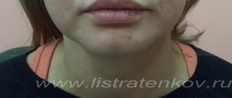
Despite the fact that the appearance of wrinkles is considered to be a sign of age-related changes and aging of the skin, in some people, due to the anatomical structure of the face or other individual factors, nasolabial wrinkles appear at a fairly early age.
Causes of early appearance of wrinkles:
- insufficient hydration and skin care;
- decreased skin elasticity;
- smoking;
- high facial activity;
- sleeping on the stomach (when a person sleeps with his face buried in the pillow).
Fortune telling by philtrum
Like other facial features, the philtrum is an object of study in physiognomy and magic. The shape of the mouth also depends on its shape, so predictors often use information about the philtrum in their predictions.
What myths are associated with the philtrum?
There is a legend that says that the philtrum is the mark of an angel's finger. When he brings a child down from heaven to earth before birth, he puts his finger to the baby's mouth. This is done so that he forgets everything he saw before birth - heaven, angels, his past lives and another world. This allows a person to live his life in blissful ignorance of his past mistakes. Screenwriter Jaco Van Doormael reflected this legend in the film “Mr. Nobody” (2009).
There are also references to the philtrum in Judaism. In the treatise Nida of the Babylonian Talmud it is said that angels teach the baby Torah while still in the womb. To erase the memories, the angel lightly hits the baby on the lips. The Jew recreates this unique part of the Torah, learned in the womb, in his memory throughout his life.
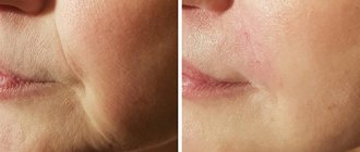
How to get rid of nasolabial wrinkles
Correction of nasolabial folds does not have strict indications based on age. The choice of technique depends on the cause of the appearance and type of wrinkles (age-related, pronounced facial wrinkles) in the nasolabial triangle.
We offer several effective techniques for eliminating nasolabial folds.
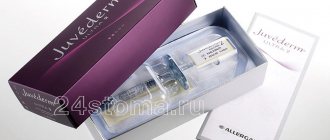
Contour plastic. Injections based on hyaluronic acid allow you to naturally fill existing wrinkles and, if necessary, recreate the missing volume in the cheekbone area to restore facial proportions. EMC doctors work only with biodegradable drugs (that is, those that “dissolve over time”) from well-known global manufacturers, with a proven clinical history and mandatory certification.
Professional skin care treatments using microcurrent therapy. The therapeutic effect of microcurrents (ultra-small amplitude current) in combination with active concentrates restores the tone of facial muscles, promotes the activation of the synthesis of collagen and elastin fibers, and has a lymphatic drainage effect.
Laser rejuvenation. Fractional photothermolysis, which underlies laser rejuvenation, helps solve a number of aesthetic problems: eliminate pigmentation, even out skin texture and tone, increase skin density and elasticity and, of course, smooth out wrinkles.
Thread lifting. Self-dissolving threads inserted under the skin tighten the area of the cheek-zygomatic area, forming a frame and fixing the skin in the desired position. Due to tissue reinforcement, wrinkles in the nasolabial triangle become less pronounced. A combination of fillers (hyaluronic acid injections) and threads gives a high aesthetic result and long-term effect.
Thermage rejuvenation. Deep thermolifting using monopolar radio wave energy allows you to achieve a pronounced effect of rejuvenation and elimination of wrinkles in just 1 procedure. Within six months after the procedure, new collagen fibers are synthesized, the skin becomes denser, and wrinkles become less pronounced.
Surgical lifting. Facelift and SMAS lifting are recommended for patients with pronounced age-related facial changes and sagging skin (ptosis). With surgical correction, you can eliminate deep wrinkles on the face, return tissues to the correct position, tighten the oval of the face, and correct the cervical-chin angle.
Why does asymmetry of nasolabial folds appear?
Physiological reasons
The faces of most people are asymmetrical, which is explained by slight differences in the structure of the right and left halves and the formation of facial wrinkles. Asymmetry of the nasolabial folds is especially noticeable in the habit of smiling at one corner of the mouth, curling the mouth to express dissatisfaction, sleeping on one side, or chewing gum on one side of the mouth. Symmetry disorders progress with age, but in the absence of other causes they do not reach the level of a noticeable cosmetic defect.
Facial neuritis
The most common neurological cause of asymmetry of the nasolabial folds is considered to be facial neuritis (Bell's palsy), accompanied by unilateral weakness of the facial muscles. The pathology occurs primarily as a result of a cold or complicates the course of the following conditions:
- Otitis media
Symptoms develop against the background of shooting pain in the ear. - Parotitis.
The appearance of asymmetry is preceded by an enlargement of the salivary gland, changes in facial contours, and signs of general intoxication. - Herpetic infection.
The manifestation of neuritis is caused by a special form of herpes zoster - Hunt's syndrome, in which ear pain, skin rashes, hearing loss, and dizziness are observed. - Facial nerve injuries.
The nasolabial fold is smoothed out due to a violation of the integrity of the nerve trunk or its compression by scar tissue. - Melkersson-Rosenthal syndrome.
Occurs with periodic relapses. Complicated by neuritis in 2% of patients. Other manifestations include dense facial swelling and a folded tongue. - Alternating syndromes.
Facial paresis in Millard-Gübler syndrome is complemented by the opposite hemiparesis, in Gasperini syndrome – strabismus, hearing loss, and sensitivity disorders. Brissot-Sicard syndrome is characterized not by paresis, but by spasm of the facial muscles with a deepening of the nasolabial fold.
Neuritis is diagnosed with tumors of the brain and the area of the facial nerve, for example, neuroma of the internal auditory canal. In addition, Bell's palsy occurs against the background of neuroinfections, which include:
- Encephalitis.
A group of diseases of fungal, bacterial and viral nature with intoxication syndrome, general cerebral and focal symptoms. - Polio.
The lesion is caused by the polio virus and is observed in the stem form of the disease. - Brain abscess.
Limited accumulation of pus in the brain tissue, accompanied by focal symptoms and severe intoxication. - Neurosyphilis.
In the early stages, focal, cerebral, and general infectious manifestations are detected. Subsequently, mental disorders, progressive dementia, and stroke-like symptoms are detected. - NeuroAIDS.
Paresis is combined with aphasia, ataxia, mnestic disorders, and psychopathological manifestations. - Botulism.
Develops acutely after eating canned food. Paresis and paralysis are typical, respiratory and cardiac problems are possible.
In the initial stages of neuritis, asymmetry appears due to smoothing of the nasolabial fold on the affected side. In the absence of treatment or inadequate treatment, patients develop contracture of facial muscles. In this case, the nasolabial fold on the affected side, on the contrary, becomes more pronounced.
Asymmetry of nasolabial folds
Cerebrovascular disorders
Asymmetry of nasolabial folds is of great practical importance in the development of acute cerebrovascular accidents. This symptom is clearly visible and detected at an early stage. Along with other signs (slurred speech, weakness of the limbs, deviation of the tongue to the side), it allows you to quickly determine the nature of the problem and promptly deliver the patient to a medical facility. It is detected in the following variants of stroke:
- transient cerebrovascular accident;
- ischemic and hemorrhagic strokes;
- migraine stroke;
- subarachnoid hemorrhage.
The frequency of occurrence of changes in the configuration of nasolabial folds in different types of stroke varies. The symptom is quite typical, but not pathognomonic for this pathology; it occurs due to damage to the parts responsible for the functioning of the facial nerve. The absence of a sign is not a basis for excluding stroke.
Traumatic brain injuries
As in the previous case, asymmetry of the nasolabial folds develops as a result of disruption of the brain centers that regulate the activity of the facial nerve. Can be observed with the following traumatic brain injuries:
- brain contusion (mostly moderate and severe);
- acute compression of the brain;
- diffuse axonal damage;
- intracerebral, subdural, epidural hematomas.
The severity of asymmetry differs significantly. The pathology is most noticeable with acute compression and is combined with a “sailing cheek” and lagophthalmos.
Innervation disorders in children
The symptom often accompanies various forms of dysarthria in children. Gross changes in most cases are observed in cerebral palsy. Slight asymmetry of the nasolabial folds is found in children with erased dysarthria associated with inadequate innervation of the tongue, lips, and soft palate. Several cranial nerves are affected, the asymmetry is complemented by limited movements of the lower jaw and tongue, hypersalivation, and impoverished facial expressions.
Dental problems
Asymmetry of nasolabial folds can be congenital or acquired. Caused by the following reasons:
- Lack of teeth.
With a long-term absence of molars and premolars on one side, the contours of the face gradually change, the nasolabial fold deepens. Bilateral absence of teeth causes deepening of the folds on both sides, the severity of the asymmetry is determined by the location of the remaining dental units. - Crossbite.
There is a crossing of the dentition when the jaws are closed. The chin moves, the lip sinks, which entails a violation of the symmetry of the lower parts of the face. - Tumors of the salivary glands.
Distortion of the nasolabial folds can be determined by adenomas, lipomas, angiomas, neuromas, sarcomas, carcinomas, and is formed secondary to compression or germination of the facial nerve passing near the salivary gland. - TMJ diseases.
Restriction of movements in the temporomandibular joint due to arthrosis, ankylosis, and contractures causes lateral displacement of the lower jaw and distortion of the face. - Tumors of the jaws.
Asymmetry becomes one of the first symptoms of a neoplasm of the upper jaw when it is located in the projection of the nasolabial fold. - Jaw defects.
Developmental defects, post-traumatic deformities, defects after tuberculosis, osteomyelitis, and removed tumors of the upper jaw lead to receding cheeks and smoothing of the nasolabial fold. In patients with mandibular defects, asymmetry is formed due to displacement of the jaw when opening the mouth. - Injuries.
With fresh jaw fractures, asymmetry is provoked by swelling and displacement of fragments. In the long term, changes in the contours of the nasolabial folds are caused by improper fusion of bone fragments and excessive formation of callus.
Consequences of aesthetic procedures and operations
The lesion is often potentiated by the introduction of fillers based on calcium hydroxylapatite, polycaprolactone, and L-lactic acid polymer. The listed agents cause increased formation of fibrin fibers and proliferation of connective tissue. Some time after the procedure, uneven fibrosis or the formation of coarse fibrous strands causing asymmetry may be observed.
Collagen-based fillers quickly dissolve, which necessitates the need to inject an excess amount of the drug and possible overcorrection of the nasolabial folds. Sometimes, after the use of such fillers, granulomas and areas of compaction appear. The formation of granulomas is also observed after the injection of the patient’s own adipose tissue.
In some patients, asymmetry occurs after ligature lifting of the nasolabial folds and the use of various methods of surgical facelift. The reasons for the changes are insufficiently careful planning or violation of the intervention technique, the patient’s failure to comply with medical recommendations, and complications in the postoperative period.
Advantages of the EMC Clinic
There are no universal solutions in cosmetology, because each patient is truly individual. Therefore, an effective solution begins with a personal consultation with a doctor.
During the consultation, the doctor will help you choose a method for eliminating nasolabial folds that will give the most pronounced effect and long-lasting results, and will give recommendations regarding skin care and recommended aesthetic procedures.
Modern technologies in the capable hands of EMC doctors allow us to demonstrate amazing aesthetic results, primarily because in our work we always rely on the needs of the patient and take into account all his individual characteristics.
Diagnostics
The reason for the change in the configuration and depth of the nasolabial folds is determined by a neurologist. According to indications, patients are referred to dentists, maxillofacial, and aesthetic surgeons. The examination program includes the following diagnostic procedures:
- Questioning, inspection.
The doctor finds out when and under what circumstances the symptom appeared, and how it changed throughout the course of the disease. Evaluates speech, memory, psycho-emotional state of the patient. Through conversation and physical examination, identifies other manifestations of the disease. - Neurological examination.
The specialist pays attention to the violation of the symmetry of different parts of the face, the size of the pupils and palpebral fissures. Examines eye movements, asks to puff out cheeks, bare teeth, frown, show tongue. Determines reflexes, sensitivity, muscle tone of various areas of the face and body. - Radiography.
In case of TBI, radiography of the skull is performed; in case of traumatic injuries and diseases of the facial skeleton, photographs of the jaws and radiographs of the TMJ are taken. - Echoencephalography.
Performed at the initial stage of the examination. Allows you to quickly detect space-occupying processes (tumors, abscesses, hematomas) that cause displacement of the midline structures of the brain. - and MRI of the brain.
They are used at the stage of clarifying the diagnosis, making it possible to clarify the nature, volume and localization of hemorrhages, neoplasms, and inflammatory processes. - Electrophysiological studies.
Electromyography, electroneurography, evoked potentials are prescribed to determine the localization of the pathological process and the severity of damage to the nerve trunk in case of neuritis of the facial nerve.
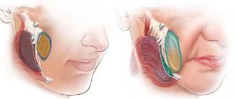
Contour plastic surgery is a method of eliminating asymmetry of nasolabial folds
How to avoid ptosis: prevention
Time cannot be stopped completely, but you can significantly slow down its pace by choosing an effective set of anti-aging procedures. They include many points that not only improve appearance, but also help maintain the overall tone of the body, and the famous saying says: “a woman is as old as she feels.”
Therefore, it is worth taking into account and using the entire arsenal of modern opportunities in order to feel great. The list includes:
- balanced diet;
- UV protection;
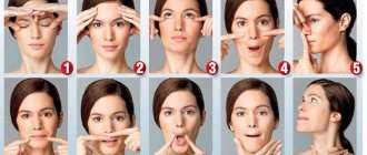
- self-massage;
- taking vitamin and mineral complexes;
- hardware cosmetology;
- good mood.
How is bullhorn cheiloplasty performed?
Considering the mass of individual features of the facial structure, each operation is unique in its own way. None of the famous plastic surgeons uses the stencil technique, but in general, three methodologies for lifting the upper lip can be distinguished.
External bullhorn is made through incisions at the base of the nose. Part of the skin and soft tissue is excised, after which the lip is placed in a higher position. In some cases, with external bullhorn, the incisions are made along the edge of the lip, but the first option is preferable, since the scars are actually hidden in the nostrils
The Italian bullhorn is a type of outdoor plastic. The difference between the method is that instead of one continuous incision at the base of the nose, two short ones are made on each side. Internal bullhorn is a fundamentally different operation. The lip is raised through cuts in the mucosa. The skin is not excised; no scars remain.
The operation takes no more than an hour and is performed under local anesthesia. The woman returns home 1-2 hours after the plastic surgery; recovery is easy and takes about 12 days. The main discomfort of the rehabilitation period is related to the fact that you cannot laugh and it is inconvenient to eat in the first few days. Pain is minimal, as is swelling after surgery.
Bullhorn plastic surgery: how to achieve harmonious proportions
According to the canons of beauty, the distance between the border of the upper lip and the base of the nose should be about 1.2 - 1.5 cm. A significant increase in this distance is a relative indication for bullhorn cheiloplasty. If the distance is less than the stated numbers, “raising” the upper lip is impractical.
Why did we call the reading relative? Because numbers taken out of context mean nothing. Attractive, feminine and sexy facial features are the result of a harmonious combination of all its elements. The subjective aesthetic perception of the mouth, lips, nose and nasolabial area is greatly influenced not only by the distance between the nose and upper lip. What matters is the height and inclination of the tip of the nose, the distance from the base of the columella to the tip of the nose, the nasolabial angle and other features of appearance, in particular the position of the corners of the mouth, the width and volume of the lips.
Bullhorn surgery is not indicated for women with a high projection of the nasal tip. In this case, the result of the correction will most likely bring only disappointment. If the distance from the columella to the tip of the nose is greater than the distance between the columella and the upper lip, it is better to refuse the operation. According to the canons of beauty, the length of the columella should not be greater than this distance, therefore, raising the upper lip even more strongly with such a facial structure is irrational.
A gummy smile and an increase in the nasolabial angle of more than 120 degrees are other “contraindications” to surgery. Of course, in this case we are talking about aesthetic contraindications. As for medical prohibitions, these include: a tendency to form rough scars, herpes infection with frequent rashes, hemophilia, neoplastic process of any localization, decompensation of endocrine and somatic pathology, pregnancy, lactation.