- Causes of human papillomavirus
- Manifestation of the disease
- Is there a difference between papillomas and warts?
- How does infection occur?
- What you need to know about HPV
- Treatment of papillomas
- HPV and cancer
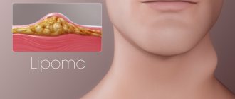
Papilloma is a benign skin tumor. Externally, it is a cylindrical growth on a stalk. The name itself comes from the Latin word papilla, which translates as “nipple”. Papilloma can be single or multiple. The growth most often appears in the armpits, on the upper eyelid, on the bends of the elbows and knees. In some cases, numerous papillomas literally cover the entire body, which creates discomfort when wearing clothes and is perceived as a serious cosmetic defect.
Causes of human papillomavirus
The cause of papillomas is a special virus. It is called human papillomavirus (HPV) or papillomavirus. Scientists know more than 100 types of HPV. Some of them are relatively harmless, while others, on the contrary, are very dangerous due to the risk of cancer.
The introduction of a virus into the body does not mean that papillomas will immediately appear on the body. As a rule, the pathogen manifests itself when immunity decreases. Growths on the skin can suddenly appear in the heat, after a person has had an acute respiratory viral infection, or after an exacerbation of chronic diseases.
Reasons for the appearance of a mole
Among the reasons for the formation of nevi are: genetic predisposition, prolonged exposure to ultraviolet radiation on the skin, hormonal pathology and other factors.
Most often, the formation of moles on the body is observed during puberty and in women with the onset of pregnancy.
This is explained by an increase in pituitary activity during this period of a person’s life.
Often people tend to consider any formation on the skin as moles, however, this is a big misconception.
Therefore, any neoplasm on the skin must be identified by a specialist.
Manifestation of the disease
As a rule, the presence of papillomavirus is indicated by growths on the skin, but this is not the only symptom.
Some types of the virus cause papillomas to affect the genitals or respiratory tract. Such diseases are not easy to diagnose, as they are similar to other pathologies.
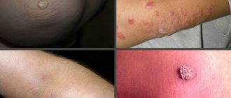
For example, when a virus affects the respiratory tract, growths appear in the nasopharynx and larynx. As a rule, a person experiences symptoms of a respiratory tract disease, so he consults an ENT doctor. It is impossible to get rid of papillomatosis in the throat with ordinary inhalations and gargles, as special therapy is required. If the papillomavirus affects the genitals, single or multiple growths also appear. They are located in the perineum and cause itching and bleeding. Some women mistake HPV for thrush and begin to treat it with appropriate medications, although they will not bring the desired effect. Depending on the type of pathogen and the morphological characteristics of the growth, experts distinguish several types of papillomas:
- Flat warts that have a rounded shape. Children, adolescents and young people are susceptible to the disease.
- Ordinary papillomas, which are called vulgar warts. They are characterized by an uneven surface and form on the arms and legs. When the growths appear on the fingers, palms, and soles, they cause considerable discomfort and interfere with walking and doing normal activities.
- Filiform papillomas are narrow growths on the neck, face, and skin folds. People of middle and mature age are susceptible to the disease. The growths are usually small, no more than 1-2 mm.
- Plantar warts are growths that appear on the plantar surface of the foot or near the nail plate. Externally, the growth resembles a callus and interferes with walking. For some people, the disease goes away on its own, but in most cases treatment is not necessary.
- Genital warts are a type of papillomas that are sexually transmitted. Externally, such growths look like a rooster's comb. They can appear on the mucous membranes of the genital organs and in the oral cavity.
- Urinary tract papillomas are more often diagnosed in men. Neoplasms form in the bladder and urethra. This leads to pain and problems with urination.
- Respiratory tract papillomas affect the larynx and nasopharynx. If the process affects the vocal cords, hoarseness and breathing problems appear.
- Papillomatosis - numerous papillomas located on the face and body.
Warts and molluscum contagiosum in pediatric practice
Warts and molluscum contagiosum are two viral dermatoses that are common in children. They usually do not present diagnostic problems, but treatment decisions may be difficult when there are multiple lesions and the child is young.
Molluscus contagiosum
Pathogenesis
Molluscum contagiosum is a benign skin tumor caused by viruses of the Proxivirus family, a bulk DNA virus of the Molluscipoxivirus subspecies. Virus synthesis occurs in keratinocytes of the epidermis. The viral genome is encoded for proteins capable of disrupting the physiological mechanisms of the host, one of the factors of which is the inhibition of T lymphocyte activation. The absence of immune cells in skin lesions explains the immune tolerance that favors this infectious agent during human infection.
Epidemiology
Infection with molluscum contagiosum is a purely human affair. Contamination most often occurs directly, but can also be indirect through contaminated objects. The presence of skin lesions may favor virus inoculation. This disease is sporadic or endemic, most often found in children's institutions. Crowding, skin-to-skin contact and a humid atmosphere, especially in swimming pools, are factors favoring dissemination, but there are also individual differences in the risk of infection. The incubation period after exposure varies from several weeks to several months, with an average period of two to seven weeks. The infection then spreads by autoinoculation, with each lesion developing over several weeks.
Molluscum contagiosum occurs quite often if there is a cellular immune deficiency, acquired or congenital, or with atypical dermatitis.
Clinic
According to the type of the most common manifestation, molluscum contagiosum appears as closed papules, in the form of pearls, hemispherical, five millimeters in diameter and with a sunken center. Pressure on the center produces a characteristic whitish discharge. Lesions are usually multiple, from several elements to several hundred; they predominate on the face and torso.
There are atypical forms. Also, molluscum contagiosum can be very small, difficult to see, or, conversely, “giant” up to a centimeter in diameter. Sometimes the umbilical depression may be absent. Inflammatory lesions sometimes take on a chaotic appearance.
Particular profuse forms have been described in atopic children, possibly facilitated by dermatocorticoids, and in immunodeficient children: with leukemia, receiving chemotherapy or in persons affected by HIV+. Molluscum contagiosum is considered an opportunistic lesion during SCID or appears to be more numerous and voluminous the deeper the immunosuppression.
Complications are rare. Inflammation ranges from simple erythema to true abscesses centered around the mollusk, corresponding to an aseptic process with a favorable course, but which may require an incision for isolation. Bacterial superinfection is possible, sometimes leaving permanent scars. An eczematous reaction adjacent to molluscum contagiosum is quite common (in approximately 10% of cases), probably secondary to a delayed delayed-type reaction directed against the Proxivirus. It often indicates regression.
Diagnostics
Clinical diagnosis. Most often it is quite simple, but requires a thorough examination to identify typical lesions. None of the paraclinical studies are helpful except in cases of clinical doubt. In these cases, histological examination is typically performed to identify intracytoplasmic eosinophilic inclusions in keratinocytes.
Treatment
Curettage
A blunt or sharp curette is used (Curette Siefel), which requires confident and precise movement. Pre-treatment with Emla anesthetic cream for an hour under a transparent film eliminates the painful “brake” of this method. Treatment often requires many sessions, depending on the number of lesions, the child's tolerance and the dose of Elma cream applied per session (depending on the weight and age of the child). In practice, based on one session per month, cure usually occurs within three to four months.
Added to this method is the simple expression of the contents of molluscum contagiosum, which often makes it possible to get rid of this lesion. In fact, minimal trauma and, a fortiori, secondary lesions during curettage, even incomplete, expose the lesions to the inflammatory process causing their disappearance.
Chemotherapy treatments are varied
Its principle is based on the induction of local inflammation, or slight superficial ulceration, by applying a caustic solution once twice a day to each molluscum contagiosum. While these treatments are sometimes effective, they are often irritating, making them difficult to use long-term. The substances used are very diverse and vary depending on fashion and schools.
Other methods
Cryotherapy and electrocautery are gradually being abandoned, as they can cause permanent scars. Immunomodulation-type treatments, such as tuberculin application, have been proposed in children previously vaccinated with BCG or isoprinosine, but their effectiveness has not been confirmed.
Molluscum contagiosum is a common pathology in children. Its benign nature orients in the direction of a rational therapeutic approach: less traumatic and less painful. In any case, based on epidemiological data (auto-inoculation, contagiousness), active management is preferred: curettage of the lesion under Elma protection in several sessions.
Special cases
In an immunosuppressed child, where molluscum contagiosum is especially profuse, the appointment of anti-HIV therapy or the administration of cidofovir (a DNA polymerase inhibitor active in the effect on cytomegalovirus) is of real interest, both in relation to the restoration of cellular immunity and in its direct antiviral effect. Currently, cidofovir is being studied for topical use.
A problem arises when molluscum contagiosum is located on eczema plaques. It is certain that treatment of eczema with dermatocorticoids favors its spread, but eczema itself contributes to its persistence and chronicity. In practice, local corticotherapy is completely useful in cases of severe eczema and does not appear to favor the proliferation of molluscum contagiosum.
WARTS
Warts are benign tumors associated with skin infection by the papilloma virus (PVH). They are extremely common in children, quite profuse, and are favored by the presence of autoinoculation. Their ubiquitous localization often complicates the choice of therapy.
Physiopathology
Human papillomavirus is a ubiquitous virus that infects humans among a large number of mammals. It has a special tropism for keratinocytes of the Malpighian epithelium. It belongs to the Papovaviridae, DNA bicathenaire viruses. These viruses are very resistant in the external environment and are not destroyed by cold, drying and detergents. . . Study of their genome revealed genes involved in viral DNA replication, proliferation and cellular immortalization. There are more than 70 human papillomaviruses, classified depending on the presence of oncogenic risk, their genotype and their tropism for the skin or mucous membranes.
Epidemiology
The incubation period is little known, ranging from a few weeks to more than a year. The prevalence of cutaneous warts is estimated at 10% of the general population, with a clear increase over two to three decades. The most common victims are school-age children and young adults, with a peak incidence at 15 years of age.
Transmission is direct or mediated by infected squamous epithelium. It can be cutaneous and mucous, transmitted sexually or not, rarely maternal-fetal, and is favored by microtraumas. Favorable factors are considered: frequent visits to swimming pools, more than once a month, a humid environment, antecedents of atopy or genetic diseases such as Wiskott-Aldrich syndrome.
Clinical forms
The clinical aspects of human papillomavirus infection are extremely diverse. Vulgar warts are most common among children. We are talking about epidermal, papillomatous and keratic tumors, the size of which varies from one to several millimeters. Their number varies from several to several dozen, sometimes merging into profuse keratized spaces. These common warts vulgaris, often located on the dorsal surface of the hands and fingers, are mainly associated with HPV-2.
Peri-ungual and subungual warts
Caused by PVH-2, more rarely PVH-1, they are located under the free distal or lateral edge and at the level of the nail matrix. They are profuse and recur in the child onychophage and are often painful. Onychodystrophy with damage to the matrix may be irreversible if treatment is too aggressive. The child's periungual (epiloia), exostosis, or other subunval tumor is removed. The disappearance of dermatoglyphs from the skin is a good argument in favor of a wart.
Filiform warts
Caused by PVH-2, they are localized at the level of the face, where they occupy a periorificial position, like perinarinal or peribuccal warts inoculated by licking, or at the level of the skin of the scalp and at the level of the neck of adolescents due to the fact of auto-inoculation during shaving.
Flat warts
These are small papules on a flat surface, protruding several millimeters, pink or light yellow, located on the face, dorsal surfaces of the hands and fingers. Their arrangement is linear or merging panels. They are associated with human papillomavirus types 3, 10 and 28; they are more common in immunocompromised patients. The profuse form makes one look for epidermodysplasia versiformis. The differential diagnosis of this type of wart should include lichen planus in pruriginous lesions and cephalic histiocytosis.
Warts on the soles
They are a common reason for seeking medical attention, either because they are painful and/or cause functional limitations or because of pressure from the school environment, particularly when the child is involved in strenuous sports activities, such as in the swimming pool. Human papillomavirus manifests itself at the level of the soles with two very different clinical pictures: myrmecies and mosaic warts. Both of these patterns rarely occur together in the same patient.
La myrmecies
Association with PVH-1 is most common. This is an endophytic wart, usually solitary, deep and painful due to the presence of pronounced hyperkeratosis. The keratized surface is dotted with black dots, which are thrombosed microcapillaries or inclusions of foreign bodies.
Mosaic warts
Less common, they are associated with PVH-2. They are located superficially, painless, multiple and merging into an extensive keratized surface. Spontaneous regression is the rule and occurs more quickly in children than in adults.
Special cases
Epidermodysplasia verrusiformis is a rare autosomal recessive genodermatosis. It is characterized by a pathological predisposition to certain types of human papillomavirus, usually pathogenic. They manifest themselves in childhood with the profusion of flat warts associated with the macula, comparable to those in pityriasis versicolor. The prognosis depends on the carcinogenic nature of individual human papillomaviruses.
In immunodeficient patients, clinical pictures are described that resemble verrusiform epidermodysplasia due to the presence of vulgar warts, especially profuse and recurrent ones, often atypical in clinical terms.
Anogenital candillomas are infections associated with human papilloma viruses with tropism for mucous membranes. They are rare in children, but their frequency increases in parallel with the frequency of condylomas acumines in adults. Anogenital warts in children are caused either by human papillomavirus types 6 and 11, or by the common human papillomavirus type 2. Although sexual transmission exists, many studies show that most often we are talking about innocent auto- or hetero- inoculation through persons dealing with children. However, in the context of abuse in an “atypical” family environment, vigilance is the rule.
Diagnostics
Clinical diagnosis. If there is any doubt, histological examination confirms viral papilloma by identifying a specific cytopathogenic effect on keratinocytes (koilocite). Virological diagnosis by molecular analysis of viral DNA is not routinely performed. PCR is the most sensitive method for determining the type of human papillomavirus and is used to identify oncogenic viruses or when there is doubt about the presence of sexual abuse.
Treatment
The most effective treatment is a host immune response induced by altering the cellular response. In an immunocompetent child, spontaneous regression rates of about 40-50% explain why therapeutic abstinence is part of therapeutic proposals.
The basis of medical treatment is to destroy the infected cells. All types of treatments used for infections with human papillomaviruses address only visible lesions, but none of them eliminates the virus that caused them. The remedies are numerous, but no study has shown the superiority of one type of treatment over another.
Physical treatment
They are represented by surgical excision, electrotherapy, CO2 laser and cryotherapy. With the exception of cryotherapy, all these methods require local anesthesia, which limits their indications.
Cryotherapy
It consists of applying liquid nitrogen to the wart using a Cotton-Tige or cryostat. The white cooling halo should remain for 15 to 30 seconds. The application is carried out every three to four weeks and recovery usually occurs within four to six months. The disadvantage of this method is pain, which limits its use in children. A recent study showed that there was no immediate or delayed effect on pain with local anesthetics such as Elma, given one hour before cryotherapy.
Chemical treatments
They are quite numerous. Keratolytics combine topical preparations based on salicylic acid and/or acetic acid, urea and glutaraldehyde. Local chemotherapy drugs including bleomycin injected into lesions, 5-fluorouracil in lipstick or podophylline have questionable effectiveness in adults and are extremely rarely used in children.
Pharmaceuticals
Currently, they are more widely used than mainline drugs. Mostly 10-60% acetylsalicylic acid is used alone, or in association with lactic acid. It is necessary to protect the skin around the wart. Colored rubber bands impregnated with salicylated petroleum jelly 15% (Transvercid) represent an interesting alternative for the child to conventional colloids, since there is a preliminary experience of pain. This should be systematically associated with weekly manual picking.
Special cases of plantar warts
Approximately 95% of plantar warts heal spontaneously within two to five years, without pain and without scarring, myrmecies more quickly than mosaic warts. In addition, children heal spontaneously much faster than adults and their horny bed is less dense, which facilitates the implementation of progressive techniques.
If the wart does not cause any functional limitations, then the natural evolution of warts and the likely time frame for spontaneous healing can be simply explained to parents. If there is a limitation, then it is most likely associated with hyperkeratosis than with the wart itself and treatment consists of mechanical pickling and/or chemical (salicylated petroleum jelly 20-50%, treated with urea 30%) under an occlusive adhesive plaster, accompanied by weekly manual pickling with a file or rasp , pumice. The adjacent skin should be protected with a bandage or varnish. Glutaraldehyde (Verutal) may also be used, but should not be associated with other treatments, especially cryotherapy due to the risk of skin necrosis.
When localized on the sole, physical treatment, cryotherapy and fortiori CO2 laser are used less and less due to the presence of pain, the functional impairment they cause and possible residual scars.
CONCLUSION
Molluscum contagiosum, like warts, are benign viral tumors that spontaneously regress over time. Contagiousness, social and school pressure, and unsightly appearance often require active treatment. In fact, it often happens that the child is deprived of the right to use the pool because of this or is asked to consult a dermatologist regarding radical treatment. In any case, in these contexts, no restrictions on the school, including all activities associated with it, are justified in these two cases.
No school restrictions if you have warts or molluscum contagiosum.
Source:
- P.Schoenlaub, P.Plantin/ Verrures et molluscums contagiosums: mise au point pratique/ Arch Pediatr ; 7; 1103-10
- Translation from French - Yu.M. Bogdanov, Department of Pediatrics, Physics and Training Faculty of the Northern Medical University, Arkhangelsk
The article was published on the website https://www.medafarm.ru
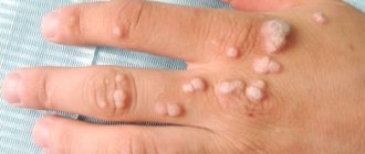
Is there a difference between papillomas and warts?
These tumors have one causative agent - papillomavirus. Externally they differ from each other. The size of warts ranges from 2 millimeters to one centimeter in diameter. The neoplasm has a round shape with clear boundaries. The surface of warts is rough and hairs can grow on it. The growth has a pronounced shade, which can be grayish, brown, or brown. Warts appear on any part of the body, but most often the growths can be seen on the limbs, face and head.
Unlike warts, papillomas have a pronounced elongated shape, the so-called stalk. They are soft to the touch. The surface of the growth is uneven, ridge-like. The color of the papilloma does not differ from the skin or has a brownish tint. In addition to the neck and face, papillomas appear in the armpits and groin area. In size, papillomas can be very small, no more than two millimeters, and larger, up to one centimeter at the base.
What is a mole
Moles or nevi (medical term) differ in shape, color and size.
They can be brown, black, purple and red.
They are located on the surface of the skin or slightly rise above the skin.
After six months of age, the first nevi may already form on the child’s body.
They can appear throughout life with varying intensity.
The formation of nevi is associated with the accumulation of melanocytes in certain areas of the skin surface.
A mole is a benign formation and does not pose a danger, but under the influence of negative factors it can degenerate into a malignant form - melanoma.
The neoplasm carries a mortal danger to humans, as it has the ability to very quickly metastasize throughout the body.
How does infection occur?
Many people believe that you can become infected with HPV through sexual contact. In fact, papillomavirus is transmitted in many ways, not just sexually. Infection can occur:
- when interacting with the carrier
- when the virus penetrates through a damaged area of skin, mucous membranes
- during sexual intercourse
- when visiting a bathhouse, sauna, public shower, swimming pool, tattoo parlor
- from an infected mother to a newborn
If one of the family members is infected with HPV, the risk of transmission of the infection is very high if people interact closely with each other in everyday life, use the same bathroom and hygiene items. The same applies to visiting facilities with a humid environment: bath and health complexes, swimming pools. If they were previously visited by an infected person, the virus can enter the body through a damaged area of skin. Warm and humid environments are ideal conditions for HPV to reproduce, but it cannot exist outside the body.
However, according to statistics, the most common route of infection is sexual. During unprotected sex, the risk of transmission of infection is 75%. If a pregnant woman has HPV, the child can become infected when passing through the birth canal.
Cryodestruction
A long-established and well-proven method of removing tumors using exposure to extremely low temperatures, in which the affected area is exposed to liquid nitrogen or other refrigerant for a short time.
The application of the refrigerant itself does not require preliminary anesthesia, because The application is painless, although the patient may feel a slight tingling or burning sensation. However, after the operation, during the “unfreezing” of the tissues, acute pain occurs, which can last up to a day. This is normal.
After a few hours, redness and swelling form around the injury, and a day later a subcutaneous blister filled with fluid forms. The rehabilitation period lasts a week, during which the swelling and hyperemia disappear, and the bladder breaks on its own. After this, it is necessary to apply an antiseptic drug prescribed by a specialist.
The death of tissue under the scab continues for three weeks, which ensures reliable destruction of infected tissue. Wound healing occurs in 1.5 months, and the mark completely disappears in six months, during which it is necessary to protect the skin from the sun and mechanical stress.
The method is successfully used to remove papillomas on the body, in the oral cavity and for the treatment of the cervix. But to remove tumors on the face and intimate area, it is better to use another method. In the first case - due to swelling and hyperemia, in the second - due to increased pain sensitivity of the genital area.
What you need to know about HPV
The penetration of papillomavirus does not necessarily mean that a person will get sick. In people with a strong immune system, the body can cope with infection on its own. It manages to gain a foothold when the body’s defenses are reduced. In this case, the virus will periodically remind itself of itself by the appearance of growths on the skin and mucous membranes. At increased risk are people with chronic diseases, women who take oral contraceptives for a long time, people who often change sexual partners, children and mature patients.
There is no drug that can completely destroy the virus. Drug therapy is aimed at increasing the body's defenses, that is, stabilizing the immune system. It will be able to resist the virus and thereby reduce the frequency of its manifestations.
A vaccine has also been invented that helps build immunity to HPV of high carcinogenic risk. These strains of the virus are capable of causing cervical cancer. It is advisable to vaccinate girls in adolescence, starting from 15 years. Older women can also be vaccinated if they have cervical abnormalities. First, treatment is carried out, which is aimed at normalizing the vaginal microflora, then vaccination can begin. In many Western European countries, HPV vaccination is mandatory. In the post-Soviet space, vaccination can be done at your own request.
Diagnosis of papillomas
No matter how much you would like to get rid of annoying papilloma quickly and at home, or even better - with the help of some folk remedies, do not rush! Self-selecting procedures can lead to disastrous results. Be sure to visit your local clinic.
In a paid clinic, for your money, they will cut/burn/freeze anything for you without any questions asked, but there is no guarantee of an accurate diagnosis, and therefore the absence of undesirable consequences.
Only a qualified specialist can make an accurate diagnosis and prescribe appropriate treatment. Be sure to take the tests prescribed by your doctor before getting rid of tumors. First of all, you need to get advice:
- a gynecologist or urologist if tumors appear in the perineal area;
- an otolaryngologist if papillomatosis of the mucous membranes of the mouth or throat is suspected;
- a dermatologist - in all other cases.
Based on the results of the clinical examination, the doctor, if necessary, may prescribe:
- collection of cells for cytological examination;
- tissue sampling for histology;
- scraping for PCR analysis;
- collecting material for the daijin test.
All these methods, in one way or another, make it possible to determine the risk of developing cancer. After removing the tumors by any method, it will no longer be possible to determine this indicator. This increases the likelihood of adverse consequences.
Treatment of papillomas
If growths appear on the skin, you should consult a dermatologist. He conducts an examination to determine the type of growth and the strain of the virus that caused the disease. Several methods are used to eliminate papillomas and warts:
- excision is a classic method that is used less and less due to its traumatic nature;
- removal with a laser or radio wave knife is an effective minimally invasive technique that is used most often;
- cryodestruction - burning out growths with liquid nitrogen;
- electrocoagulation - removal using electric current.
Laser removal of warts and papillomas is the most effective. This is a painless process in which the growth is completely removed and leaves no traces. After the procedure, a small crust remains at the site of treatment, which disappears after a few days.
Treatment of moles and papillomas
Therapy will depend on the type of tumor.
- 1. After removal of a mole, as a rule, no treatment is required, unless it is malignant. In case of degeneration, the patient will continue to be treated by the oncologist.
- 2. If the PCR analysis confirms the presence of the virus, this means that the neoplasms were provoked by papillomavirus. After they are removed, the patient will be prescribed antiviral therapy. The results of the analysis will also answer the question regarding the possibility of degeneration into an oncological disease.
The use of medications to remove papillomas is possible only with the approval of a doctor.
Since the prognosis of the disease with unauthorized removal of growths can be sad.
Of the products that are sold in the pharmacy chain for the removal of papillomas, the following are very popular: Salicylic acid, Podophyllin, Feresol, Superclean.
If there is no effect, more radical methods are used to remove growths.
When growths degenerate into a malignant neoplasm, the method of surgical excision is used.
HPV and cancer
The biggest danger of HPV infection is cancer. However, don't be afraid. Most strains of the virus have a low carcinogenic risk and therefore do not pose a threat to life. Strains 16, 18, 31, 33, 45, 51, 52, 56, 58, 59, 67 have oncogenic potential. According to statistics, most people have strains 6 and 11, which are low-risk and lead to the formation of genital warts on the genital mucosa organs. It has been proven that varieties of papillomavirus with high oncogenic potential cause cervical cancer in women. This is a long-term result of a viral infection in the body. The disease can develop 10-15 years after infection. Before this, precancerous diseases may occur: cervical erosion, dysplasia, chronic inflammatory processes in the cervical canal.
In case of such pathologies, a woman is prescribed an HPV test. To do this, a smear is taken from the cervical canal and the presence of infection and the strain of the virus are determined.
At a young age, the body actively resists infection, and as a woman gets older, she should pay special attention to her health. Cervical screening should be done once every three to five years. This is an effective prevention against diseases caused by HPV infection.
What is the difference between a mole and a papilloma
The main distinguishing feature of neoplasms is the cause of origin.
Papillomas are formed as a result of infection of the body with papillomavirus, and nevi are formed as a result of the accumulation of melanocytes.
To understand what kind of tumor this is, you need to pay attention to the following signs:
- Localization location. Warts form in places where skin friction and sweating most often occur, as well as on mucous membranes. A nevus can form on any part of the skin surface. No factors can influence the location of the mole.
- The edge of a healthy mole is always smooth, the formation has a symmetrical shape.
- The size of the wart is small; if damaged, it can rapidly increase in size. Nevus can have different sizes: from small to very large.
- Presence of pigmentation. The mole can have a color: black, brown, red, purple. Papilloma always has a light pink tint.
- Warts are characterized by a loose structure, while nevi are dense, hard formations. It should be noted that these formations do not always meet these criteria.
- The mole does not contain any vessels. Papilloma is equipped with capillaries.
- Nevi can be inherited, but papillomas cannot.