ATTENTION! From January 2022, a new medical center will open for you at the address: Dmitrovskoye Shosse, 81 (5 minutes walk from the Selegerskaya metro station), where you can receive a wide range of medical services, including CT (computer tomography).
- home
- Surgery
- Fibroma removal
KDS Clinic offers its clients operations to remove skin fibroids in an outpatient setting. The best doctors in the field of surgery and dermatology will conduct a full examination and help you get rid of the tumor quickly and safely.
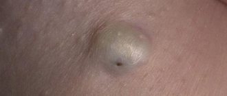
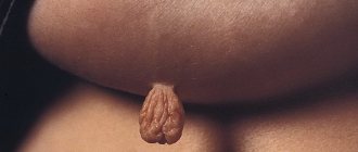
Skin fibroma is a benign tumor that forms from mature connective tissue cells. It can be localized on any part of the body, but mainly affects the face and neck.
Preparing for surgery
As such, no preparation is required for surgery to remove fibroids on the leg, arms or face. During the initial appointment, the doctor examines the patient, determines the volume of the affected area and prescribes a standard examination:
- clinical examination of blood and urine;
- blood for Rh factor and group, tests for RW, HIV, hepatitis, as well as biochemical research;
If a tumor is suspected of being oncogenic, a consultation with an oncologist and cytological tests will be prescribed.
Diagnostics
Initially, experienced specialists - oncologists or multidisciplinary surgeons - conduct a detailed examination of the patient, record all the complaints that exist, palpate the formation and make a preliminary diagnosis. Then you need to determine the nature of the tumor. After this, you need to conduct an ultrasound examination of the tissues where the tumor is located to determine its size, nature, and changes in the tissues. If necessary, an X-ray examination is performed. If questions remain, a biopsy of the suspicious area should be performed to rule out a malignant process.
Fibroids can be detected by palpation (feeling with fingers or hands) performed as part of a pelvic examination, or diagnosed through imaging procedures such as ultrasound, computed tomography (CT), and magnetic resonance imaging (MRI).
Doctors surgeons
Dzhadzhiev Andrey Borisovich Surgeon
Cost of admission 1500
₽
Make an appointment
Marynich Alexander Alexandrovich Vascular surgeon, phlebologist, angiologist
Cost of admission 1500
₽
Make an appointment
Myshlyaev Anton Vsevolodovich Surgeon coloproctologist
Cost of admission 2000
₽
Make an appointment
Operating principle
The procedure in the clinic is performed on an outpatient basis, under local anesthesia, and takes 10-15 minutes. After antiseptic treatment of the skin, the doctor adjusts the laser taking into account the individual characteristics of the patient, the size and nature of the tumor, and removes the skin formation layer by layer.
Fractional CO2 laser has an ultra-thin beam that acts precisely and does not affect surrounding healthy areas of the skin. The laser has sterilizing properties and prevents infection of the wound. Minimal heating of the wound surface creates favorable conditions for rapid tissue healing without the formation of a pronounced scar.
After the procedure, the doctor treats the removal site with an antiseptic and applies a sterile bandage.
The procedure is well tolerated by patients - only a slight tingling or warmth is felt.
Contraindications
Despite the fact that the operation is considered minimally invasive, it still has some contraindications:
- herpes in the acute stage;
- acute infectious and autoimmune lesions of tissues and internal organs;
- low blood clotting;
- malignant formations;
- epilepsy;
- type 1 diabetes mellitus.
Such restrictions apply to any type of surgery, including laser removal of fibroids. If the patient has a history of one of the contraindications, specialists from KDS Clinic will select an alternative treatment method with high efficiency.
Prices for radio wave removal of skin lesions on a wide base
| Removal of 1 milia (grass, wen) in the eye area | 950 rub. |
| Removal of 1 wide-based skin lesion (digital dermatoscopy 750, removal 2150, anesthesia 550, histology 1500, free consultation on the day of treatment) | 4950 rub. |
| Removal of 2 wide-based skin lesions (digital dermatoscopy 750, removal 4300, anesthesia 1100, histology 3000, free consultation on the day of treatment) | 9900 rub. 9150 rub. ( 8% discount |
| Removal of 3 broad-based skin lesions (digital dermatoscopy 750, removal 6450, anesthesia 1650, histology 4500, free consultation on the day of treatment) | 14850 rub. 13350 rub. ( 11% discount |
If you are reading these lines, then you probably have a formation (or several) on your skin and you want to get rid of them. keratoma pigment nevus fibroma milia (also called millet or wen)
hemangioma (angioma) dermatofibroma
- Do you think that keratomas or milia
on your face make you less attractive? - Because of a nevus
in your armpit, are you now wielding a razor like a professional hairdresser? - Are fibroids on your back
making it difficult to wear a bra? - Instead of playing sports and active recreation, are you thinking about accidentally damaging the hemangioma
? - At the beach, do you cover dermatofibromas
on your skin with a band-aid and are afraid to sunbathe? - Tired of touching the wart with your hand
or clothes, and are you thinking about getting rid of it for the hundredth time?
Read this text to the end and you will learn how to get rid of unwanted formations on the skin in one visit without scars and 100% safely.
Operation
At the outpatient clinic of our medical center, fibroids are removed from the face, neck or other part of the body using the most progressive and effective techniques. The operation is performed under local anesthesia in several ways:
- surgically - involves removing fibroids using a medical scalpel, followed by the application of sutures or staples;
- laser removal of fibroids - using a laser scalpel, tissue burning and coagulation;
- radio wave removal - using radio waves in the high frequency range;
- cryodestruction - freezing the formation with liquid nitrogen.
The duration of the operation depends on the chosen method and usually ranges from 20 to 40 minutes.
Features of laser removal
Initially, the main method was traditional surgery, which was accompanied by pain and long recovery. The transition to laser removal of skin tumors has reduced these shortcomings to a minimum.
The beam of the device works pointwise, burning only “excess” tissue. And damaged capillaries are sealed at high temperatures. In addition, ultraviolet radiation only affects the surface of the skin, without affecting the body itself. All this minimizes trauma from the procedure.
Laser equipment is optimal for eliminating skin defects in hard-to-reach and delicate areas:
- earlobe;
- areas around the eyes;
- areas with mucous membranes, including the tongue.
Problems can only arise when treating the feet. Tumors in this area often grow not only in breadth, but also in depth: the laser power may not be enough. Another point is that the device literally evaporates pathogenic cells. If the fibroma requires additional examination, it is removed using Surgitron or a scalpel.
Possible complications
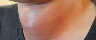
Fibroma turns into a malignant form, therefore low-risk. The exception is location in the friction zone of clothing, or traumatically dangerous places, since the likelihood of violating the integrity of the cover and twisting of the growth can lead to swelling, inflammation, bleeding and the addition of a secondary infection.
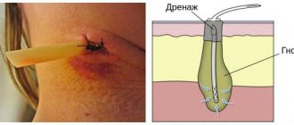
After surgery, in 97% of cases, the problem is completely eliminated without the risk of relapse. But, if the wound is not properly cared for, infection and blood poisoning can develop. Therefore, you should strictly adhere to all the recommendations of our specialists.
Benefits not related to removal technique
- Honest prices.
No hidden fees or intrusive services. Either consultation or removal is paid, strictly according to the price list. You can always check the price with me on VKontakte by sending a photo with a ruler in the background. - Free information support before and after surgery.
If you have any doubts before your visit, you can easily and free of charge by mail or VKontakte. After the operation, you will have my personal phone number, by which you can ask me at any time about what is bothering you. - Fast and convenient.
Removal is carried out within a few minutes, in one visit. - Online appointment booking and removal without calls or waiting
- No preliminary tests, dressings, or suture removal required
- Histology results by email
. You can read them on these sites: docplanner.ru (more than 40 reviews), napopravku.ru (24 reviews), vk.com (more than 150 reviews)
Causes and symptoms of skin fibroids
Scientists do not yet know the exact reason for the formation of skin fibroids. But there is a risk group of patients who may develop a growth:
- women during pregnancy;
- diabetes mellitus type 2;
- heredity and genetic predisposition;
- obesity;
- patients over 40 years of age.
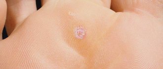
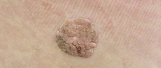
In appearance, fibroma resembles:
- a small growth on the leg or tightly fitting;
- can be localized throughout the body in several places at the same time;
- They have different colors from flesh to black.
If you experience symptoms indicating the development of skin fibroids, you should immediately consult a doctor for advice. The price for fibroid removal at KDS Clinic depends on the chosen method.
Types, characteristics of fibroids
There are more than 10 different morphological variants. Pathologists give a description of what each form looks like and how it differs after histological examination - elastrophibroma, soft, dense fibroma.
- Dermatofibroma is a single or multiple formation of intradermal localization, mobile and painless when palpated with unchanged color.
- Desmoid fibroma has another name - aggressive fibrosis, refers to mesenchymal tumors originating from excess collagen fibers and differentiated fibroblasts. They often progress and recur, forming in soft tissues, the retroperitoneum or on the abdominal wall.
- Chondriomyxomas are rare tumors that affect bone tissue - the edges of long bones, pelvis, ribs, vertebrae, feet, metatarsus.
- Angiofibriomas in the cheek and nose area, containing small tubercles with connective tissue fibers.
How long does it take to recover after removal of uterine fibroids?
It is difficult to determine in advance how long rehabilitation will last, since this parameter depends on many conditions: from the patient’s age to the treatment method. For example, the early postoperative period after hysterectomy is 10-14 days, and recovery after laparoscopy for uterine fibroids usually does not exceed 5 days. The late and late periods also differ from each other in duration.
In addition to the type of intervention, the period determines the age, general health of the woman and the quality of the selected treatment. As a rule, complete recovery in the absence of serious complications takes about a year.
results
The effect after the procedure is visible immediately - the skin is without pathologies and neoplasms. At the site of the removed mole or other benign neoplasm, a crust forms, which should come off on its own.
For 2 weeks after the procedure, you cannot visit the sauna, swimming pool, bathhouse, or gym. It is prohibited to sunbathe in the sun or in a solarium. After the crust comes off, young skin needs to be protected from ultraviolet radiation, and before going outside, apply sunscreen cosmetics.
It should be borne in mind that there are contraindications for laser removal of tumors: acute inflammatory diseases, fever, cancer and pregnancy.
Indications for removal
Doctors warn that independent removal of benign tumors leads to complications. Therefore, if a fibroma appears on the body, you must immediately contact a surgeon.
There are several indications for tumor resection:
- if the fibroma is located on the genitals or face, its removal is required to achieve a cosmetic effect;
- with suppuration;
- if the tumor is on the uterus, this is a relative contraindication for bearing a fetus;
- when wearing clothes or combing hair, the formation is constantly exposed to trauma, so its removal is required;
- tumor resection is also required if it is localized on the mucous membranes, for example, on the eyelids or the inner lining of the oral cavity, as well as on the tongue;
- If severe pain occurs, the fibroma increases in size, itching and bleeding are observed, that is, the symptoms of the pathological process increase, you should immediately seek help from a doctor.
Fibroid removal takes place on an outpatient basis using local anesthesia.
What is fibroadenoma?
Fibroadenoma is a benign tumor that develops against the background of inadequate production of sex hormones, so it is predominantly found in young women. The peak incidence occurs at the time of entry into menopause - approximately 45 years. The end of menstruation leads to involution of the glandular tissue, and benign processes are becoming less common, and cancer is developing much more often.
All hyperplastic processes in the organs of the female reproductive system are controlled by the ratio of estrogen and progesterone, therefore, when a woman has a hormonal imbalance, mastopathy, endometrial hyperplasia, uterine fibroids and benign breast tumors often coexist in one set or another.
To the touch, fibroadenoma has clear boundaries, an even and smooth surface, and dense. It never fuses with the skin, being located inside the gland tissue, it is easily displaced and painless. The size of the tumor node is variable; as a rule, in a small breast it is possible to feel a centimeter-long nodule.
Options for breast fibroadenoma:
- inside the duct - intracanacular, which began to grow in the wall, gradually narrows the duct, involving nearby glandular lobules;
- near the duct - the pericanacular grows around, “twisting” the layers like the rings of a tree trunk, while the lumen of the duct does not change;
- mixed - the vast majority, almost eight out of ten fibroadenomas.
Different tumor variants do not differ in symptoms or course, and therefore have no practical significance in practical medicine.
A benign tumor does not threaten to degenerate into cancer, but breast cancer can develop, masquerading as a fibroadenoma. Early cancer and a small benign nodule are difficult to distinguish even on a mammogram, so all neoplasms in the mammary glands must be subjected to a fine-needle biopsy and removed if there is doubt about the benignity of the microscopic picture.
