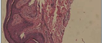
Lichen planus
Cutaneous manifestations of lichen planus are a rash that looks exactly the same, located on various parts of the body. The lesions have the form of peculiar compacted elevations on the skin, which have an irregular shape; their surface appears shiny when examined. They have a pinkish-purple color and a slight depression in the central part. Sometimes the color of the formations can be crimson-red. Their sizes are most often small - from 2 to 3 mm. The shine of the surface of the lesions is the most characteristic feature. It is very visible if you look at the patient's skin at a slightly angle, directing the light from the lamp in the same way.
Although the size of the lesions is relatively small, they may increase in the future. Individual lesions are capable of merging with each other, resulting in large plaques forming on the patient’s skin. Their surface begins to peel off. The scales are small in size, which is a fairly characteristic feature. For a more accurate diagnosis, there is also a simple test. Its essence is that a small layer of vegetable oil is applied to the skin in the area of pathological lesions. As a result, visible signs appear on the surface of the affected skin. Some of them are small white dots. Others look like white stripes, which seem to be located in the thickness of the skin and shine through it. After the name of the scientist who discovered it, this phenomenon is called Wickham’s sign. It is due to the fact that in the area of pathological formations there is a thickening of the stratum corneum of the skin, but this occurs unevenly. After the patient recovers and the lesions disappear, more intensely colored areas of skin remain in their place. They are quite persistent and can last for a long time. Skin rashes with lichen planus are accompanied by subjective sensations on the part of the patient. This manifests itself in the form of a feeling of itching and pain. The pain syndrome is constant and can be intense, as a result of which sleep, appetite and daily life are disrupted, and non-vrosis-like symptoms can develop.
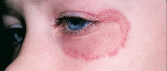
Lichen planus has, so to speak, a “favorite” location on the skin, which is located in the area of the flexor surfaces of the forearms. Other common sites of localization may be the areas of the wrist joints, the inner surfaces of the thighs, and the extensor surfaces of the legs. Often plaques are found in the groin, armpits, and oral cavity. The skin of the face and scalp, palms, and soles, on the contrary, is almost never affected. In some cases, the elements of the rash are located in an interesting way: they appear as stripes that alternate with areas of normal skin. This is most often observed on the extremities. The appearance of the patient's skin in these cases is so unique that it is almost impossible to confuse the pathology with any other. Sometimes there is not a single pathological focus on the patient’s skin, while the mucous membranes are affected to a very significant extent. This occurs in a quarter of all patients with lichen planus.
The mucous membranes of various locations are affected: in the mouth, glans penis in men and the vestibule of the vagina in women. Lesions on the mucous membrane of the cheeks look characteristic. Here they have a grayish color and are represented by very small nodules, almost the size of a pinhead. They are always arranged in groups that have the most bizarre shapes: in the form of rings, nets, openwork lace. The lesions located on the surface of the tongue are completely different in appearance. They are white and similar to the rashes associated with leukoplakia. These are plaques protruding above the surface of the mucous membrane, which have clearly defined jagged contours. Rashes with lichen planus can also be located on the red border of the lips. In these cases, the color of the lesions is purple; they also appear as plaques with a scaly surface. Another characteristic feature is that the plaques are covered with a grayish-white mesh. The lower lip is affected much more often than the upper lip. Some patients with lichen planus have changes in the nail plates. A clearly visible longitudinal striation appears on them, which is sometimes so pronounced that it looks like scallops. The nail bed becomes red due to an inflammatory reaction of allergic origin. Spots of cloudiness form on the nail plates of the fingers and toes.
Another argument in favor of the allergic origin of the disease is that lesions of lichen planus appear mainly after exposure to irritating factors on the skin. Moreover, the focus is formed strictly in the place where the impact was applied. For example, often lesions resulting from scratching have the appearance of characteristic stripes.
The course of the disease is long, sometimes even for years and months. Sometimes the lesions occupy not only the above areas, but also spread to all skin integuments. This form of the disease is called generalized. This is a severe and long-lasting pathology, which is often complicated by the addition of another disease - secondary erythroderma.
The so-called typical course of lichen planus was described above. However, making a diagnosis is a simple task due to the extremely characteristic manifestations of the pathology. But this does not happen in all cases. There are a number of atypical forms of the disease that develop as new types of allergens, in the presence of concomitant diseases of the skin and other organs, and in cases of disorders in the immune system and metabolism in the body.
Hypertrophic warty form. As the name implies, this form of the disease is based on the appearance of warty growths of the skin. Plaques appear that are purple or brownish-brown in color. Their surface is covered with a large number of small warts, in the area of which there is a thickened stratum corneum of the skin. Around these plaques and warts there may be small nodules quite typical for ordinary lichen planus. The process most often affects the front surfaces of the legs. Lesions can also occur in other places, but this is much less common. Atrophic and sclerotic forms of the disease. At the very beginning, the disease proceeds quite typically, with plaques and nodules characteristic of lichen planus appearing on the skin. However, later, when they disappear, areas of atrophy and thickening (sclerosis) of the skin remain on the patient’s skin in these places. A very unique and interesting picture develops when the process is localized simultaneously on the scalp. In this case, spot baldness develops, which may resemble some other pathologies in appearance. In this case, compaction and thickening of the stratum corneum of the skin on the extensor surfaces of the extremities is almost always observed. This peculiar picture was named after the authors who discovered and described it, the Little-Lassouer symptom.
Pemphigoid, or vesicular, variety. This is a very rare form of the disease. It manifests itself in the form of the formation of lesions on the skin in the form of small bubbles filled with clear liquid, sometimes mixed with blood. The bubbles are small in size, often no larger than a pea or cherry. Blisters can appear on the skin either without prior visible irritation or against the background of nodules, plaques, etc. The process is located in most cases in the area of the legs or feet. In some cases, lesions characteristic of both the simple form of lichen and the blistering form may appear simultaneously on the skin of the same patient.
Moliniform variety of lichen planus. This form of pathology is also rare. Quite large lesions appear on the skin, which are arranged in chains and seem to be strung in the form of a necklace. They are most often the size of a cherry pit. The lesions are nodules. They rise dome-shaped above the skin and have a rounded appearance. Sometimes they are quite small, dense, located close to each other in the form of strips, which can create the appearance of linear scars. In some cases, they clearly resemble beads in appearance. Such rashes usually affect large areas of the skin. There are especially many of them in the forehead, on the back of the ears, on the neck, in the area of the elbows, on the back of the hands, on the stomach, and in the buttocks. In most patients, the lower areas of the face - cheeks, nose - remain completely unaffected; areas of skin above the collarbones, between the shoulder blades. The palms and soles, as well as the genitals, are never affected. A number of authoritative researchers are inclined to consider this pathology not as a type of lichen planus, but as a completely independent disease that has its own causes.
Pointed, or perifollicular, form. With this type of lichen planus, lesions characteristic of its simple form, and others that are slightly different, appear on the patient’s skin. The nodules have a conical shape and contain a small spike at their top. This picture develops when the hair follicles are predominantly affected by the pathological process. It is clear that the most common localization of rashes is the scalp. After the lesions disappear, small scars remain in these places, which seem to sink in comparison with the surrounding skin.
Ring-shaped variety of the disease. It develops when the lesions of lichen planus very quickly increase in size, at the same time they begin to heal from the central part. The result is like a ring. In the center after healing there is always a brighter colored area of skin. In addition to rings, pathological formations on the skin in this form can be in the form of garlands, arcs, or half rings. They can occur both during the growth of one lesion, and during the merging of several. This type of disease develops much more often in males, and the formations are located mainly in the genital area. Sometimes the lesions can very closely resemble in their appearance papules of syphilitic origin, which leads to an incorrect diagnosis and incorrect therapy.
Linear lichen planus and zosterimorphic lichen planus . They are very similar forms of the disease to each other, so they are combined into one group. In this case, the lesions are located on the skin along the course of large nerves and resemble herpetic rashes.
Erosive-ulcerative form. The disease affects the mucous membranes. This is the most severe and difficult to treat type of lichen planus. Defects appear on the mucous membranes of the oral cavity and on the lips, which in some cases can even turn into ulcers. Around these formations there are areas of redness of the skin, in the area of which lesions typical of lichen planus occur. Defects in the mucous membranes have irregular contours and are often covered with films and plaques. If these films are removed with a spatula, bleeding easily occurs. In most patients, these lesions are few in number and small in size, while the patient does not experience any subjective sensations or make any complaints. The difficulty of treating this form is that during the therapy itself, most ulcers disappear on their own without a trace. However, after stopping treatment, the rash almost always develops in the same or different place. The process can continue for a long time.
Shingles: “adult” version of chickenpox
The first signs of herpes zoster, or “herpes zoster” in everyday life, begin not with the skin, as one might assume, but with a sharp pain in the ribs, often at chest level. Patients with this symptom are initially examined for cardiac pathologies and neuralgia. Whereas specific signs of the disease appear somewhat later.
Varicella variant
Few people know, but chicken pox and herpes zoster are different “masks” of the same virus.
Varicella-zoster (or herpes type 3) provokes chickenpox in children, and more often shingles in adults.
The peculiarity of the virus is that once it enters the body (for example, with chickenpox in childhood), the pathogen never leaves it. And it remains to “live” forever in the nerve fibers of the infected person, albeit under the strict supervision of immune forces.
If the immune system is intact and adequate, herpes may never manifest itself again. But weakening the body literally removes the guards from the “detention cell” and allows the virus to run rampant.
Signs of reactivation
The “awakened” herpes virus begins to actively multiply, provoking the immune system to “remember” its responsibilities.
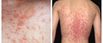
This causes a classic inflammatory process, accompanied by sudden pain, often along the ribs, and after 2-3 days - redness of the skin and the appearance of “classic” herpetic blisters.
The blisters open over time and can merge with each other, forming a large wound surface. Which, in conditions of weakening of the body, is accompanied by prolonged healing and, often, the addition of a bacterial infection.
Such “stripes” of lesions are always one-sided, which is a distinctive feature of “lichen.” But the geography of location may vary.
Most often, the intercostal spaces of the chest or, less commonly, the lumbar region are hit. However, herpetic blisters may appear in the ear canal and on the face, indicating damage to the nerve fibers of the brain (trigeminal, facial nerve and others) and a high risk of severe neurological complications.
Diagnostics
In most cases, the characteristic symptoms of lichen allow a diagnosis to be made without any additional tests.
However, if in doubt, ask your doctor, or if you want to be examined yourself, you will need a blood test for antibodies.
Detection of IgA antibodies to the Varicella-Zoster virus confirms the reactivation of the pathogen.
And the presence of IgG is a marker of chickenpox and the presence of the virus in the body.
Other research methods, such as PCR diagnostics of rash elements, are not suitable for the examination itself, since they have a number of important nuances when collecting material. And only a dermatologist or venereologist can take the material correctly.