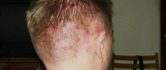
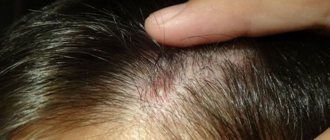
Moles on the head are not uncommon. Even if they are quite large in size, they are hidden under the hair and do not pose an aesthetic problem. However, when visiting a hairdresser, even a small mole can cause discomfort. It’s good if the hairdresser is your own and trusted, most likely he already knows where all the moles are and delicately avoids traumatizing them. Unfortunately, it is not always possible to find such a master. And so, when once again a mole in the hair is touched by simple combing, its owner (or owner) thinks about removing it.
What should I do? Will you have to shave your entire head first and then, after removal, walk around with receding hairline?? Let's deal with this and other difficult questions.
What types of moles are there on the head?
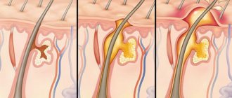
The most common type of moles growing on the scalp are pigmented nevi with papillomatosis
. They appear in childhood or adolescence and slowly increase in size. At first they appear as a small flesh-colored bump that protrudes at the level of the surrounding skin and is covered with hair. Then, as it grows, the mole becomes more and more elevated and acquires the appearance characteristic of this type of nevus.
At later stages of development, this type of nevus becomes covered with something like small papillae. This is where its name comes from - nevus with papillomatosis
(from Latin papilla - nipple). These moles may appear large, but at the same time, they are in contact with the scalp over a much smaller area than it appears from above (see picture below). This makes them easier to remove - the smaller the contact area, the easier it is to remove the mole.
Intradermal nevus
A type of ordinary nevi that have a dome-shaped shape. Such moles rarely appear in childhood, but they are extremely common in the adult population. Up to ten such formations can appear on the body at the same time. Such neoplasms got their name due to the fact that they are located under the upper layer of the epidermis. Often such moles are difficult to notice; they hardly stand out above the surface of the body, and their shade is close to the surrounding tissues.
Nevus can appear in almost any area. Favorite places for neoplasms are the upper part of the arms, eyelids, neck, face, etc. The maximum size of moles is up to 1 centimeter. When they appear in childhood, nevi are practically invisible, but with age they begin to darken and often acquire a convex shape. If the mole persists, then after 70 years it will gradually begin to discolor.
There are several reasons why intradermal nevi can form on the human body:
- Hereditary factors
. If the baby’s parents have more than 50 ordinary moles on their body, then the child’s nevi will also be multiple in nature. - Exposure to sunlight
. Ultraviolet radiation negatively affects the skin, causing damage. Moles often appear in place. People with too light skin are at risk. - Decreased immunity
. Most often, nevi begin to form against the background of reduced immunity, including after taking drugs that suppress the immune system.
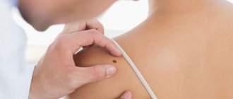
Even ordinary moles that have a symmetrical appearance and do not increase in size are important to constantly monitor. It is better to have a checkup with a dermatologist at least once a year, this way you will be able to identify the problem at an early stage of its occurrence. It is worth planning a visit to a specialist outside of the schedule if the mole begins to grow, changes its shape, acquires a different shade, or begins to bleed.
A dermatologist may prescribe a procedure for excision of a mole if it causes cosmetic discomfort to the wearer, if there is a risk of the neoplasm degenerating into melanoma, if the surface of the nevus is constantly damaged during the day.
Are moles in hair dangerous?
Moles located on the scalp are very rarely dangerous. As a rule, they accompany a person for a significant part of his life and hardly change at all, with the possible exception of slow growth, no more than 1-2 mm per year. Sometimes even benign nevi of this type can become covered with small crusts. These crusts periodically fall off and appear again. It is important to note that this is a normal occurrence for pigmented nevus with papillomatosis. If it is not accompanied by bleeding, weeping of the surface of the mole, change in color or rapid growth, such formations are most likely not dangerous.
Kinds
Experts identify several types of moles that can appear on the head.
- Large nevi are congenital and gradually increase in size. It is these neoplasms that occur most often and require medical intervention.
- The convex formations are hemispherical in shape and protrude strongly above adjacent areas of the skin. Dermatologists recommend removing such moles, since they can be easily injured when combing your hair or over-dried with a hairdryer.
- Flat nevi are the most common and safest nevi. As a rule, they do not cause discomfort, do not grow and do not require special care.
- Blue neoplasms have an unusual color: the shade varies from light blue to purple. Usually they protrude slightly above the surface of the skin.
- Hanging formations are quite rare on the head. But if they occur, immediate removal is required.
Nevi of all types are prone to malignancy when injured. To avoid dangerous complications, it is necessary to constantly monitor them and properly care for them.
How are moles located under the hair removed?
The type of operation and what happens after it depends on one single factor - the area of contact of the mole with the skin:
- If the area is no more than 15 mm.
These moles are the easiest to remove.
Modern high-tech methods are suitable - laser and radio wave surgery. With this type of removal, no preparation is required and the mole is removed in one visit,
within 10-15 minutes
.
It is as if it is “cut off” from the skin and the wound heals like a simple deep abrasion. - From 15 to 20 mm.
To remove such a mole, you need a scalpel and preliminary tests, the application and removal of sutures, regular medical supervision and dressings are required. - More than 20 mm.
Skin grafting may be required to remove such a mole. This is because it is very difficult to match the edges of the wound on the head for suturing. The defect has to be covered with a “patch” made from the skin of the owner of the mole. The skin for the “patch” is taken from another area of the body (most often, the inner surface of the shoulder or lower abdomen).
Congenital melanocytic nevus is a benign proliferation of cutaneous melanocytes that is clinically observed at birth or appears during the first weeks of life. Congenital nevi vary in size, macroscopic appearance and histological characteristics [1] and appear in approximately 1% of newborns. Most nevi are small in size, but there are large congenital nevi (LCN) and giant congenital nevi (GCN), which have a significant area and sometimes extend to an entire segment. GVNs are relatively uncommon congenital malformations that, when examined macroscopically, are usually characterized by intense pigmentation and can often have variable hair patterns [2].
In histological examination, GVN are characterized by an abundance of small and medium-sized “nevus cells” (melanocytes), with a relatively small amount of weakly oxyphilic cytoplasm, moderately basophilic nuclei of predominantly irregular ovoid shape. In the upper layers of the dermis (the area of the papillary layer), nevus cells often form clearly demarcated, irregularly rounded nested clusters of different sizes and contain a finely clumped brown pigment (melanin) in the cytoplasm. In the deep layers of the dermis, nevus cells of the HVN extend up to the border with the hypodermis and can penetrate into it, as through fibrous septa separating the cells of the subcutaneous fatty tissue, and appear directly in its tissue. An increase in the number of macrophages in nevus tissue and their increased activity, expressed, in particular, in the increased absorption of melanin pigment by macrophages, is reported [2].
Congenital giant pigmented nevi are often a significant cosmetic problem and a significant psychological traumatic factor for the child. The particular significance of giant pigmented nevi is also due to the fact that they are characterized by the potential for transformation into a malignant neoplasm - melanoma [2-4]. There is an opinion that malignant melanoma, developing from other types of nevi, usually arises in places of the epidermal-dermal junction (in the superficial layers of the skin), while melanoma from the GVN originates mainly in the deep layers of the dermis [2-4], which is obvious , is a condition that preserves the possibility of malignancy of the remnants of the GVN when it is incompletely removed. The reasons for the emergence and development of GVN are discussed, although there is a fairly well-established opinion according to which GVN develop from primitive cells of neuroectodermal origin (from neural crest cells) that are delayed during migration and subsequently proliferate [5]. There is also evidence that these tumors differ in a variety of morphological manifestations and clinical behavior [6]. Due to the fact that most congenital giant pigmented nevi undergo early surgical and therapeutic treatment for cosmetic reasons and for prophylactic purposes, the true incidence of their malignancy is difficult to determine [2].
There is also no consensus on the need to remove nevi and an unambiguous view on the choice of treatment method. Currently, several basic methods for eliminating WMH are described in the literature and used in practice: staged excision with plastic surgery using a free split or full-thickness autograft, the use of flaps obtained by tissue dermatosis, dermabrasion, curettage and laser [7]. A number of authors [8–10] report on surgical treatment of GVN using microsurgical techniques.
The purpose of the study is to study the clinical and histological features of nevus tissue, which are important for the choice of surgical intervention technique, immediate and long-term results.
Material and methods
During 2008–2010, we examined and operated on 10 patients with VVN and GVN of different localization. The age of the children ranged from 9 months to 7 years. When formulating the diagnosis, we used the classification proposed by foreign authors, according to which a nevus constituting less than 1% of the body surface area on the face and less than 2% on the body is considered BVN, and more than 1 and 2%, respectively, is considered to be GVN [1, 8].
All admitted children were registered with an oncologist and had a conclusion that there were no contraindications to surgical treatment. As part of the preoperative examination, all children were examined by a neurologist and a neurosurgeon. According to indications, magnetic resonance imaging of the affected area was performed. The examination did not reveal any neurological abnormalities or concomitant developmental anomalies.
Surgical treatment included staged excision of the nevus with replacement of the defect with local tissues in the form of rotational flaps, thick split or full-thickness skin autograft. The choice of donor site depended on the location of the surgical wound. For facial transplantation, full-thickness autografts from the inner surface of the shoulder were used. The donor wound was closed with a split autograft from the anterior inner surface of the upper third of the thigh. For plastic surgery of defects of other localization, excess skin obtained by the method of expander dermatension and split autografts from the anterior inner surface of the thigh were used. Removed skin fragments from GVN and BPN were studied using morphological research methods: macroscopic description, light microscopy, morphometric calculations of certain indicators and, in some cases, using immunohistochemical (IHC) reactions.
results
The main clinical symptoms of nevi (except for the presence of the GVN itself with its macroscopic characteristics) were a feeling of itching in the affected area, a periodic burning sensation and peeling. Itching most often bothered patients in the area of nevi with hairline, was obsessive in nature and was accompanied by scratching and trauma. The most pronounced hair growth was observed when the nevus was localized in the lumbosacral region (Fig. 1).
Figure 1. GVN in the lumbosacral region. In 2 patients, new small pigment spots were noted on different parts of the body within 1-1.5 years after birth, which may indicate the continued mitotic (proliferative) activity of melanocytes [11].
In case of multiple nevi, surgical intervention was primarily performed on the GVN and BVN, located in areas of the body that experience increased stress and are most susceptible to trauma (hands, feet, Achilles tendon area; Fig. 2).
Figure 2. BVN located on the thighs, legs, Achilles tendon area, and feet. Priority removal was also given to the GVN and BVN, which were located in open areas of the body most susceptible to solar insolation (face, cranial vault, upper limbs). Then the largest nevi of another location or nevi that bothered the children were removed. Since warty nevi were the most frequently traumatized, they, in our opinion, must be removed regardless of size and location (Fig. 3).
Figure 3. Warty nevi.
When choosing a method for closing a wound defect, the age and constitutional characteristics of the child, the thickness of the skin, and the severity of the subcutaneous fat layer were taken into account. In children under 3 years of age and with an asthenic constitution, large-volume expanders were placed on the cranial vault and chest area to avoid deformation of bone structures, the development of trophic complications over the domes of the expanders, and limiting respiratory excursions of the chest.
At the first stage of surgical treatment, we tried to maximize the use of local tissue reserves to close the defect formed after excision of the nevus area.
The next stage was planned depending on the localization. If the nevus was located in an area suitable for placing an expander, expander dermatension was performed, followed by using the resulting supply of integumentary tissue to close the defect after removal of the nevus. In other cases, free full-thickness or split-thickness autografts were transplanted.
Already during the surgical intervention, significant differences in the appearance of intact skin and nevus tissue were noticed. The latter was characterized by a loose consistency and differed from leather in its reduced strength and reduced elasticity. The subcutaneous fatty tissue also differed from the usual: it was more yellow in color, easily fragmented and was quite flabby.
During macroscopic examination of the surgical material, in almost all cases, changes were observed that were characteristic of nevi in general and of GVN and BVN in particular. The skin fragments were of different sizes and shapes. The surface of the removed skin flaps had a gray-brown, brown or dark brown color. Already at the stages of visual assessment of the material, it was noted that nevi (small and relatively medium-sized and even some large ones) were removed within intact tissues: around the nevi there was a clearly visible, narrow or narrow grayish rim of little changed skin. Fragments of especially large nevi and GVN in a number of cases were removed within the altered tissues. Some of the skin flaps had a slightly raised outer surface that was quite smooth, in some cases it appeared finely tuberous, sometimes with small papillary formations. In some cases, the surface of the nevus was rough and dirty gray in color. On the section, most often the superficial layers of the dermis and the epidermis region (total thickness from 0.03 cm to 0.08-0.1 cm) had a gray-brown color, the deeper areas of the dermis were whitish. The remains of yellowish subcutaneous fat retained their cellularity.
During microscopic examination, almost the same type of changes were observed in all preparations from fragments of nevi with subcutaneous fatty tissue.
The epidermis retained a fairly well-defined stratification; in some places, moderately pronounced hyperkeratosis was noted (Fig. 4, c),
Figure 4. Giant pigmented nevus (hematoxylin and eosin staining). a — “nest” accumulations in the papillary layer of the dermis of melanocytes containing brown lumpy pigment (×100); b - continuous extensive “fields” of nevus cells in the reticular layer of the dermis (×50 and ×200); c — moderately severe hyperkeratosis (×100); d — small papillomatous structures (×50). Small papillomatous structures were often encountered (Fig. 4, d).
Melanocytes of the basal layer of the epidermis and Langerhans cells were present in the usual numbers and in the usual proportion with the cells of the stratified squamous keratinizing epithelium. During an IHC study, a pronounced positive expression of antibodies to S-100 antigens was observed in these cells (Fig. 5, d),
Figure 5. Giant pigmented nevus: IHC study. Positive expression of the S-100 antigen in nevus cells of the dermis (a, b), in the connective tissue layers in the subcutaneous tissue (c) and in Langerhans cells of the epidermis (d). CD1a (Fig. 6, b),
Figure 6. Giant pigmented nevus: IHC study. Negative expression of CD1a antigen in nevus cells of the dermis (a) and its positive expression in Langerhans cells of the epidermis (b); brown clumpy pigment (melanin) in nested clusters of nevus cells in the papillary dermis (c). and langerin (Fig. 7, b),
Figure 7. Giant pigmented nevus: IHC study. Negative expression of Langerin antigen in nevus cells of the dermis (a) and its positive expression in Langerhans cells of the epidermis (b); brown clumpy pigment (melanin) in nevus cells in the papillary dermis (c). and in melanocytes of the basal layer - also Melan A (Fig. 8, b)
Figure 8. Giant pigmented nevus: IHC study. Positive expression of the Melan A antigen in nevus cells (a) and in part of the epithelial cells of the basal layer of the epidermis (b). and MITF (Fig. 9, b).
Figure 9. Giant pigmented nevus: IHC study. Positive expression of the MITF antigen in nevus cells (a) and in part of the epithelial cells of the basal layer of the epidermis (b). Quite thin acanthotic cords extended from the epidermal layer into the dermis.
The papillary layer of the dermis was often noticeably thinned and smoothed, its papillae preserved only in some places. The reticular layer of the dermis appeared significantly thickened. In both the papillary and reticular layers of the dermis, an abundance of nevus cells—melanocytes—was observed. Melanocytes (Fig. 4, b) are homogeneous, rather large cells, with moderately expressed light eosinophilic cytoplasm and relatively large, light, irregularly round, irregularly oval or “bean-shaped” nuclei. Some nuclei had small single nucleoli.
In the papillary layer, nevus cells form clearly defined “nest” clusters of varying sizes. Most of the cells in them contain a lumpy brown pigment in the cytoplasm - melanin (Fig. 4, a). In the reticular layer of the dermis, the cells are located compactly, in continuous extensive “fields” (Fig. 4, b), and only in some places (mainly in the marginal zones of the nevus) they form numerous clearly defined clusters, separated by fibrous layers. The ratio of nevus cells and collagen fibers of the stroma is sharply shifted towards nevus cells. In the deep layers of the dermis, down to the subcutaneous tissue, nevus cells also form continuous “fields”, but are located less compactly (Fig. 10, a):
Figure 10. Giant pigmented nevus (hematoxylin and eosin staining). The abundance of nevus cells in the deep layers of the dermis, directly adjacent to the subcutaneous tissue, and in the fibrous layers of adipose tissue (a; ×100) and small accumulations of melanocytes in the cells of subcutaneous fat (b; ×100 and ×200). When morphometrically calculating the proportion of stroma in the upper 2/3 of the dermis, it was revealed that the connective tissue stroma on average occupied only 5.4% of the section area, while the average proportion of stroma in the lower 1/3 of the dermis reached 44.4%.
Often, during histological examination of GVN and large nevi, a vertical distribution of melanocytes was observed in depth down to the subcutaneous fatty tissue and along the fibrous layers (Fig. 10, a), extending from the reticular layer of the dermis, further into the subcutaneous tissue. Individual nevus cells were also found directly between lipocytes in the cells of the subcutaneous tissue (Fig. 10, b). The prevalence of melanocytes in small and small nevi rarely reached the hypodermis and even the deep layers of the dermis.
During an IHC study, a pronounced positive expression of antigens S-100 (Fig. 5, a-c), Melan A (Fig. 8, a) and MITF (Fig. 9, a) was observed in all nevus cells, which indicated their melanocytic nature.
The morphological characteristics of some melanocytes (an oval or bean-shaped nucleus, a longitudinal cleft or feature that makes the nuclei of some cells look like coffee beans) suggested that at least some of the cells in the nevus tissue would express antigens characteristic of Langerhans-type cells. However, the reaction of antibodies to CD1a (Fig. 6, a) and langerin (Fig. 7, a) antigens in melanocytes was almost completely negative. At the same time, pronounced expression of these antigens was observed in Langerhans cells of the epidermis (Fig. 6b and 7b, respectively).
During an IHC study, only single nevus cells with positive expression of the Ki-67 antigen were found among the melanocytes of the GVN (Fig. 11, a),
Figure 11. Giant pigmented nevus: IHC study. Positive expression of the Ki-67 antigen in single nevus cells of the dermis (a), its positive expression in some epidermal cells (b) and in epithelial cells of the hair follicles (c). which, along with the virtual absence of mitotic figures in melanocytes, confirms their low proliferative activity in the studied material. More often, positive expression of the Ki-67 antigen was observed in the cells of the stratified squamous epithelium of the epidermis (Fig. 11, b) and in the epithelial cells of the hair follicles (Fig. 11, c).
The cellular subcutaneous fatty tissue removed along with the nevus was formed from highly differentiated mature lipocytes. Its cells are separated by fibrous cords (septa) extending from the dermis, in which numerous nevus cells were often detected. Also, nevus cells could be seen directly between lipocytes in fiber cells (Fig. 10, b).
Thus, histologically, the nevus tissue had significant differences from normal skin. In all sections, weak expression of stromal elements and the absence of the usual appearance of collagen and elastic fibers were noted. In a number of cases, nevus cells spread along the septa extending from the dermis into the subcutaneous fatty tissue, which, according to K. Kishi et al. [7], causes an increased risk of repigmentation (i.e., continued growth of remaining tumor fragments and, accordingly, their possible malignancy). The histological data we obtained indicate that the spread of nevus cells into the subcutaneous tissue is mainly characteristic of BVN and GVN, while medium and small nevi often do not extend beyond the dermis. The identified trend is consistent with the observations of a group of foreign authors [7]. We observed one case of repigmentation after staged removal of the facial vein and replacement of the defect with a thick split autograft. To prevent this complication, it is recommended to perform curettage of subcutaneous fat tissue in order to remove remaining nevus cells; however, for obvious reasons, this manipulation is not recommended on the face, so we consider the maximum use of local tissue on the face to be optimal.
Signs of cellular atypia characteristic of a malignant tumor were not found in any case. According to visual control carried out intraoperatively, nevi were removed along the periphery and in depth within intact tissues. However, in some cases, the spread of nevus cells along the dermal septa into the subcutaneous fatty tissue (hypodermis), revealed during histological examination, cast doubt on the radicality of excision of the nevus tissue.
The described clinical and histological features of the pathologically altered dermis, in particular a significantly reduced content of stromal elements (collagen fibers), indicate an increased risk of developing failure of the intradermal surgical suture when performing staged excision of a large and/or giant nevus, which dictates the need for careful suturing of the surgical wound and ensuring rest of the operated area in the postoperative period.
When the nevus is localized in the middle and lower parts of the face, its staged excision with the application of surgical sutures between the changed and intact skin often leads to failure of the suture or to a gradual expansion of the scar line within 6 months after surgery (Fig. 12, a, b).
Figure 12. Failure of the suture (a) and gradual expansion of the scar line (b) within 6 months after staged excision of the nevus with the application of surgical sutures between the changed and intact skin (localization of the nevus in the middle and lower parts of the face). This is due to gravitational ptosis of the lower edge of the suture and, in some cases, the inability to provide rest to the operated area. The cosmetic effect can be improved with the help of a compression mask, however, as practice shows, the child does not always behave adequately to medical recommendations. The use of combined skin grafting (thick split-thickness or full-thickness autograft) in the middle and lower parts of the face is also associated with an increased risk of developing wide scars due to differences in the thickness and elastic properties of local and grafted tissues.
Excision of a large area of nevus followed by simple suturing of a wound on the face is impossible due to the risk of developing asymmetry and deformation. With this localization of the nevus, it is necessary to strive for the most complete excision of the nevus using large rotational flaps from the zygomatic, cheek, parotid and submandibular areas with sutures between healthy tissues, including using an intradermal suture.
The use of an intradermal suture allows one to achieve good cosmetic results provided that the nevus is removed within intact tissues. If a suture is placed in a functionally active area, we recommend immobilizing the segment with a plastic splint until reliable consolidation of the suture edges occurs. In case of multiple nevi, in case of their radical removal, it is necessary to strive for the maximum use of intradermal technique when suturing the wound.
We used excision of the BVN and GVN followed by combined skin plasty when the nevus was localized in the upper parts of the face - the frontal and paraorbital regions (Fig. 13, a-d).
Figure 13. Excision of the BVN and GVN (localization of the nevus in the upper parts of the face - in the frontal and paraorbital region) followed by combined skin grafting: good immediate and long-term cosmetic results. a, c - before surgery; b, d — after surgery. Good immediate and long-term cosmetic results were obtained.
Thus, when performing surgery, we recommend:
1) priority removal of nevi in functionally active areas;
2) maximum use of local tissues on the face in the form of rotational flaps with intradermal sutures;
3) removal of BVN and GVN within normal skin around the circumference and up to the fascial layer in depth;
4) immobilization of the limbs in young patients when applying intradermal sutures on the limbs and during staged excision of the nevus, when the remaining nevus tissue is included in the suture.
All operated children were observed by us for 2 years. Functional and aesthetic results remained stable.
What happens after removing a mole on the head?
After the first two types of operations
the results are very good. There are no bald spots or bald spots, the hairline is completely preserved. When, after the radio wave removal of the mole, I invited Anna for a second examination, we did not immediately find the place where the mole was:
This result is caused by the fact that the hair follicles are located deeper than the mole itself. After radio wave removal, hair freely finds its way to the surface of the skin and thereby seriously complicates the search for the removal site. Moreover, skin cells located in hair follicles freely migrate to the wound surface. This leads to a reduction in healing time and an improved cosmetic result when removed on the head - compared to areas of the body without hair.
Unfortunately, when a mole is removed with a skin graft, a hair defect is formed. Men who cut their hair short suffer more from this. In women with long hair, even large defects in most cases remain invisible to others.
Borderline nevus
The formations got their name due to the fact that they often develop into malignant formations. It is important to constantly monitor such moles and contact a dermatologist if any changes are detected.
Such moles are hidden under the stratum corneum of the skin; they have a uniform brown color and clear boundaries. The following factors should cause concern in the patient and provoke a visit to a dermatologist:
- the mole begins to increase in size;
- changes its color, becomes darker or lighter;
- pigment spots of various types began to appear around the adjacent area;
- the mole has become dense, standing out above the surface of the skin;
- Soreness appeared in the mole, the surface was very itchy.
Briefly about the main thing:
If the mole is located on the head and the area of its contact with the skin does not exceed 15 mm, it can be removed quickly and with an excellent cosmetic result. Laser and radio wave surgery are the best options for this. These methods also make it possible to send the material for histological examination. If the area of contact with the skin is from 15 to 20 mm, a scalpel is best suited. If the area is 20 mm or more, skin grafting will most likely be required. In the hands of a skilled surgeon, even such operations are performed with good cosmetic results.
sign up for mole removal on your head here
Localization value
Astrologers and fans of esotericism believe that any marks on the body indicate previous or future changes in fate and tell about the individual character of a person. To determine the value, the location of the spot, shape, color, and size are taken into account.
Nevus on the right side of the head. Promises success in a political career. The owner can achieve heights and climb the social ladder. Green or red color - signs of ministers, presidents.
Birthmark on the left temple. The person has a meek character. If the mark is on a man, then the head of his family will be his wife. She will control your life, ultimately destroy your reputation, and provoke a quarrel with friends. Perhaps life will be well-fed, rich, but without fame in high circles.
In the middle of the head (crown). The fly will give you enormous wealth. The personality is quite active, purposeful, with strong leadership abilities. You have your own view on any events; disputes are inappropriate.
Occipital zone, crown. It grows in people with opposite prejudices and a closed character. The bearer of the stain is gloomy, modest, and does not like to work.
The described characteristics are generalizations and are not always true.