What is pemphigus? Symptoms, causes and treatment
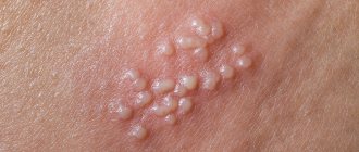
Pemphigus is a chronic autoimmune disease characterized by the appearance of a special type of blisters on the surface of previously healthy skin and mucous membranes. Among the types of pemphigus can be distinguished: vulgar, vegetative, erythematous and foliate.
Pemphigus can be diagnosed if acantholytic cells are detected, which are detected in a smear taken or as part of blisters in the epidermis itself (during histological examination). To treat pemphigus, glucocorticosteroids are first used (a whole course of treatment is prescribed). The latter always goes well with extracorporeal hemocorrection (plasmophoresis, cryoapherosis, hemosorption).
Causes
The reasons for the development of pemphigus have not yet been fully studied. One of the main causes of pemphigus is a violation of autoimmune processes, thereby the cells become antibodies to the immune system.
Violation of cell structure is subject to the influence of external factors, as well as aggressive environmental conditions. As a result, the communication between cells is disrupted, which leads to the formation of bubbles. The incidence rate in people with a hereditary predisposition is much higher.
Interdigital fungus: causes, symptoms and prevention
- Causes of fungus between toes
- Symptoms of fungus on the feet between the toes
- Disease prevention
- Photo of fungus between fingers
The overwhelming majority of cases of fungal diseases begin with damage to the skin between the toes. Here infectious agents find the most favorable conditions for life and reproduction. Anyone can become infected if several conditions are combined.
Causes of fungus between toes
The outer layer of skin (epidermis) is covered with a thin layer of fat from the sebaceous glands. It contains bacteria, enzymes and acids (the pH of this emulsion is 4 - 5.7). This is a barrier to infections. It can be destroyed by 3 factors:
- Microtrauma of the skin due to abrasions, diaper rash or inflammation. In such a place, the fungal spores will “take root” firmly;
- High humidity in skin folds. Between them, in the sweat and stratum corneum, there is a rich nutrient medium for fungus;
- Warm. A greenhouse microclimate is formed inside the shoes.
The presence of physiological, pathological health characteristics and habits in a person increases the likelihood of fungal infection of the interdigital spaces.
- Physiologically, some people have dry or very moist skin, which is conducive to fungal infections;
- The same picture emerges for those whose skin is always hot. They do not freeze, the surface of the body is of normal humidity, but excess heat provokes the appearance of a fungal infection on the toes;
- Women during menopause have periods of physiologically justified active sweating.
- Infants and the elderly are susceptible to infection due to weak immunity;
- Heart failure provokes swelling, making it almost impossible to hygienically treat the skin between the fingers and in the folds under them. Vascular disorders create stagnation of blood in the legs, which leads to trophic complications (dryness and thinning of the skin, the appearance of small ulcers on it);
- Sick kidneys do not remove metabolites from the blood. The mycelium uses “slags” for its nutrition;
- Infectious diseases weaken the immune system (HIV - the human immunodeficiency virus destroys it) and cause increased sweating;
- Oncological diseases and methods of their treatment (radiation exposure, chemotherapy) also greatly reduce immunity - a barrier to fungal infections of the skin of the feet.
Symptoms of fungus on the feet between the toes
The onset of the disease is characterized by slight redness and slight itching between the toes. More often, in intervals of 3-4-5 fingers, counting from the “big one”. The manifestations do not cause significant discomfort, so the patient often ignores them.
The itching intensifies, cracks and areas of swelling appear on the skin. In some places it changes color to bright red or yellowish. Peeling and burning sensation occurs. Callous seals form between the fingers.
At the next stage, the cracks deepen and erosion appears. The associated bacterial infectious process reveals itself with a strong unpleasant odor. Pain and itching of greater intensity interfere with night rest and torment during the day.
The last stage is characterized by worsening symptoms. The itching does not stop, severe pain and deep erosions between the bases of the fingers. The skin is red all over the foot. The appearance of extensive blisters with watery contents is added.
You can read about treating fungus between the toes here.
Disease prevention
It is necessary every day, during evening bath procedures, to examine the skin, especially the spaces between the fingers. Any damage found must be treated with a disinfectant (chlorhexidine, octeniderm, miramistin). If itching or peeling appears in this area, see a doctor. Take precautions.
- Wear shoes of a suitable size and comfortable width made from natural materials. Do not give your shoes to anyone and do not use someone else’s;
- Never walk anywhere barefoot;
- Carefully monitor the cleanliness and dryness of your feet;
- At work, wear light, replaceable shoes that are as open as possible;
- Change socks once, and in case of severe sweating - several times a day;
- Dry your shoes using shoe dryers with ultraviolet lamps.
You can read more about fungal prevention here.
Mechanism of bubble formation
Human skin can be figuratively described as a water-spring “mattress” covered with a kind of “wall”. The “mattress” does not participate in the formation of bubbles - only the top layer, the epidermis, suffers.
The epidermal layer consists of 10-20 cell layers, which look like bricks under a microscope. The “bricks” of the second layer of the epidermis are connected to each other by peculiar “bridges”. On top of the “wall” there are layers of cells that are no longer quite similar to cells, reminiscent of applied cream. These are scales, corneocytes, necessary for protection from mechanical, chemical and physical damage.
If, under the influence of internal or external causes, antibodies are formed that destroy the “bridges” - desmosomes between the cells of the basal layer (this is called acantholysis and can be seen under a microscope), this is true pemphigus. If tissue fluid penetrates between the basal and upper layers of the epidermis without destroying the “bridges,” it is pemphigoid. Viral pemphigus also occurs without destruction of desmosomes.
Classification
Types of non-acantholytic pemphigus:
- Non-acantholytic pemphigus is benign. Pathological elements are formed exclusively in the human oral cavity. Upon examination, inflammation of the mucous membrane, as well as its slight ulceration, can be detected.
- Bullous form of non-acantholytic pemphigus. This is a benign disease that develops in both adults and children. Blisters form on the skin, but there are no signs of acantholysis. These pathological elements can spontaneously disappear without scarring.
- Cicatricial non-acantholytic pemphigus. This pemphigoid is called pemphigus of the eye in the medical literature. Most often it is diagnosed in women who have crossed the 45-year age limit. A characteristic symptom is damage to the visual apparatus, skin and oral mucosa.
Classification of true pemphigus:
- Erythematous form. This pathological process combines several diseases. Its symptoms are similar to seborrheic dermatitis, an erythematous variant of systemic lupus, as well as true pemphigus. Erythematous pemphigus in adults and children is very difficult to treat. It is worth noting that the disease is diagnosed not only in people, but also in some animals. A characteristic symptom is the appearance of red spots on the skin of the body and face, covered with crusts on top. Simultaneously with this symptom, seborrheic manifestations appear on the scalp.
- Pemphigus vulgare. This type of pathology is diagnosed in patients more often. Blisters form on the skin, but there are no signs of inflammation. If pemphigus is not treated on time, pathological elements can spread throughout the entire skin. It is worth noting that they can merge and form large lesions.
- Pemphigus foliaceus. This form received its name due to the characteristics of the pathological elements. Blisters form on the human skin, which practically do not rise above the epidermis (not tense). Crusts form on top of them, which tend to layer on top of each other. The effect of sheet material folded in stacks is created.
- Brazilian pemphigus. There are no restrictions regarding gender and age. Cases of its development have been recorded in both young children and elderly people aged 70 to 80 years. It is also possible that it may progress in middle-aged people. It is worth noting that this variety is endemic and is therefore found only in Brazil.
Manifestations of the disease
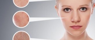
The symptoms of hemorrhagic vasculitis are quite varied. The most characteristic external sign is skin damage, manifested in the form of so-called purpura - a bright rash in the form of small purple-bluish spots, symmetrically appearing on the legs and feet, and with the development of the disease rising higher, to other parts of the body. In addition to purpura, other types of rash may appear on the patient’s body - erythema, petechiae, vesicles. In severe cases, necrotic areas of skin may appear.
In addition, more than 2/3 of patients have damage to the joint surface. It is accompanied by pain and swelling, which can last for several days. The most severe symptom of hemorrhagic vasculitis is abdominal pain - a consequence of hemorrhagic damage to the intestinal wall. The nature of the pain is similar to manifestations of appendicitis, peptic ulcer or intestinal obstruction. In some cases, the presence of blood is detected in stool and vomit. Some patients develop glomerulonephritis, cough, and shortness of breath.
There is a definite difference between the manifestations of the disease in children and adults.
Hemorrhagic vasculitis in children is characterized by striking symptoms, including:
- acute onset and course of the disease;
- the presence of fever in about a third of patients;
- loose stools streaked with blood;
- abdominal syndrome;
- manifestations of glomerulonephritis with the presence of protein and red blood cells in the urine.
Cases of hemorrhagic vasculitis in adults, as a rule, are characterized by blurred, insignificant manifestations at the onset of the disease. Abdominal syndrome is present in no more than half of patients, mostly without nausea and vomiting. When the kidneys are damaged, chronic glomerulonephritis and chronic renal failure almost always develop.
Are you experiencing symptoms of hemorrhagic vasculitis?
Only a doctor can accurately diagnose the disease. Don't delay your consultation - call
Symptoms
Considering that experts have identified several different types of this pathology, the symptoms of each of them will be very specific. Of course, there are a number of general trends and signs inherent in all types of the disease. This may include, for example, the wave-like course of the pathological process.
Periods of exacerbation alternate with the transition of pemphigus to a calmer stage, when the main symptoms subside or completely disappear. An important factor for the patient will be the fact that in the absence of timely diagnosis and prescription of an effective course of treatment, there is a high risk of developing severe conditions aggravated by concomitant diseases.
- The presence of crusts, ranging from pale pink soft to red dense, reminiscent of lichen;
- There is a deterioration in the general condition;
- Decreased immune response of the body;
- Formation of bubbles of varying densities;
- Also, in severe cases, separation of the layers of the epidermis is noted, and it can occur both in the lesion and away from it.
- Damage and ulcers of the mucous membrane of the mouth, nasopharynx or genitals;
- Pain when performing the act of swallowing or when eating;
- Bad breath, indicating damage to the mucous membranes;
- Hypersalivation or, in other words, increased salivation;
- In the seborrheic form, characteristic yellowish or brown-brown crusts appear on the scalp.
- Bubbles vary in appearance, ranging from flat to thin-walled, which burst with a slight touch. In their place, erosions and, subsequently, crusts form.
- In severe cases, an eroded surface of the skin may form in place of the blisters. Their feature is a tendency towards peripheral growth. Over time, such erosions occupy a large surface of the skin, causing pain and inconvenience to the patient.
- In children, manifestations of pemphigus are localized over the entire surface of the skin, including the limbs.
Experts say that with this disease, both a pure form of the pathological process and mixed forms that smoothly transform into one another can be observed. Therefore, the symptoms and signs of pemphigus in a given person may vary and indicate the presence of several types of disease.
Diagnostic methods
Since in children the disease manifests itself with bright, acute symptoms from the very beginning, there are, as a rule, no problems with making a diagnosis. Diagnosis of hemorrhagic vasculitis in adults is much more difficult, especially in the absence of a characteristic rash at the onset of the disease. As a rule, it is based on laboratory tests, which include:
- blood tests - general, biochemical, coagulogram;
- urine examination for hematuria, proteinuria, cylindruria, Nechiporenko and Zimnitsky tests, biochemical analysis;
- stool test for the presence of blood.
An important stage of diagnosis, which allows us to establish the degree of damage to internal organs, is instrumental studies - ultrasound of the abdominal cavity and kidneys, ultrasound of the renal vessels, gastroscopy. In severe cases of the disease, a biopsy of the skin and kidneys is prescribed to determine the size of immunoglobulin deposits and the permeability of the vascular wall.
Diagnostics
Experts say that a correct diagnosis can be made based on a comprehensive examination of the patient, which includes several important stages:
- Examination of the patient for the presence of a clinical picture. At this point, the doctor establishes the nature of the lesions, their localization, the degree of development of the disease, etc.
- Cytological analysis necessary to establish the presence of acantholic cells in smears of biomaterial.
- Carrying out the Nikolsky test, which allows to differentiate pemphigus from similar pathological processes.
- Method of direct immunofluorescence. This study allows us to detect the presence of immunoglobulin in the intercellular substance of the epidermis.
- A histological study, which is based on a technique for detecting crevices and other damage within the epidermis.
Only the totality of all the results makes it possible to make an accurate diagnosis and prescribe an effective course of treatment, leading to the patient’s recovery.
What kind of disease is this
This disease is of an infectious-allergic nature: the main cause of hemorrhagic vasculitis is the presence in the blood of patients of immune complexes and active components of the protective system of proteolytic enzymes. Immune complexes accumulate in the bloodstream, and with an excessive amount of antigens or a lack of antibodies, protein formations are deposited on the endothelium of the microvascular wall. Factors contributing to the development of the disease are:
- bacterial and viral respiratory infections suffered 2-4 weeks before the onset of the disease, which account for up to 60-80% of all cases of the disease;
- foci of chronic infection (sinusitis, tonsillitis, caries, etc.);
- some medications, mainly of antibiotic action;
- potentially allergenic foods (strawberries, citrus fruits, chocolate, eggs, etc.);
- administration of a vaccine or serum;
- hypothermia and dampness or prolonged exposure to the sun.
There is evidence of a hereditary predisposition to the disease. In some cases, it is impossible to find out which factor served as the “trigger” for the patient’s body.
Treatment of viral pemphigus
Treatment of viral pemphigus involves the use of the following systemic drugs:
- cytostatics stop the division of immune cells: Sandimmune, Azathioprine, Methotrexate;
- antiviral: Viferon, Laferon, Cycloferon;
- glucocorticosteroids: Dexamethasone, Prednisolone;
- antipyretics: Ibuprofen, Paracetamol, Nimesil, Mefenamic acid;
- antihistamines relieve itching: Cetrin, Diazolin, Fenistil.
For external treatment of affected skin areas, the following may be prescribed:
- antimicrobial local anesthetics for irrigating the oral cavity if viral pemphigus has affected the child’s mucous membranes: Forteza, Orasept;
- antiseptics: Chlorhexidine, Methylene blue, Miramistin;
- combination preparations of antiseptics and anesthetics: Oflokain, pharmaceutical talkers;
- antipruritic lotions made from nettle juice, aloe, and walnut oil.
Since children with this diagnosis are usually treated in a hospital setting, to enhance the therapeutic course, therapeutic procedures can be carried out aimed at clearing the blood of antibodies:
- plasmapheresis - replacement of the liquid part of the blood with similar solutions without microbes, immune complexes and antibodies;
- hemosorption using a carbon filter.
Only a doctor can tell how to treat viral pemphigus, because in each individual case it can acquire some special features. As for other forms of pemphigus, the therapeutic course for them is also determined individually.
How to treat other forms of pemphigus?
The treatment process for pemphigus is quite complicated. Therefore, self-medication of this type of disease is under no circumstances acceptable. The disease progresses rapidly, affecting large areas of the skin, which leads to disruption of the internal organs.
Treatment of pemphigus is mandatory in a dermatological hospital. First of all, corticosteroid drugs, cytostatics and other drugs are prescribed to alleviate the course of the disease and the life expectancy of patients.
The drugs must first be taken in large doses. At the same time, pay attention to blood and urine sugar levels, monitor blood pressure and observe personal hygiene rules. With frequent changes of bed linen and underwear, secondary infection is prevented.
Causes of redness of the skin between the fingers
Have you been trying to cure FUNGUS for many years?
Head of the Institute: “You will be amazed at how easy it is to cure fungus by taking it every day...
Read more "
Redness between the fingers is very often noted by parents in their children. As you know, children are very curious; they learn about the outside world by touching various objects. When playing on a playground where there is dirty sand, picking up twigs and pebbles from the ground, a huge amount of pathogenic bacteria gets on their hands, which can cause redness and rash. There are other causes of red spots between the fingers; they can be of viral etiology or be the result of insufficient hygiene. A doctor can determine exactly what caused the redness between the fingers based on a visual examination and a series of laboratory tests.
Causes of the pathological process
If your child's skin is red and itchy between his fingers, an allergic reaction can be diagnosed. It forms after a child touches an irritant, for example, a cat, a plant or washing powder. The doctor’s task is to determine the provocateur of the body’s unusual reaction, and the parents’ task is to avoid this irritant in the future.
A special feature of contact allergies is that in the absence of prolonged contact with the irritant, redness and itching between the fingers goes away on its own, but you should not let the situation take its course.
OUR READERS RECOMMEND!
Our readers successfully use Tinedol to treat nail fungus. Seeing how popular this product is, we decided to bring it to your attention.
Atypical dermatitis has similar symptoms to an allergic reaction, but between the fingers, redness and peeling gradually affects new areas. In addition, fluid-filled blisters are often noted. From the bursting bubbles, the liquid enters new areas and infects them. Bubbles need to be treated as quickly as possible before they burst on their own. If the color of the fluid in the blister turns yellow, it is likely that there is a bacterial infection. If the formation of ulcers and erosions is observed between the fingers in places of redness, this is evidence of weeping erythema - a consequence of advanced contact dermatitis.
Irritation between the fingers can be caused by streptococci. The parent himself can recognize streptococcal infection, as it is accompanied by:
- increased temperature;
- sore throat;
- decreased appetite;
- irritability;
- the skin in places of redness peels and swells;
- At the site of redness, a thick yellow crust forms over time.
In kindergartens and schools, parasitic infestations are very often observed, as well as scabies, caused by a skin mite, which is transmitted through touch. Parasites that have begun to multiply in the body provoke redness between the fingers, impaired bowel movements, increased temperature, and also reduce general well-being, the child becomes lethargic and indifferent.
It will be interesting to know that redness and peeling between the fingers does not require treatment if it is caused by nervous shock or neurosis. Some children, after stressful situations, experience red spots of different locations. As soon as the child calms down, they disappear.
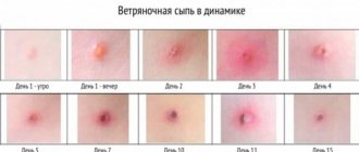
Irritation of the skin between the fingers can be a symptom of infectious diseases such as rubella, measles and chicken pox. All of the listed ailments are characterized by a period in which there are no rashes yet, but the skin in the places of their future development turns red. Adults should carefully monitor new symptoms that appear at this time, and if the child’s well-being worsens, immediately go to the hospital. Infectious diseases of this type are characterized by many complications, and it is not always possible to eliminate them later.
There are many photos of symptoms of diseases of viral and infectious etiology on the Internet. Parents can compare the current picture with them and draw certain conclusions about what could have caused the disease. Taking tests and going to the doctor will be a bit of a hassle in any case, but you will already know which direction to move in, describing to the doctor possible provocateurs.
If the skin between your fingers peels off, you can assume a fungal infection or lichen. The doctor will decide what type of lichen the baby has by taking a scraping from the affected area of the skin.
Medicines for the treatment of pemphigus
The patient is advised to take glucocorticoids in high doses. The following drugs can be used for this:
- Metipred;
- Prednisolone;
- Dexamethasone;
- Polcortolon.
When symptoms begin to regress, the doses of these drugs are gradually reduced to the minimum effective. Patients with pathologies of the gastrointestinal tract are prescribed long-acting glucocorticoids:
- Metipred-depot;
- Diprospan;
- Depo-Medrol.
Treatment with hormonal drugs can cause a number of complications, but they are not a reason to discontinue corticosteroids. This is explained by the fact that refusal to take them can lead to relapses and progression of pemphigus.
Possible complications during treatment:
- acute psychosis;
- arterial hypertension;
- depressive states;
- insomnia;
- increased excitability of the nervous system;
- steroid diabetes;
- thrombosis;
- obesity;
- angiopathy;
- erosions or ulcers of the stomach and/or intestines.
If the patient’s condition sharply worsens while taking corticosteroids, the following measures may be recommended:
- diet: limiting fats, carbohydrates and table salt, introducing more protein and vitamins into the diet;
- drugs to protect the gastric mucosa: Almagel, etc.
In parallel with glucocorticoids, cytostatics and immunosuppressants are prescribed to increase the effectiveness of therapy and the possibility of reducing doses of hormonal agents.
The following medications can be used for this:
- Sandimmune;
- Methotrexate;
- Azathioprine.
To prevent electrolyte imbalance, the patient is recommended to take calcium and potassium supplements. And for secondary infection of erosions - antibiotics or antifungal agents.
The ultimate goal of drug therapy is to make the rash disappear.
Preventive measures
There are no specific measures to prevent the development of pathology. The higher the level of immune protection, the less chance of dermatological diseases.
Important:
- control the nature of chronic diseases;
- strengthen immunity;
- maintain personal hygiene;
- Healthy food.
Measures to prevent pemphigus in newborns:
- change your underwear more often;
- Caring for newborns with pustular skin lesions is prohibited;
- Take regular care of your child’s skin;
- strengthen the immune system of weakened children;
- daily wet cleaning and ventilation of the room are required.
If you notice any rashes on the skin, the formation of pustules and blisters, immediately contact a dermatologist.
Forecast
The prognosis for acantholytic pemphigus is conditionally unfavorable. On the one hand, in the absence of effective treatment, there is a high probability of complications and death.
On the other hand, patients with pemphigus are forced to take glucocorticosteroids for a long time, and sometimes for life, which is fraught with the development of side effects. But hasty refusal of drugs leads to immediate relapse of the disease. Glucocorticosteroids do not eliminate the cause of the disease, but inhibit the pathological process and prevent its progression.