SPECIALISTS Gynecologist Gynecologist-endocrinologist Pediatric gynecologist Mammologist-oncologist Dermatologist Hirudotherapist Intimate plastic surgery Doctor Contour plastic doctor Ultrasound doctor SERVICES AND PRICES Gynecology Mammology Ultrasound diagnostics Paid tests Intimate surgery Contour plastic Treatment for women PROMOTIONS AND DISCOUNTS Students Teams Friends and subscribers am For residents of the region For pensioners Promotions in clinic
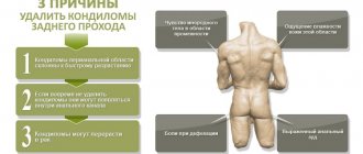
Have growths on the labia, rashes and pimples near the vagina? Maybe there is a feeling of “something extra” near the anus and there is an itching of the anus? What is it, the reasons and how can a woman get rid of genital warts? Cauterization of condylomas is a procedure for eliminating viral skin lesions. Removal is associated with physical discomfort and some nuances, so it should take place in a medical center under the supervision of a gynecologist.
Why are genital warts dangerous?
The content of the article
A cervical infection caused by an oncogenic type of HPV, if left untreated, causes cervical cancer. Various factors, especially immunosuppression, increase the persistence and severity of infection and may contribute to the development of cancer.
A very effective vaccine has been developed to prevent these HPV-related diseases. The quadrivalent vaccine protects against high- and low-risk HPV. This article reviews the prevention, diagnosis, and treatment of HPV infections and their clinical manifestations, with special attention to acute condylomata.
How does the virus enter the body?
The content of the article
The main route of infection is sexual. Moreover, regardless of the form of sexual contact - anal, vaginal or oral. A condom reduces the risk of infection, but it is not a panacea.
With a much lower probability, HPV can enter the body through contact and household contact - through touching, hugging, personal hygiene products, manicure tools, etc. Most often, danger awaits in public places - bathhouse, swimming pool, sauna, bus, etc.
The presence of damage and microcracks on the skin favors infection. A vertical route of infection is also possible - from mother to child. The virus can be transmitted to an infant during fetal development or when passing through the birth canal.
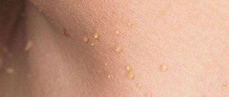
Symptoms - how to identify condylomas
The primary manifestations of the disease are a small red growth. In the case of hyperkeratosis, the lesion becomes whitish. After a short time, new growths appear, together forming an unpleasant-looking cauliflower-shaped wart. Thus, the lesion is complex because it consists of several individual papillomas.
Typical places with favorable conditions for the development of papillomas:
- the anal area, where large cauliflower-like lesions are most often observed due to the warm and humid environment;
- in women - labia minora, vaginal opening and vagina;
- in men - the philtrum, the head and the opening of the urethra.
The lesions inside the body are usually not condylomas, but rather ordinary flat warts. Associated medical conditions may predispose to infection: inflammation of the anus, chronic vaginal discharge, or phimosis.
The growth rate of genital warts varies. The influence is caused by systemic immunodeficiency, skin maceration, pregnancy, which can also increase the spread of rashes. Small rashes are often asymptomatic, but larger rashes usually cause pain, bleeding and itching.
Genital warts Genital warts
Types and symptoms of warts, papillomas, condylomas and molluscum contagiosum
The disease does not have any clear clinical symptoms; sometimes the patient does not even suspect that he is infected with the HPV virus. Women may experience spotting after sexual intercourse, and during examination, the doctor may notice signs of cervical erosion and ask about the most typical symptoms for this type of disease.
But at the same time, there are many signs by which one can judge the presence of HPV:
Genital warts (or genital warts)
Flesh, pinkish or reddish papillary-shaped growths that appear on the skin or mucous membranes of the genital organs (external genitalia, vaginal and cervical mucosa, around the anus or external urethra). They can be formed either individually or in groups.
Genital warts can be a symptom of cancer development, that is, they are characterized by moderate oncogenic potential. And, of course, you need to get rid of them, and the sooner the better, but treatment should only be carried out by a high-level specialist.
Flat condylomas
This type of tumor protrudes above the surface of the skin to a lesser extent (unlike the pointed form), but is characterized by a higher level of development of cancer. For high-quality and effective treatment, more complex diagnostics are required.
Warts
One of the common types of clinical manifestations of HPV. Warts appear in children and adults on the fingers and toes, on the feet, on the face (on the lips) and neck and other parts of the body. They come in different types, which differ in color, size and location.
Molluscum contagiosum virus
Another type of clinical manifestation of HPV is in the form of small whitish pimples. Sizes can range from a millet grain to the average size of a pea. The virus can infect a large surface of the skin, but does not penetrate into the deeper layers of the dermis.
In children, the molluscum contagiosum virus is diagnosed more often than in adults, and it brings a lot of inconvenience, not only physical, but also moral, since it is very unaesthetic in appearance. Sometimes pimples can be accompanied by itching and bother the baby.
A child can become infected in any public place (in a sandbox, in a kindergarten, in a swimming pool) and at the same time become a spreader of the disease.
Wen, or lipoma
This is a neoplasm that appears in the form of a small compaction, soft to the touch, with well-defined defined boundaries. The tumor can move if you put a little pressure on it. Over time (without treatment) it may increase in size.
Typically, lumps do not cause pain or symptoms such as fever or chills. The localization of neoplasms varies - from the crown to the fingertips.
Reasons for the development of condylomas
The etiological factor of genital warts is the viruses HPV6, HPV11, HPV42, HPV43, HPV44, which lack oncogenic properties. They are also responsible for the formation of laryngeal papillomas, giant Buschke-Levenshtein condylomas and flat condylomas of the cervix. HPV 16 and HPV 18 remain associated with cervical cancer.
According to available data, the most important risk factor for developing cervical cancer is infection with oncogenic HPV. However, in men, persistent long-term infection with an oncogenic HPV type predisposes to the development of penile intraepithelial neoplasia (PIN) and penile cancer.
How are condylomas transmitted - how does infection occur?
Genital warts are highly contagious. Infection most often occurs through sexual contact. It is now one of the most common diseases transmitted this way—approximately 1% of sexually active adults under the age of 50 have had or currently have genital warts. The highest frequency of their occurrence is observed in women and men aged 18 to 28 years. The risk of infection increases with the number of sexual partners and the frequency of sexual intercourse, especially with an already infected partner. The largest independent risk factor is the number of sexual partners in the last 2 years.
Humans are the only known reservoir of the papilloma virus. These viruses multiply in differentiating squamous epithelium, so they infect only active, proliferating cells of the skin and mucous membranes. The incubation period is 1-6 months, although incubation time may be longer.
Viruses enter the basal cells due to trauma to the epithelial cells of the penis or vulva (eg, an abrasion).
Condyloma removal
Prognosis and prevention of pathology
It is impossible to completely eliminate the virus; it can only be deactivated for a long time.
But with a decrease in immunity, a relapse of the disease occurs. With timely treatment, the prognosis of the disease is favorable, and doctors are able to eliminate the growths. If there is no treatment for some reason, the prognosis will be poor; often, a person develops an oncological tumor as a complication. As a preventive measure, men are advised to get rid of bad habits, lead an active lifestyle, eat right, and avoid the appearance of provoking factors. The sexual partner must be alone on a permanent basis. Today, a vaccine against HPV has been developed in medicine, but it does not guarantee that the virus will not manifest itself fully.
Diagnosis of condylomas
HPV infection of the mucous membranes can occur in latent, subclinical and clinical forms.
- Clinical form
. Papillomas are visible to the naked eye, while hidden infections can only be detected using an HPV DNA test. - Subclinical form
. Colposcopic and cytological studies play an indispensable role in the diagnosis of subclinical infections. - Latent form.
After sexual intercourse, the virus often enters a latent phase, which can last from several months to several years, histological changes are not visible, and HPV DNA remains in the nuclei of the basal cells. Most HPV infections are latent. The action of certain stimuli, such as microtrauma or infection with other pathogens, leads to stimulation of keratinocyte proliferation and activation of HPV replication.
Molecular testing detects HPV DNA on the mucous membranes of 15-20% of people without clinical signs of infection. Specific antibodies against viral capsid proteins exist in more than 50% of cases. This clearly indicates that only 25% of the population has no clinical, virological or immunological evidence of genital HPV infections.
Condylomas can be recognized with the naked eye, and clear visualization of the rash is achieved using a 3-5% acetic acid solution, which lightens the mucus and dehydrates the cells, which makes the areas of dysplasia and neoplasm whitish for a short time.
In addition to the above methods, tests used to diagnose changes associated with HPV include histopathological examination and rectoscopy. It makes it possible to detect lesions of the rectum, which are extremely common in perianal rashes. If the area of the urethral orifice changes, urethroscopy is mandatory.
Complications
Possible complication of anal condylomas
If not treated in a timely manner, single condylomas may begin to grow, covering new areas of the skin or epithelium. This often leads to growths closing the anus and urethra, causing problems with urination and defecation. When injured, warts can bleed, creating good conditions for infectious bacteria to enter the body.
In some cases, the development of a purulent process, the appearance of fistulas, and sepsis are possible.
Factors that increase the risk of developing condylomas
HPV infections themselves do not cause malignant transformation of the affected cells. Cofactors play a significant role - ultraviolet radiation, smoking, folic acid deficiency, pregnancy, immunodeficiency, Chlamydia trachomatis infection, herpes virus (HSV2).
Patients undergoing immunosuppressive therapy and patients with defective cellular immunity, especially those infected with HIV, have higher rates of HPV infections and respond poorly to treatment.
On the other hand, in immunocompetent individuals there is a clear tendency for spontaneous recovery. It is highly likely that hormonal contraceptives also act as a cofactor and can enhance the cancer process.
The significant role of coccinogens is indicated by the significant time interval between the peak of HPV infections (18–25 years) and the peak incidence of cervical cancer (45–50 years). The process of tumor transformation is very slow and requires the presence of additional favorable factors.
The epidemiological relationship between infection with oncogenic types of HPV and the development of neoplasms of the genital organs is very high and in the case of cervical cancer is 95-99.7%, and in the case of anal canal cancer - 88%. Cervical cancer is the second most common malignancy in women. Every year there are 500 thousand new cases of the disease, from which 50% of sick women die.
Effectiveness of the HPV vaccine
Given these alarming data, a vaccine against genital HPV types offers great and justifiable hope. Its structure is based on the recombinant L1HPV protein (VPL) and does not contain any elements of the viral genome. The vaccine works by inducing specific antibodies by targeting HPV capsid antigens. Thus, it limits HPV infection and reduces the risk of precancerous changes.
The first studies, published in 2002, were already promising. The efficacy of a monovalent HPV16 vaccine administered in 3 doses was analyzed. The research group included 2392 people (women 16-25 years old). As a result of vaccination, 99.7% of women experienced seroconversion.
In the group of vaccinated women, after 17 months of observation, not a single case of CIN was detected (22). Subsequent studies were conducted on a group of 1113 women aged 15-25 years and showed the high effectiveness of the bivalent vaccine against HPV 16 and HPV 18 in preventing incidental (91.6%) infections, persistent (100%) and cytological lesions (93.5%). %).
The currently introduced quadrivalent vaccine has demonstrated 100% clinical efficacy against HPV6/11/16/18 in studies, preventing the occurrence of genital warts and cervical lesions. This was proven by the FUTURE II study published in 2005.
The quadrivalent HPV vaccine is used to prevent high-to-moderate cervical dysplasia (CIN2/3), cervical cancer, high-to-moderate dysplastic vulvar lesions (VIN2/3), and external genital warts. It has been found that it is most beneficial to vaccinate women before potential exposure to HPV infection, that is, before sexual activity begins.
Thus, routine administration of the vaccine to girls aged 11–12 years is recommended, although it can be administered earlier - to girls from 9 years of age. and for boys 9-15 years old. The full course of vaccination includes intramuscular administration of 3 doses of the vaccine according to the scheme 0 + 2 + 6 months.
Women with a positive or equivocal cytology test result, a positive Hybrid Capture 2 molecular test, and patients with existing condylomata may also be vaccinated.
Clinical trial data do not indicate that the quadrivalent vaccine has a therapeutic effect on current epithelial lesions, current HPV infection, or existing genital warts. However, a complete preventive effect can be expected against the remaining HPV types included in the vaccine formula.
To date, the longest period of observation of vaccinated patients is 5 years - during this period the full effectiveness of the vaccine has been demonstrated. It is unclear whether booster doses will be required.
An inherent aspect is the potential adverse effects of HPV vaccination. The vaccine dramatically reduces the risk of developing cervical cancer, but does not eliminate it completely. The essence of this phenomenon lies in its valency - neoplastic changes caused by HPV16 and HPV18, which are responsible for 70% of cervical cancers, are prevented, but there is still a fairly significant reserve of cancers associated with other types of the virus.
They can be prevented through regular preventive examinations (cytology), which, however, can be neglected due to an illusory sense of security among vaccinated people.
In the context of the currently implemented HPV vaccinations, the sociological aspect is interesting. Resistance by parents of adolescents to vaccination against sexually transmitted diseases, which may expose their children to risky sexual behavior, is seen as a major barrier. Empirical evidence does not support these concerns.
The motivation of patients for vaccination is noteworthy - they emphasize the importance of protection against genital warts, which are perceived as a real risk (peak incidence is 20-25 years), in contrast to possible long-term cervical cancer. .
In terms of prevalence, human papillomavirus infection (PVI) ranks first among sexually transmitted infections. Anogenital warts are the most common clinical manifestation of low-oncogenic risk PVI. Human papillomaviruses (HPV) types 6 and 11 are the cause of 99.8% of cases of anogenital warts [1]. According to the World Health Organization (WHO), more than 42 million cases of anogenital warts are observed annually in the world. The average time between infection and the development of anogenital warts is 11-12 months in men and 5-6 months in young women. In rare cases, they can progress to malignancy (Buschke-Levenshtein tumor). This pathology received its name after the authors A. Buschke and L. Löwenstein, who first described giant genital warts of the penis in 1925. In Russia, among people aged 18 to 29 years, the pathology accounts for up to 65% of the structure of sexually transmitted infections. According to the Federal State Statistics Service, the frequency of anogenital condylomas, according to the Ministry of Health of Russia, in 2001 was 26 per 100 thousand population, in 2009 - 34.4, in 2012 - 25.9 per 100 thousand population, and in 2015 - 21.2 [2].
In Germany, the incidence of condylomas in women and men aged 22-29 years in 2006 was 462 and 345 per 100 thousand population, respectively [3], in England (2000) in women aged 20 and 24 years - 715 and 699 per 100 thousand population, respectively [4]. On average, the prevalence of anogenital condylomas, established according to the results of an all-Russian study, in both women and men aged 18 to 60 years was 9.2%. The maximum prevalence was observed in young women aged 18–24 years and reached 14.5% [2].
There is no specific therapy for the treatment of anogenital warts associated with HPV. Methods of cryodestruction and surgical removal are effective, but in the presence of PVI they do not prevent the development of relapses. HPV types 6 and 11 can also cause a rare condition known as recurrent respiratory papillomatosis, in which warts form in the larynx (on the vocal cords) or other parts of the respiratory tract. Recurrent respiratory papillomatosis is observed mainly in children under 5 years of age (juvenile type) or in people in their third decade of life (adult type). Women with genital PVI can transmit the virus to their newborn during childbirth. The result of infection is the growth of warts in the larynx, and therefore children undergo multiple surgical interventions to remove them. Recurrent respiratory papillomatosis can lead to airway obstruction, even death.
Giant Buschke-Levenshtein condyloma is wart-like elements of the papillomas type, merging with each other and forming a lesion with a wide base infiltrating the underlying tissue. The neoplasm has locally invasive growth, but in most cases is histologically a benign tumor, and malignancy is rare. Their occurrence and possible further relapses are associated with a weakening of general and local immunity. Genital warts usually occur in areas that are traumatized during sexual intercourse, but the area around the anus is not usually associated with anal intercourse. The fact is that HPV does not affect a limited area, but large areas of skin (as a rule, this is the entire skin of the genital organs, groin area, perineum, skin around the anus). From the moment of infection to the appearance of genital warts, it can take from several weeks to several years. The course of genital warts depends on the state of the immune system. The following options are possible: gradual growth of condylomas (both size and number); no changes (for a long time); self-resolution. Genital warts can be injured and bleed, interfere with normal sexual life, cause concern as a cosmetic defect, cause psychological discomfort and decreased self-esteem, and interfere with normal childbirth. If genital warts are suspected, a number of diseases should be excluded, such as molluscum contagiosum and condylomas lata (manifestations of syphilis). All patients with genital warts are required to be examined for the presence of syphilis and HIV. It is also advisable to screen for other sexually transmitted infections, to determine HPV of both high carcinogenic risk (HCR) and low carcinogenic risk (LCR). It is mandatory to take material for oncocytological examination using a liquid or traditional method in order to exclude cervical pathology. The question of morphological classification has not yet been finally resolved. Many authors classify this pathology as giant condylomas (benign tumors), while others, on the contrary, classify Buschke-Levenshtein tumor as a transitional form with malignancy into carcinoma. During pregnancy, anogenital condylomas, especially in combination with genitourinary infections, pose a high risk of premature birth, the development of fetoplacental insufficiency, infection of the fetus and newborn, complications during labor and the postpartum period. Therefore, the issues of examination, active treatment tactics and methods of delivery are relevant.
There are many treatments for genital warts (see table). The goals of treatment are the destruction of anogenital warts, preventing the development of complications, reducing the number of relapses and improving the quality of life of patients. Currently, the effectiveness of treatment methods for patients with anogenital condylomas, even taking into account repeated courses, is 60-80% [7]. Relapses in 25-50% of cases occur during the first months after treatment and are most often caused by reactivation of the virus. For example, the cost of treatment in the United States annually is $176.4 million [6]. Treatment of anogenital warts, like other STIs, should be carried out by both sexual partners, and during the period of treatment they should observe sexual rest.
Treatment methods for anogenital warts and their effectiveness [6]
A combination of conservative treatment (viferon therapy in this situation, both before and after surgery, as well as anti-inflammatory and antibacterial therapy) and surgical (radio wave excision of anogenital warts using the Surgitron apparatus) is considered more universal.
Viferon - a drug of recombinant interferon-alpha-2b (IFN-alpha-2b) in combination with highly active antioxidants (vitamins E and C) - is widely used for the treatment of infectious and inflammatory diseases of the urogenital tract. Currently, some experience has been accumulated in the management of pregnant patients infected with HPV. Adequate treatment of patients with urogenital infection during pregnancy, including pathogenetically based administration of interferon therapy (Viferon), improves the condition of the cervix of pregnant women, prevents the progression of clinical manifestations of PVI, reduces the incidence of gestational complications, and improves perinatal outcomes [10].
A good therapeutic effect in the form of an increase in the frequency of virus elimination, regression of low-grade squamous intraepithelial lesions of the cervix and a decrease in the frequency of infection of newborns is achieved using a combination of local application of human recombinant interferon - Viferon gel in combination with systemic use of this drug in the form of rectal suppositories [9]. Rectal administration of combined IFN allows one to achieve a higher concentration of interferon and a longer stay in the blood than with parenteral methods of administration.
Highly active antioxidants - vitamin E and ascorbic acid, which are part of the drug Viferon, have a membrane-stabilizing effect and also have a potentiating effect on the production of its own interferon, which not only leads to predictable clinical results, but also ensures an increase in inter-relapse intervals. One of the important features of the drug - the base in the form of cocoa butter - provides a number of the following advantages:
— due to the presence of stearic acid in its composition, it has excellent sliding properties, ensuring painless and atraumatic insertion;
- due to the presence of linoleic and oleic acids, it has an anti-inflammatory and wound-healing effect, activates lipid metabolism;
— polyphenols have an antiallergic effect, while, unlike cocoa powder, butter does not contain natural allergens;
— melts quickly, causing no discomfort from the presence of a foreign body in the rectum;
— ensures rapid absorption of all components of the suppository and, unlike various synthetic fats with dehydrating properties and a high melting point, does not have an irritating effect.
Destructive methods of choice for the treatment of pregnant women with genital warts
is radio wave therapy and the use of chemical coagulants - solcoderm, trichloroacetic acid. It is possible to use laser therapy, electrocoagulation, surgical methods and cryotherapy. The most acceptable method from the perspective of gynecological oncology is the radio wave method. Removal of condylomas by radio waves, or radio wave surgery, is carried out using the Surgitron apparatus. This device is a generator of high-frequency waves emitted with variable power and a constant frequency of 3.8-4.0 MHz. At a frequency of 4.0 MHz, the absorption of radio waves by adipose tissue is 12 times greater than by moist skin, so subcutaneous fat retains radio waves that freely penetrate through the upper layers. High-frequency radio waves - 4.0 MHz - perform a “cold” tissue cut at a temperature of 38 to 70 ° C (“The higher the frequency, the lower the temperature effect”), resulting in the absence of deep necrosis and injury. The healing process accelerates (proliferation is observed from the second or third day) and occurs without the formation of a rough postoperative scar with minimal pain and a high cosmetic effect. This technology allows us to obtain high-quality, most accurate histological tissue samples. Equally significant advantages are the sterilizing effect of high frequency radio waves (4.0 MHz) during manipulations and the “dry” (bloodless) incision, which makes it easier for the surgeon to view the surgical field. Surgitron high-frequency radio wave generators significantly facilitate and speed up surgical procedures. After surgical operations performed with a high-frequency radio wave generator, there are practically no such unpleasant postoperative consequences as pain, swelling and wound infection. Cosmetic results far exceed those obtained with traditional electrosurgical and laser techniques.
I would like to remind you that condylomas themselves are a manifestation of the human papillomavirus. This virus may pose a risk of fetal infection and laryngeal papillomatosis in the unborn child. Studies do not confirm that 100% of women who carry the virus become infected with the fetus. Unfortunately, it is impossible to predict whether a child will become infected with the virus during pregnancy and childbirth. Undoubtedly, the presence of condylomas increases the risk of infection, so it is better to remove the tumors.
The need for delivery by cesarean section in women with condylomatosis is decided individually. Spontaneous birth is preferred, even despite the presence of formations in the area of the external or internal genital organs that are present before birth. There have been cases where children born by cesarean section to pregnant women with HPV carriage suffer from laryngeal papillomatosis [9]. Caesarean section is performed for pregnant women with extensive condylomatosis of the external genitalia, when this prevents vaginal delivery, or in combination with other complications and diseases during pregnancy.
However, after any type of treatment, the risk of relapse is about 30%, but what is important is the possibility of childbirth per vias
naturalis
.
Clinical observation.
Patient
S.Yu.A.
, 33 years old.
Diagnosis upon presentation:
pregnancy 21-22 weeks, giant anogenital perineal condylomas, colpitis.
Anamnesis of life:
heredity is not burdened. Past infections: chicken pox, acute respiratory viral infection. There are no chronic diseases.
Gynecological history:
menstruation from the age of 14, regular, 4 days, every 28 days, moderate, painless. Sexual life from the age of 18. Pregnancy 1, spontaneous.
Gynecological diseases:
acute adnexitis at the age of 20, inpatient treatment was carried out.
Contraception:
interrupted sexual intercourse.
History of the disease.
In 2011, I noticed the appearance of warty pink formations on the labia majora. The dimensions of the elements ranged from 1 mm to several centimeters, which gradually increased. I did not contact a gynecologist. With the onset of pregnancy, I noted a sharp increase in the intensity of growth of these formations. She continued to be sexually active. I went to the doctor late, only when registering for pregnancy. Upon examination by a doctor at the place of residence, giant exophytic condylomas of the vulva were diagnosed. Immediately after the discovery of condylomas, she was sent to the Moscow Regional Research Institute of Obstetrics and Gynecology.
Complaints at the time of contact:
growth of formations in the area of the external genitalia, sensation of a foreign body, discomfort when walking, inability to sit, a pronounced unpleasant odor from the vagina, difficulties during sexual intercourse, cosmetic defect, psychological discomfort.
Inspection.
General condition is satisfactory. The skin is pale pink, clean, moderately moist. Blood pressure - 115/70 mm Hg. The abdomen is enlarged according to the stage of pregnancy. The uterus is in physiological tone, painless in all parts. Feels fetal movements well. The fetal heartbeat is clear, rhythmic up to 142 beats/min.
Gynecological status.
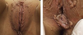
The external genitalia are represented by tumor-like formations covering the entire anogenital area on both sides of the vulva, labia majora and minora, 30 cm long, 10 cm high and 10 cm wide (Fig. 1 and below). Giant condylomas were also identified in the perianal area, and the anus was difficult to see. The structure of the formations was typical of giant Buschke-Levenshtein condylomas, which looked like cauliflower and completely covered the external genitalia. There were no areas of necrosis. The border of exophytic formations is at the entrance to the vagina. The vagina is free, the walls are not changed. During colposcopy: the cervix without pathological changes (conclusion: satisfactory colposcopic picture 3T-I, normal colposcopic picture). The discharge is copious, mucopurulent in nature with a putrid odor.
Rice.
1. Giant Buschke-Levenshtein condylomas in patient S. Inspection using mirrors. The vaginal mucosa is pink, without pathological changes, the cervix is conical, the vaginal part is up to 3 cm long, the external pharynx is closed.
Extended colposcopy (Fig. 2): the cervix is covered with stratified squamous epithelium with smooth relief, cyanotic, columnar epithelium is visualized around the external pharynx - without pathological changes after treatment with acetic acid. When stained with Lugol, the mucous membrane of the cervix is evenly stained, except for the area around the external pharynx.
Rice. 2. Results of extended colposcopy of patient S.
Preliminary diagnosis:
pregnancy 21-22 weeks. Giant Buschke-Levenshtein condylomas. An elderly primigravida. HPV type 6.
As a result of the examination, it was established: RW-negative, HIV not detected, hepatitis virus not detected.
Biochemical and clinical blood tests are within normal limits. General urine analysis without pathological changes.
Pseudomonas
aeruginosa
(105 CFU/ml) was isolated in urine culture
HPV type 6 was detected in the discharge from the surface of condylomas and the cervix using polymerase chain reaction (PCR). HCV HPV was not detected. When tested for sexually transmitted infections using the PCR method, chlamydia, herpes simplex viruses, and cytomegaloviruses were not detected. Mycoplasma and ureaplasma were detected. A serological study using an enzyme immunoassay for sexually transmitted infections revealed positive immunoglobulins G for cytomegalovirus and Epstein-Barr virus. When examining a smear for the degree of purity from the vagina: leukocytes 3-4, rod flora, Trichomonas were not detected (II degree of purity). A bacteriological examination of the mucus of the cervical canal revealed ureaplasma (104 KOE/ml), mycoplasma (103 KOE/ml). Cytological examination of material from the endo- and exocervix did not reveal any atypical cells. LSIL HPV+ was established.
Cytological examination of the vulva: NILM (Negative for intraepithelial lesion or malignancy).
Cytological examination of the perianal area: NILM.
Ultrasound: one living fetus is detected in the uterine cavity, the size of which corresponds to 20-21 weeks of pregnancy. No developmental defects were identified.
Diagnosis.
Pregnancy 21-22 weeks. Giant Buschke-Levenshtein condylomas. HPV type 6. Chronic cytomegalovirus infection and Epstein-Barr virus infection. An elderly primigravida.
The patient was prescribed a course of complex anti-inflammatory, antibacterial and antiviral therapy. Treatment was carried out with chlorhexidine and epigen spray locally 3 times a day. Vilprafen 500 mg 2 times a day for 10 days.
After obtaining the patient's prior written consent, it is recommended to use the drug Viferon in the form of rectal suppositories of 1 million IU, 1 suppository 2 times a day for 10 days. Examination and treatment of the sexual partner. Sexual rest.
Given the intensive growth of giant Buschke-Levenshtein condylomas, surgical treatment was performed. At 24-25 weeks of pregnancy, together with proctologists, a two-stage operation was performed under local infiltration anesthesia. Radio wave excision of anogenital condylomas within healthy tissue was performed using the Surgitron apparatus (Fig. 3).
Rice. 3. Removal of giant Buschke-Levenshtein condylomas in patient S. a - stage I; b, c — stage II.
Stage I consisted of removal of giant anogenital condylomas Buschke-Levenshtein of the labia minora and majora on both sides (see Fig. 3, a). Under aseptic conditions under local anesthesia (Ultracaine D 10%), loop radio wave excision of condylomas in the area of the labia majora and minora on both sides and radio wave hemostasis with preliminary application of clamps to the stalk of the condyloma were performed. Treatment with chlorhexidine. Blood loss was 10 ml. A Foley catheter was installed.
In the postoperative period, the wound surface was treated with chlorhexidine 5-6 times a day, treated with Viferon - 1 million IU 2 times a day per
rectum
, cefotaxime 1 g 2 times a day intravenously.
After 3 days, stage II of the operation was performed (see Fig. 3, b, c). Giant anogenital condylomas of Buschke-Levenshtein of the perianal area were removed. Loop radio wave excision of giant anogenital condyloma in the perianal area, radio wave hemostasis and ligation were performed under aseptic conditions under intravenous anesthesia. Blood loss was 20 ml. There are no condylomas in the anal canal. A Foley catheter was inserted.
The postoperative period was without complications. In a hospital setting, the anogenital area was treated with chlorhexidine 5 times a day. A repeated course of viferon therapy was also administered, 1 million IU once a day at night into the rectum. Antibacterial therapy (cefotaxime 2 g 2 times a day intravenously for 7 days). In parallel, topical application of Viferon gel (36,000 IU IFN alpha-2b) was carried out 2 times a day for 30 days. The drug was applied in a thin layer to the skin and mucous membrane of the external genitalia 2-3 times a day, resulting in the formation of a thin film covering the affected areas, as well as nearby healthy tissue. The local effect of the drug made it possible to exclude possible re-infection of the basal layers of the epithelium with the virus, and also blocked the assembly of viral particles in the surface layers of cells.
According to the conclusion of the histological examination of the material obtained at stage I, the sent material contained multiple (6) large fragments (from 2.5 cm to 5×3×2 cm) with structural features of mild squamous intraepithelial lesion (LSIL, condyloma acuminata), underlying stroma with chronic inflammatory infiltration and focal hemorrhages.
Histological conclusion of the study of stage II material: the sent material contains multiple (8) large fragments (from 3×2×1 cm to 9×6×4 cm) with structural features of mild squamous intraepithelial lesion (LSIL, condyloma acuminata), underlying stroma with chronic inflammatory infiltration and focal hemorrhages.
The patient was discharged at 25-26 weeks of pregnancy in satisfactory condition. It is recommended to continue using the drug Viferon 500 thousand IU, 1 suppository 2 times a day, local treatment with epigen spray and treatment of the wound surface with chlorhexidine.
Follow-up examination at 28 weeks (Fig. 4).
Rice. 4. Follow-up examination of the patient at 28 weeks of pregnancy.
Dynamic ultrasound: Pregnancy 26-27 weeks. No congenital malformations were detected in the fetus. There are no hemodynamic disturbances. According to the cardiotachogram, no signs of fetal distress were identified.
Status
localis :
there is no edema in the perianal area and vulva, epithelization is complete. The discharge is light, mucous, odorless.
Colposcopic conclusion: Satisfactory colposcopic picture (ZT 1).
Suppositories with methyluracil rectally, 1 at night, Viferon 500 thousand IU 2 times a week, epigen spray topically 2 times a day are recommended.
The subsequent course of pregnancy was uneventful. Hospitalized for delivery at 38 weeks of pregnancy at the 2nd Obstetric Clinic of the Moscow Regional Research Institute of Obstetrics and Gynecology. In the hospital, clinical, laboratory and instrumental examinations and preparation of the body for childbirth were carried out. During control analysis for HPV types 6 and 11 (using the PCR method): HPV type 6 was detected.
At 40 weeks' gestation, regular labor spontaneously developed. Childbirth was carried out through the natural birth canal with drug anesthesia. A live full-term girl was born weighing 2840 g, height 49 cm (head circumference - 33 cm, chest circumference - 30 cm) in satisfactory condition with an Apgar score of 8 and 9 points. Meconium amniotic fluid. Activities carried out in the delivery room: contact with the mother, sanitation of the upper respiratory tract, breastfeeding. Noteworthy was the decrease in the subcutaneous fat layer and peeling of the skin. Condition at birth was satisfactory. Diagnosis: Chronic intrauterine hypoxia. Intrauterine growth retardation I degree, hypotrophic form. The child was classified as at risk of developing an intrauterine infection. In the early neonatal period, manifestations of conjugation jaundice of the first degree were noted (the level of indirect bilirubin on the 4th day of life was 258 μmol/l, phototherapy was performed), dryness and flaking of the skin persisted. The child's condition was assessed as satisfactory. There were no pathological changes in the neurological status or internal organs. She was breastfed. The maximum weight loss on days 3–4 of life was 170 g (6.1%). The child underwent a clinical and laboratory examination: a clinical blood test and a general urine test revealed no pathological changes. In a biochemical blood test on the first day of life, the level of CRP was 0.9 mg/l, the level of total IgG was 12.21 g/l. Cultures of throat mucus and anal swabs taken immediately after birth showed no growth of microorganisms. Chlamydia, ureaplasma, mycoplasma, herpes simplex viruses, and cytomegaloviruses were not detected in the throat swab using the immunofluorescence reaction. In a child, in a scraping of the buccal epithelium and epithelium from the vulva, HPV 16, 18, 31, 33, 35, 39, 45, 51, 52, 56, 58, 59, as well as types 6 and 11 were not detected by PCR. discovered.
In the maternity hospital, the child received therapy, including intramuscular administration of Vikasol, rectal administration of Viferon suppositories 150,000 IU twice a day for 5 days. Vaccination against hepatitis B and tuberculosis was carried out in accordance with the preventive vaccination calendar; no post-vaccination complications were noted.
Histological conclusion of the placenta
. A placenta with a normal weight of 467 g and a placental-fetal coefficient of 0.16 with low weight-height indicators for the period of pregnancy of 2840 g/49 cm. The villous tree is mature, with persistence in some fragments of growth zones, unevenly vascularized, over a larger extent within normal limits , with angiomatosis, obliterative angiopathy, numerous syncytial nodes, intervillous fibrin deposits, placental septal cysts, focal lymphocellular infiltration in the basal plate and membranes.
The postpartum period proceeded without complications. The patient was discharged with the child on the 4th day. Repeated colposcopy, cytological examination 8 weeks after birth, and examination of the child by a neonatologist at the Moscow Regional Research Institute of Obstetrics and Gynecology are recommended.
Thus, the analysis of the clinical observation conducted indicates a favorable outcome of pregnancy, childbirth and the early neonatal period in a pregnant woman with giant Buschke-Levenshtein condylomas and carriage of HPV type 6. Timely hospitalization, complex therapy, including antibacterial, immunomodulatory therapy, both systemic and local, in combination with surgical treatment during pregnancy, allowed not only to improve the patient’s quality of life, but also to deliver through the natural birth canal.
The authors declare no conflict of interest.
How are condylomas treated?
The requirements for treatment methods for condylomas are clearly defined. But none of them are 100% effective. Treatment should not lead to scarring, and should not be too painful or invasive. Only lesions with a risk of cancer progression - Buschke-Loewenstein tumors, warts in people with reduced immune system efficiency - are treated with increased intensity.
Current treatment methods include pharmacological and surgical methods. The results of clinical studies and medical practice clearly indicate imiquimod therapy and laser therapy as the most effective, with an improved safety profile and characterized by the lowest relapse rate.
Condyloma acuminata
Electrocoagulation
The method is aimed at cauterizing the growth using electric current. With its help, you can remove papillomas and condylomas on any part of the body, including delicate ones. However, for the urogenital area and eyelids, it is better to give preference to methods that are less painful.
Electrocoagulator
The formation is destroyed under the influence of high temperature, due to which the surrounding tissues are also damaged. In addition, patients note poor healing, and sometimes scarring and relapses. The advantages of this type of removal are sterility, bloodlessness, the ability to adjust the current strength and control the depth of exposure.
Treatment of condylomas with imiquimod
Action of Imiquimod:
- stimulates local production of interferon alpha and TNF alpha (tumor necrosis factor);
- activates Langerhans cells;
- stimulates cytotoxic NK cells (natural killer cells) and macrophages to secrete cytokines - IL 1, 6, 8, GM-CSF (granulocyte-macrophage colony). stimulating factor)), MIP-1 (macrophage inflammatory protein) and MCP-1 (macrophage chemotactic protein);
- reduces the expression of viral oncoproteins E6 and E7 and mRNA encoding the capsid protein.;
- influences angiogenesis depending on factors produced by neoplastic cells.
Imiquimod 5% is used 3 days a week for 16 weeks. The cream is left overnight. After topical application, less than 0.9% of the dose is absorbed through the skin. During treatment, local inflammatory reactions may occur - redness, burning, erosion. Then you should not apply the cream until the lesions have healed.
After treatment, discoloration or discoloration may occur, and very rarely, scarring. This drug can be used in children after the dose has been adjusted.
In patients after surgical removal of condylomas from the anal canal, rectal tampons soaked in imiquimod were used 3 times a week for 3-4 months. In a follow-up study after 9 months, no relapses were observed in any of the 10 patients.
Laser removal of warts
Laser therapy is one of the most effective methods of treating condylomas. It is considered a simple, safe and highly effective method. This is due to the unique operating characteristics of the laser. Its main advantages:
- possibility of precise tissue removal;
- control of the depth of the zone of thermal tissue damage;
- regeneration of collagen fibers after the procedure.
There are two types of lasers currently in use. CO 2 laser is mainly used to treat lesions of keratinizing squamous epithelium. Most condylomas can be removed by evaporation; others can be removed using a laser beam. A slight charring of the tissue appears at the site where the laser is applied, so the wound remains closed and the risk of infection is minimal. The wound heals in about 10 days.
The second type of laser used in the treatment of acute condyloma, the Nd:YAG laser, is used primarily in the area covered by non-keratinized squamous epithelium and on the mucous membranes. The laser head can be moved along a fiber optic channel and used in endoscopy. This allows you to treat lesions located in hard-to-reach places.
Laser therapy is one of the few methods that allows intervention during pregnancy. Using a CO2 laser, teaching. Gay et al performed lesion evaporation procedures on 18 pregnant patients (15–38 weeks of gestation). There were no side effects such as miscarriage, bleeding, infection or premature birth.
Relapses occurred in only 2 patients. As a result of the use of laser therapy, 72-97% of therapeutic successes were achieved, and the relapse rate was 25-39%.
It is also possible to use cryotherapy during pregnancy. The method is effective mainly for not very extensive changes. Freezing with liquid nitrogen up to 1-2 mm thick can be repeated 2-3 times with an interval of 1-2 weeks. The therapeutic success rate is 70-96%, and the relapse rate is 25-39%.
Alternative medicine
Often, treatment of condylomas is carried out using traditional medicine, in particular iodine and celandine. Burning out warts with these means must be done very carefully and carefully.
Preparation based on celandine juice
They are applied to the growth itself, without affecting healthy areas of the body, so as not to provoke burns. These folk remedies can be effective only in the presence of single formations, but they will not bring the desired result with numerous growths.
Pharmacological methods of treating condylomas
Among the pharmacological methods, the best known is podophyllin and its purified derivative, podophyllotoxin in the form of a 0.5% solution. It is obtained from plants of the Coniferae and Berberidaceae groups (e.g. Juniperus and Podophyllum). It inhibits the mitosis of metaphase cells and causes necrosis of epithelial cells. A 20% solution is applied to condylomas, while protecting healthy skin. The substance is washed off after a few hours. The procedures are repeated once a week for 4-6 weeks.
Podophyllin is well absorbed through the skin, providing a general effect. If applied to an area greater than 10 cm2 it may cause bone marrow suppression and is also considered mutagenic. Its effectiveness is 38-79%, relapse rate is 21-65%.
A 0.5% solution of podophyllotoxin is much less toxic. It is used 2 times a day 3 days a week, has an antimitotic effect and has a cytolytic effect on lesions. The application of the drug was facilitated by the appearance of a 0.15% cream form.
Both podophyllin and its derivatives are strictly contraindicated during breastfeeding and pregnancy; there is a risk of miscarriage. Therapeutic success is achieved in 66-88%, relapses in 16-34%.
The cytostatic drug 5-fluorouracil, which interferes with the synthesis of DNA and RNA, leads to impaired growth, damage and death of cells, is rarely used. It operates primarily in the S phase of the cell cycle. Available in the form of a 5% ointment, applied once a day.
It cannot be used on large surfaces, on children or during pregnancy - it is teratogenic. During treatment it is necessary to use contraception. In addition, gastrointestinal disorders, neutropenia and thrombocytopenia were observed. Due to its dangerous consequences, 5-fluorouracil therapy is considered a last-line treatment. The therapeutic success rate is 68-97%, and the relapse rate is 0-8%.
80-90% trichloroacetic acid works by chemically coagulating cells. It is not absorbed through the skin, so the drug can be used by pregnant women. But relapses occur in 35% of cases.
In the case of genital warts that are resistant to other treatment methods, local injection of a 0.1% bleomycin solution into the lesions is possible. Injections are repeated several times every 3 weeks.
Bleomycin inhibits DNA synthesis by damaging the nucleic acid chain. When the bleomycin-iron ion complex binds to DNA, the strands are cleaved and the cell cycle is inhibited in G2 and S phases, resulting in neoplastic cell death. This drug has a mutagenic effect and is contraindicated during pregnancy and breastfeeding.
Alpha interferon therapy
Alpha-interferon therapy is carried out in the form of injections, especially in the presence of condylomas that cannot be treated with other methods. Interferons are glycoproteins produced by cells that have antiproliferative effects on some cells, influencing cell differentiation and stimulating the immune system. They inhibit the formation of blood vessels.
The antiviral and antitumor activity of interferons is due to several factors:
- blocking protein synthesis;
- stimulation of the immune system by increasing the phagocytic activity of macrophages and enhancing the specific cytostatic effect of lymphocytes on target cells;
- direct inhibition of some oncogenes.
Naturally produced interferon alpha is produced by white blood cells and consists of many subtypes. Treatment uses genetically engineered drugs containing one subtype, for example alpha-2a, alpha-2b.
The treatment regimen is as follows: 0.1 ml of a solution containing 1 million IU is injected into the base of the lesion 3 times a week, every other day for 3 weeks. Up to 5 condylomas can be treated in one session. The maximum weekly dose is 15 ml. After 4-8 weeks of treatment you can expect visible results.
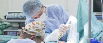
Surgical removal of condylomas
This method is used especially in case of extensive lesions or in case of relapses. The procedure is preceded by local anesthesia using EMLA cream or, rarely, performed under general anesthesia. An incision is made with scissors in the reticular layer of the skin. Thanks to the abundant blood supply, wounds heal quickly without leaving scars.
Sources
- Koutsky L.A., Kiviat N.B.: Genital human papillomavirus. 1999.
- Sonnex C. Infection due to human papillomavirus, with special reference to genital diseases. 1998.
- Bonnez V, Reichman RK: Papillomaviruses. 1990.
- Fazel N, et al: Clinical, histopathological and molecular aspects of cutaneous human papillomavirus infections. Dermatol.1999.
- Carr J, Györffy T: Human papillomavirus. Epidemiology, transmission and pathogenesis.2000.
- Shah K.V.: Human papillomaviruses. 1998.
- Sedlacek T.V.: Advances in the diagnosis and treatment of infections caused by the human papillomavirus. 1999.
- Müller U et al: Laser therapy for genital warts associated with human papillomavirus. 2001.
- Ferley J, et al: Cancer incidence, mortality and prevalence worldwide. IARC Oncology Base No. 5 version 2.0, IARC Press, Lyon 2004.
- Coatsie LA: Prophylactic quadrivalent human papillomavirus (HPV) (types 6, 11, 16, 18) L1 virus-like particle (VLP) vaccine (Gardasil). Congress of the Infectious Diseases Society of America, 2005
- Reed R: Physical and surgical principles of carbon dioxide laser skin surgery. 1999.
- Beutner KR, Ferenczy A.: Therapeutic approaches to genital warts. 1997.