It's all the virus's fault!
The term "stomatitis" is derived from the merger of two Greek words: stoma (mouth) and itis (inflammation). There are a great variety of different types of disease - serous, aphthous, allergic, etc. The most dangerous is herpetic, or cold sore, stomatitis caused by a virus. Its main manifestations are painful ulcers covering the oral mucosa. The trigger mechanism of the disease is the activation of herpes simplex virus types 1 and 2. First of all, the disease threatens those who have a weakened immune system and, as a result, the body simply does not have the strength to give a worthy rebuff to viruses.
Forms of the disease
Lightweight
It is considered the most beneficial for the body. In this form, people with high immunity suffer from herpes stomatitis. It flows without temperature. It is distinguished by single rashes that do not cause discomfort and disappear on their own without consequences.
Average
General disorders are added: weakness, drowsiness, fatigue, loss of appetite. Rashes appear in several places at the same time. The temperature rises to 37-37.6°C.
Heavy
This form of stomatitis indicates extremely low immunity. The rashes are multiple and painful. Severe headache, chills, and vomiting appear. The temperature exceeds 38oC.
If the disease is mild, the patient may not notice any external signs!
How to distinguish herpes from stomatitis?
Many patients try to find an answer on the Internet to the question: “Do I have stomatitis or herpes? How to recognize? Herpes stomatitis can be easily distinguished from ordinary stomatitis by 3 key signs.
- With herpes infection, the rash is localized in the gum area. Whereas with stomatitis - on the soft tissues of the oral cavity (tongue, cheeks).
- A herpes rash first appears as blisters, which then ulcerate, while stomatitis begins with the appearance of ulcers.
- Herpetic stomatitis is characterized by a stable appearance of the rash in the same places, and with ordinary stomatitis its location often changes.
Herpetic or aphthous?
It will be somewhat more difficult to distinguish between herpetic and aphthous stomatitis. The latter got its name from the Greek term “aftha”, which means “ulcer”.
If with herpetic stomatitis there are many ulcers, but they are small, then with aphthous stomatitis there are few of them, and the size can reach 7–8 mm.
The second important distinguishing feature is the absence of swelling of the gums with aphthous stomatitis.
If you are looking for differences between herpetic and aphthous stomatitis, then the third thing you should pay attention to is the localization of the rash. Aphthous is characterized by the appearance of ulcers in the oral cavity, while herpes infection can spread to the border of the lips.
Symptoms of inflammatory diseases
Inflammation in the oral cavity is manifested by both general and local symptoms [1]. Even before the onset of local manifestations, general malaise, pain, irritation, rashes in the mouth, increased body temperature, and decreased appetite are noted [1]. In addition, inflammation in the mouth itself can act as a symptom of another disease or general pathology [1].
Symptoms of certain inflammatory diseases
Stomatitis is a wide group of diseases that can be caused by individual pathogens, for example, fungi (candidal stomatitis) or the herpes simplex virus (herpes stomatitis), and systemic diseases, for example, chronic diseases of the gastrointestinal tract (aphthous stomatitis) [2] .
Most often, stomatitis appears as rashes (ulcers, blisters or other forms) on the oral mucosa. The nature of the rash is a striking distinctive feature that plays an important role in diagnosis. For example, with aphthous stomatitis, the ulcers never appear on the outer surface of the lips, as is the case with lesions caused by the herpes simplex virus [2].
Herpetic stomatitis, as a rule, accompanies a general infection of the body and is characterized by rashes on the inner surface of the cheeks, tongue, palate, and lips [2].
With insufficient oral care, bacterial microflora may develop and cause deeper lesions of the mucous membrane.
If the necessary treatment of acute herpetic stomatitis is not carried out, a recurrent form occurs, which is accompanied by regular rashes on the oral mucosa of vesicles and aphthae "Microbiology, virology and immunology of the oral cavity", ed. Doctor of Medicine V. N. Tsareva.
Catarrhal stomatitis occurs quite often and develops due to the lack or poor hygiene and the presence of chronic foci of infection in the oral cavity. Symptoms of this type of stomatitis are swelling of the mucous membrane, the appearance of plaque on it, first white, then brown, and bad breath [1].
Candidiasis (candidal stomatitis, thrush) is caused by fungi of the genus Candida, which are normally always present in the mouth in small quantities [2]. Factors contributing to the sharp growth of fungi are long-term use of antibiotics and some other drugs, hypovitaminosis, endocrine and other disorders.
A striking symptom of candidiasis is the appearance of a white or yellowish-white coating on the tongue and the mucous membrane of the cheeks due to the development of inflammation in the mouth. The plaque is easily removed, revealing reddened, inflamed and eroded areas of the oral mucosa underneath.
Leukoplakia refers to chronic inflammatory diseases. It develops in response to constant irritation of the oral mucosa, for example, from a part of a denture, a sharp edge or chipped tooth, hot drinks or food, alcoholic beverages, or smoking [1]. Leukoplakia manifests itself in the form of whitish thickenings, usually in the cheek area along the line of closure of the teeth, in the corners of the mouth, on the back and on the lateral surfaces of the tongue.
How to distinguish herpes sore throat from stomatitis?
Despite the fact that these medical terms have the same grammatical root, herpetic sore throat and herpetic stomatitis are two different diseases. Unlike stomatitis, herpes sore throat occurs not due to the penetration of the herpes virus, but as a result of an adenovirus infection (in particular, the Coxsackie A virus). Children are more likely to suffer from this disease than adults. The rashes are localized, for the most part, on the soft palate and tonsils. Typical symptoms of stomatitis include pain in the abdomen and abnormal bowel movements.
Attention!
The disease begins acutely, with a jump in temperature to 40 degrees, and is severe, so differential diagnosis should only be carried out by a doctor.
Ulcers as local manifestations of general diseases on the oral mucosa
- Oral tuberculosis
is usually a secondary manifestation of pulmonary tuberculosis. It occurs as a result of penetration of tuberculosis bacteria into the oral mucosa through damaged epithelium. The membranes of the cheeks, tongue, and floor of the mouth are affected. First, typical tuberculous tubercles are formed, and then, after their disintegration, small ulcers are formed, which increase in size over time. The ulcer itself is not deep, with a loose bottom, which is covered with easily bleeding granulations (young tissue), uneven edges are observed, soft to the touch. With this disease, there is a sharp pain in the ulcer. In addition to the local manifestation in the form of an ulcer, there is a general deterioration in the well-being of patients: emaciation is noted, the amount of plaque on the tongue increases, sweating and body temperature increase. General treatment of tuberculosis of the oral mucosa is carried out in specialized anti-tuberculosis institutions. As for local treatment, in this case, sanitation of the oral cavity is carried out during the period of remission (weakening of the disease), treatment of the mucous membrane with antiseptic and anti-inflammatory agents. - Syphilis
is a chronic infectious disease caused by the so-called Treponema pallidum. All periods of development of this disease (in addition to the incubation period, which lasts 21 - 24 days) are characterized by the presence of ulcers in the oral cavity. At the initial stage of development of the disease, the presence of a painless ulcer is observed, which has a round or oval shape with raised, smooth edges and a cartilage-like specific infiltrate. The bottom of the ulcer is bright red, shiny or covered with a grayish-dark coating. The ulcers heal in 3 to 12 weeks with or without scar formation. Even with tertiary syphilis, there is no sharp pain, as, for example, with a tuberculous ulcer. The ulcer is surrounded by a powerful infiltrate, which is a dense bluish-red ridge that rises above the level of the mucosa. Its edges are smooth, bright red, covered with granulations, and bleed easily. After the ulcer heals, a retracted star-shaped scar forms. This process lasts 3 - 4 months. After the ulcers heal, scars remain, which are a sign of previous syphilis. General treatment of syphilis is carried out in a venereology hospital, local treatment is carried out during the period of remission or recovery (sanitation of the oral cavity, elimination of local traumatic factors). - Acute necrotizing gingivostomatitis
is a viral infectious disease. Most often, ulcers are localized on the mucous membranes of the gums, cheeks, soft palate, arches and tonsils. Favorable factors for the development of this disease are a decrease in the overall resistance of the body, a violation of the integrity of the oral mucosa, and a deficiency of vitamins in the body. There are also cases of this disease occurring against the background of cooling or overwork. It can also be a complication of viral infections, as well as allergic stomatitis. Usually young people (under 30 years of age) are affected, more often men.
Among the symptoms it is necessary to highlight: pain when eating; extremely unpleasant odor from the mouth; increased salivation; elevated body temperature. The gum mucosa becomes swollen, painful, and bleeds when touched. The epithelium of the gingival margin and gingival papillae becomes cloudy. The surface of the gingival margin is covered with a grayish-yellow coating, which is easily removed. The ulcers have soft, uneven edges and are covered with a dirty green coating (with a foul odor), which is easily removed. In this case, a loose bleeding bottom is detected. The surrounding tissues are swollen.
Treatment of necrotizing gingivostomatitis is carried out in accordance with the general condition of the body, taking into account its location and severity of the lesion. In moderate and severe stages of the disease, broad-spectrum antibiotics are prescribed, as well as drugs that prevent or reduce the manifestation of allergies. At any stage of development of the disease, vitamins C and P, high-calorie foods, juices are prescribed, and in some cases, according to indications, taking cardiac medications.
Local treatment is carried out under anesthesia (removal of necrotic tissue). The oral cavity is treated with warm solutions of antiseptics and anti-inflammatory drugs. The ulcer is also sprinkled with white clay powder. After acute inflammation has been relieved, professional oral hygiene is carried out.
Mouth ulcers can also form due to HIV (gum ulceration occurs in approximately 30% of people infected with HIV). How to cure mouth ulcers in this case? Treatment is specific and carried out by infectious disease doctors. Dental care is provided in all dental institutions with careful adherence to safety precautions.
Prevention of all of the above oral diseases consists of eliminating the causes of their occurrence. For example, to prevent infectious diseases that manifest themselves on the oral mucosa, such as syphilis, measures are necessary that prevent infection from entering the body. In other cases, wellness measures are very important, which include systematic independent and professional oral hygiene, which is offered by almost all dental clinics in Moscow.
Bacterial or viral?
In addition to viral origin, the disease can be caused by bacteria: streptococci and staphylococci are normally present in the microflora of the oral cavity and begin to multiply uncontrollably during the inflammatory process. The latter may be caused by caries or periodontitis (read more about the disease in the article).
How to distinguish viral stomatitis from bacterial one? It is extremely difficult to do this at home, so it is better to consult a doctor. The main differential feature is the localization of the rash. With viral herpetic stomatitis, vesicles with transparent contents first appear on the tongue (its tip, along the side surfaces and under it), and then can even spread to the pharynx and tonsils.
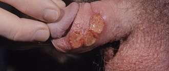
For bacterial stomatitis, the location of the rash on the gums and those areas where the skin borders the mucous membrane (for example, on the red border of the lips) is more common. Also, with a disease caused by streptococci, “jams” are often observed - pustules on the corners of the mouth, which quickly begin to bleed, become covered with a crust, crack and cause constant discomfort while eating and talking.
Types of diagnostics
An experienced doctor can identify herpetic stomatitis in adults during an initial examination, relying on only two methods.
- Clinical picture.
Based on the totality of the patient’s specific complaints and distinctive external signs, the dentist will not only assess the severity of the disease, but also differentiate it from ordinary stomatitis, candidiasis, etc. - Immunofluorescence.
Express microscopy method, the most accurate for diagnosing acute herpetic stomatitis.
The occurrence of ulcers due to diseases/damage to the soft tissues of the mouth and tongue
1. Recurrent
aphthous stomatitis is a chronic inflammatory disease that is characterized by periodic eruptions of aphthae (small ulcerations) on the oral mucosa. Aphthae can be localized on the tongue, buccal mucosa, hard and soft palate, and also on the mucous membrane of the lips. They are painful. In some cases, with constant injury, aphtha can turn into a long-term non-healing ulcer, after epithelization of which a scar is formed.
Patients with recurrent aphthous stomatitis usually suffer from colitis. Nervous strain, minor injuries to the mucous membrane, for example, when brushing teeth, as well as menstruation, can also predispose to the onset of the disease. The aphtha heals within 7 - 10 days. In case of complications of the disease, the number of ulcerations may increase, then the healing period is extended by 2 - 4 weeks.
2. Herpetiform stomatitis
characterized by the appearance of numerous small ulcers. In appearance they resemble the sores of herpes simplex. As a rule, they occur in women under 30 years of age. They are localized mainly on the lower surface of the tongue and in the area of the floor of the mouth. They do not have clear boundaries, the base is gray. The healing process is completed largely without scarring within 7 to 10 days. In simple forms of stomatitis, white ulcers in the mouth are observed (or rather, the center of the ulcer is covered with a thin, loosely fitting white or grayish film). Also, a white ulcer in the mouth can form in children in cases where there is candidal, or fungal, stomatitis.
As for the treatment of stomatitis, in this case the form of the disease and the severity of the lesion are taken into account. General and local treatment depends on this. But for almost all forms of stomatitis, vitamin C is prescribed to improve immunity.
3. Recurrent necrotizing peryadenitis
(Setton's aphthae) is characterized by the formation of a compaction in the submucosa, then painful ulcers with raised and compacted edges form in this place, as well as the presence of an inflammatory infiltrate (accumulation of cellular elements mixed with blood and lymph). Ulcers are localized on the upper and lower lips, cheeks, and lateral surfaces of the tongue. Eating food becomes extremely difficult for many patients, even to the point of giving it up. The same severe pain can be observed when talking. The ulcers do not heal until several months, and the disease lasts for years.
4. Afty Bednar
occur exclusively in children and are defined as traumatic erosions (ulcerations). The cause is poor oral hygiene or rough mechanical rubbing of the mucous membrane of the palate (this is where they are located). Covered with a whitish-yellow coating.
5. Traumatic ulcer
in the mouth is most often the result of physical impact, hence its name. As a rule, such an ulcer occurs as a result of an accidental or intentional bite of the mucous membrane, damage by a toothbrush. The presence of a traumatic ulcer in the oral cavity can provoke dental treatment (with careless use of dental instruments or, for example, with pointed temporary crowns).
Also among traumatic ulcers are the so-called prosthetic ulcers, which arise from exposure to complete or partial removable dentures, the dimensions of which are larger than necessary, or their surface is poorly processed and has sharp edges. Such ulcers can be located directly under the prosthetic structure. As a rule, healing takes place within 10 - 14 days, provided that the traumatic factor is eliminated. Treatment may not be necessary, since traumatic ulcers are often painless and small in size. In the case of the opposite, antimicrobial and anti-inflammatory drugs are prescribed, and the use of an ultrasonic brush is recommended, which not only cleans, but also has an antibacterial effect.
Also, the presence of ulcers can be caused by the effects of alkalis, acids, and certain medications on the oral mucosa.
Causes of herpes stomatitis
There are two types of herpetic stomatitis: acute and chronic. Acute herpetic stomatitis, according to Dr. Komarovsky, occurs only in children under 3 years of age, when for the first time the child’s body, already deprived of antibodies to the herpes virus received from the mother, is first exposed to a viral attack from the outside. Moreover, the source of infection, as a rule, is the parents themselves - carriers of the virus, who kiss the baby or lick his pacifier or feeding spoon.
Recurrent or chronic stomatitis is already the lot of adults. The disease is recurrent in nature as soon as the body’s immune forces are weakened. In this case, primary infection can occur either through airborne droplets (sneezing), or through household contact (for example, through the use of the same dishes with a virus carrier) or hematogenous (through blood during injections, etc.). The incubation period of the disease can last up to two weeks depending on the state of the immune system.
Attention!
The pathogenesis of the disease in dentistry is still unknown. But if the body is weakened, any injury to the palate or gums can provoke activation of the herpes virus types 1 and 2!
Causes of candidiasis
Thrush in the mouth of an adult is a lesion of the mucous membrane, which may indicate serious health problems. For the fungus to multiply, special conditions are required. Most often, the disease occurs in patients who neglect oral hygiene. The presence of caries and inflammatory gum diseases increases the chances of developing fungal inflammation. This is explained by the fact that a large number of pathogenic microorganisms depletes the defense mechanisms.
The second group of reasons is weakened immunity due to a number of diseases and conditions:
- HIV, diabetes mellitus;
- oncological diseases;
- dystrophy, deficiency of vitamins and minerals;
- previous operations, severe infections, etc.
There are also specific reasons for the development of thrush. It can appear after prolonged and powerful antibacterial therapy. The use of antibiotics leads to the destruction of beneficial flora and imbalance. This causes active reproduction of Candida.
Oral candidiasis also develops while taking inhaled corticosteroids. Usually the lesion has the appearance of erythema and appears in areas where the medicine came into contact with the mucous membrane: on the palate, tongue.
Dietary features affect the likelihood of developing candidiasis. Thus, the predominance of carbohydrates predisposes to fungal activity. The growth of Candida and its attachment to the mucosa are enhanced in the presence of sugars.
Bad habits increase the chances of developing leukoplakia, lichen planus and other diseases. This is especially true when it comes to smoking. Candidiasis often develops in patients with tongue piercings.
The presence of removable dentures is also a risk factor if the patient does not comply with hygiene rules. In the absence of high-quality cleansing, the prosthesis becomes covered with a biofilm, which contains a lot of fungi. Disinfection is the main measure of disease prevention and part of complex treatment for progressive oral candidiasis. If the patient does not remove the structure at night, this also increases the likelihood of developing the disease. The mucous membrane remains without oxygen for a long time and is not washed by saliva - these conditions are suitable for the development of fungi and anaerobic microorganisms. The prosthesis can injure the mucous membranes if it does not fit. Microtraumas weaken local defenses and contribute to the onset of fungal infection. Injuries can also be associated with sharp chips of teeth and fillings, chemical and thermal burns.
Dry mouth due to decreased salivation, changes in saliva viscosity and its composition is one of the causes of candidiasis. This can be caused by other diseases, so it is important to find out the causes of dryness in order to effectively combat the consequences.
Candidiasis in the mouth of a child is more common. The immaturity of the immune system and the colonization of the oral cavity by Candida from the mother’s vaginal canal during natural childbirth lead to the development of the disease in early infancy. However, older children can also suffer from the disease, which is associated with weakening immune forces.
Ask a Question
Treatment of herpetic stomatitis
How to treat herpes stomatitis? Unfortunately, the herpes virus, once entered into a person’s blood, remains in a “dormant” state for the rest of his life. But treatment of viral stomatitis is possible provided that you do not delay visiting the dentist when the first signs of the disease are detected. During the period of therapy, in order to avoid infecting loved ones, you should eat and drink from separate containers and avoid kissing. The patient is recommended a diet that excludes spicy, smoked, sour and salty foods, which can irritate the damaged oral mucosa, and an increased drinking regimen (up to 2.5 liters per day), aimed at combating the manifestations of general intoxication of the body.
Ulcerative stomatitis - symptoms and treatment
The main goal of therapy is to reduce pain and speed up healing. First of all, it is necessary to eliminate irritants that cause discomfort to the patient. The next step is to reduce symptoms as much as possible.
To treat stomatitis, rinsing with solutions of furatsilin, miramistin, chlorhexidine, and hydrogen peroxide is prescribed. Rinsing will reduce pain and itching, and cleanse the oral cavity of food debris that injures the damaged areas.
In case of severe pain, applications with painkillers are prescribed - “Kamistad”, “Lidocaine Asept”. Treatment for different forms of stomatitis will differ.
Infectious stomatitis. When treating, it is first necessary to understand what problem with the immune system caused this condition and to strengthen the body’s defenses. To do this, Prodigiosan is administered intramuscularly, the oral cavity is treated with antiseptics, proteolytic enzymes, and UV therapy is prescribed [11].
Vesicular stomatitis. To reduce symptoms, proper rest, drinking plenty of fluids, and taking antipyretic medications will be helpful. Also, the oral mucosa is treated with antiseptics (Suprastin, Hexetidine, Pipolfen) and antiviral ointments are used - rhiodoxol and tebrofen. Antiherpetic drugs are often prescribed - Famciclovir, Acyclovir, Valacyclovir. If you follow the doctor’s recommendations, the symptoms of the disease quickly go away and the patient recovers.
Vincent's ulcerative necrotic stomatitis. During treatment, dental plaque is carefully removed, the oral mucosa is treated with antiseptics, and multivitamins are prescribed (for example, Complivita).
Allergic stomatitis. Treatment consists of eliminating the cause of the allergy and taking oral antihistamines. In severe cases of allergic stomatitis, sodium thiosulfate solution is administered intravenously. During treatment in a hospital, drip infusions of hemodez, isotonic sodium chloride solution, polyglucin, and corticosteroids are prescribed.
Chronic recurrent aphthous stomatitis. With local treatment, traumatic factors are eliminated, the mouth is rinsed with tetracycline (250 mg per 5 ml of water 4 times a day for 5-7 days), applications are made with corticosteroids and antibiotics, and painkillers are prescribed. For deep ulcers, proteolytic enzymes are used.
General treatment includes taking medications:
- tetracycline, rifampicin (two capsules twice a day);
- “Tarivid” (one tablet twice a day for 20 days);
- sodium thiosulfate (10 ml of 30% solution intravenously once a day or 1.5-3 g orally);
- “Prodigiosan” (start with 15 mcg once every five days and increase the dose to 100 mcg);
- "Levamisole" (50 mg three times a day two days in a row a week or 150 mg once);
- "Delagil" (one tablet once a day);
- "Colchicine" (one tablet twice a day for two months);
- "Aevit" (1 ml once a day intramuscularly for 20 days);
- “Histaglobulin” (2 ml subcutaneously once every three days) [11].
Stomatitis due to intoxication with salts of heavy metals and chemical solutions. If a chemical gets on the mucous membrane, you must immediately wash it off with a neutralizing solution. Further treatment of patients with chemical burns is carried out with painkillers and antiseptics and drugs that accelerate the restoration of the epithelium. A high-calorie diet is also important. For extensive scars, surgical intervention is indicated [15].
Drug therapy
Rinse
To stop the spread of infection throughout the oral cavity, as well as to prevent the occurrence of sore throat, anti-inflammatory drugs such as Stomatidine and Miramistin are prescribed. The greatest effect is achieved by repeating the rinsing procedure every 3 hours, strictly adhering to the instructions for medicinal solutions.
Antiviral drugs
To suppress the reproduction of the herpes virus, the patient is advised to take “Immudon” and “Acyclovir” orally according to the scheme, and for external use - “Viferon” ointment or “Silicea” gel.
Vitamin therapy
Tablet vitamin complexes such as “Complivit” help improve immunity.
Attention!
Before use, consultation with a specialist is recommended!
Treatment at home with folk remedies
On the Internet you can often come across the question: “How to treat herpetic stomatitis with folk remedies?” Let’s make a reservation right away: when treating this disease, you cannot rely solely on traditional medicine methods. Therapy must be comprehensive, and only a doctor can choose it correctly.
An effective folk remedy for rinsing the mouth is propolis tincture diluted with boiled water 1:3. In case of pronounced “jams” in the corners of the mouth and painful ulcers inside, applications with natural sea buckthorn oil have an analgesic and wound-healing effect.
Features of treatment
The basis of treatment is systemic and local antifungal drugs. Today they are widely represented on the pharmaceutical market, but it is important to know that every year the level of resistance of Candida fungi to basic drugs increases. For example, resistance to drugs such as Fluconazole is almost complete. Previously, this remedy was used in almost all cases of the disease, but today doctors are forced to reconsider standard treatment regimens.
Treatment of oral thrush in adults is selected individually. The choice of systemic antifungal agent is based on the type of pathogen, the patient’s condition and the individual characteristics of his health. There are drugs to which the infection has minimal resistance. The doctor may prescribe drugs based on nystatin, imidazole derivatives, etc.
Additionally, local remedies must be used:
- solutions for rinsing the mouth;
- gels and suspensions for application to affected areas;
- local tablets and lozenges;
- Irrigation solutions and aerosols;
- ointments for placing in the oral cavity on a cotton-gauze swab, etc.
A specialist may prescribe pharmaceutical antiseptics or weak saline solutions for rinsing. Typically, solutions based on iodine, chlorhexidine, potassium permanganate, gentian violet, sodium tetraborate in glycerin are used. Your doctor may recommend placing some tablets in your cheek.
Prevention measures
- Strengthening the immune system.
Avoid excessive exercise and stress. Take a multivitamin in the fall and spring. Regular exercise and hardening increase the vitality of the body. - Healthy lifestyle.
Get rid of bad habits: scientists have proven that excessive smoking and alcoholic beverages can become a catalyst for the activation of the herpes virus in the body. - Timely treatment of chronic diseases.
Do not forget that simple stomatitis, if you do not seek medical help in a timely manner, can develop into herpetic stomatitis. Chronic caries and acute respiratory viral diseases suffered “on the legs” also seriously undermine the body’s immune defense. - Maintain personal hygiene.
According to statistics, it is poor oral hygiene that most often opens the way for herpes infection. - Avoid oral trauma.
Even a minor microcrack from a prick with a fish bone or careless use of a toothpick can become an “entry gate” for the herpes virus.
Attention!
With reduced immunity, herpes stomatitis becomes chronic and relapsing: the disease can recur 2-6 months after recovery.
Possible reasons
- Mycoses. Most often, red dots in the mouth are symptoms of oral candidiasis. Fungi of the genus Candida are considered opportunistic. They live in the intestines and do not cause harm to the body. However, when they enter the oral cavity, they quickly multiply and provoke inflammation of the mucous membrane. Spots of different shades of red can form on the gums, palate, cheeks, tongue, and tonsils.
- Bacterial infections. One of the main pathogens is streptococci. They change the structure of the mucous membrane and lead to inflammation of the soft tissues. They provoke the development of pharyngitis and tonsillitis.
- Viral diseases. Acute respiratory viral infections can also be accompanied by the formation of red spots on the oral mucosa. Pathogens enter the oral cavity through the nose. The gums and palate become inflamed, local hyperemia appears. If the spots become covered with a white coating, we can talk about a fungal or bacterial infection.
- Neoplasms. Suspicious spots on the gums may turn out to be benign tumors. The most common include papillomas and granulomas. Also, the risk of malignant neoplasms cannot be excluded.